往期刊物2021

卷册: 11, 期号: 15

left

Dec

20

Dec

5

Nov

20

Nov

5

Oct

20

Oct

5

Sep

20

Sep

5

Aug

20

Aug

5

Jul

20

Jul

5

Jun

20

Jun

5

May

20

May

5

Apr

20

Apr

5

Mar

20

Mar

5

Feb

20

Feb

5

Jan

20

Jan

5

right

生物化学

利用Ribo Mega-SEC高效快速分析细胞和组织中的多聚体和核糖体亚基

Efficient and Rapid Analysis of Polysomes and Ribosomal Subunits in Cells and Tissues Using Ribo Mega-SEC

利用Ribo Mega-SEC高效快速分析细胞和组织中的多聚体和核糖体亚基

HY Harunori Yoshikawa
RS Ramasubramanian Sundaramoorthy
DM Daniel Mariyappa
HJ Hao Jiang
AL Angus I. Lamond
5104 Views
Aug 5, 2021
Polysome profile analysis is a popular method for separating polysomes and ribosomal subunits and is typically achieved using a sucrose density gradient (SDG). This has remained the gold standard method since ribosomes were first discovered; however, this method is time-consuming and requires multiple steps from making the gradient and long ultracentrifugation to collecting and analyzing the fractions. Each of these steps in the SDG workflow can introduce potential technical variation that affects the reproducibility of gradient profiles between samples. To address these limitations, we have developed a flexible, alternative approach for analyzing polysomes and ribosomal subunits based on size-exclusion chromatography (SEC), termed ‘Ribo Mega-SEC.’ In comparison with the SDG method, Ribo Mega-SEC involves a single step using ultra-high-performance liquid chromatography (uHPLC). The entire workflow, from injecting the lysate to collecting the fractions, can be performed in as little as 15 min, with high reproducibility. By varying the pore size of the SEC column, polysomes and ribosomal subunits can be separated using extracts from either human or mouse cultured cell lines or from tissue samples, Drosophila embryos, or budding yeast. The resulting separated fractions are suitable for analysis using a wide range of subsequent analytical techniques including mass spectrometry (MS)-based proteomics, RNA-Seq, electron microscopy (EM), and multiple biochemical assays.
用转位酶Oxa1L纯化HEK细胞线粒体核糖体

Purification of Mitochondrial Ribosomes with the Translocase Oxa1L from HEK Cells

用转位酶Oxa1L纯化HEK细胞线粒体核糖体

HY Hanting Yang
ND Nirupa Desai
4102 Views
Aug 5, 2021
Mitochondrial ribosomes (mitoribosomes) perform protein synthesis inside mitochondria, the organelles responsible for energy conversion and adenosine triphosphate (ATP) production in eukaryotic cells. To investigate their functions and structures, large-scale purification of intact mitoribosomes from mitochondria-rich animal tissues or HEK cells have been developed. However, the fast purification of mitoribosomes anchored to the mitochondrial inner membrane in complex with the Oxa1L translocase remains particularly challenging. Herein, we present a protocol recently developed and modified in our lab that provides details for the efficient isolation of intact mitoribosomes with its translocase Oxa1L. We combined the cell culture of PDE12-/- or wild-type HEK293 cell lines with the isolation of mitochondria and the purification steps used for the biochemical and structural studies of mitoribosomes and Oxa1L.Graphic abstract:Schematic procedure for the purification of mitoribosomes from HEK cells. The protocol described herein includes two main sections: 1) isolation of mitochondria from HEK cells; and 2) purification of mitoribosome-Oxa1L from mitochondria. RB: Resuspension Buffer (see Recipes) (Created with BioRender.com).
HEK293细胞表达的重组野生型和突变型Ryanodine受体的纯化

Purification of Recombinant Wild Type and Mutant Ryanodine Receptors Expressed in HEK293 Cells

HEK293细胞表达的重组野生型和突变型Ryanodine受体的纯化

YH Yifan Hu
KI Kavita A. Iyer
AN Ashok R. Nayak
NK Nagomi Kurebayashi
TM Takashi Murayama
MS Montserrat Samsó
2932 Views
Aug 5, 2021
High quantities of purified ryanodine receptor (RyR), a large (2.26 MDa) intracellular homotetrameric membrane protein, can be obtained from heterologous expression in HEK293 cells and used for structure determination by cryo-EM. The advantage of using recombinant protein is that the variability due to post-translational modifications can be minimized, to which the high resolution of up to 2.4 Å achieved for RyR2 can be attributed (Iyer et al., 2020). In addition, recombinant protein expression enables the study of mutations that are deleterious when expressed homozygously in animals. Protein purification was achieved using two strategies, sucrose density gradient and affinity chromatography, which have previously been used for purification of RyR from tissue. The sucrose gradient method was developed from (Lee et al., 1994) and later adapted for cryo-EM (Samsó et al., 2005). The affinity chromatography method takes advantage of the high affinity of RyR for its ligand FKBP12/12.6, by using a construct between FKBP and streptavidin binding protein (SBP) (Cabra et al., 2016). While the sucrose gradient method can yield a higher protein concentration (≥ 2 mg/ml), the affinity purification method is faster. Both methods are suitable and applicable to the purification of recombinant proteins and were successfully used in the first 3D near-atomic reconstructions of RyRs purified from cells expressing disease mutants (Iyer et al., 2020). This purification protocol is also suitable for functional studies, such as single-channel analysis, that require pure RyR protein.

生物物理学

植物线粒体核糖体的纯化和低温电镜分析

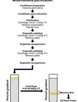
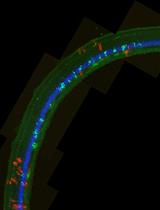
Purification and Cryo-electron Microscopy Analysis of Plant Mitochondrial Ribosomes

植物线粒体核糖体的纯化和低温电镜分析

FW Florent Waltz
PG Philippe Giegé
YH Yaser Hashem
3007 Views
Aug 5, 2021
Plants make up by far the largest part of biomass on Earth. They are the primary source of food and the basis of most drugs used for medicinal purposes. Similarly to all eukaryotes, plant cells also use mitochondria for energy production. Among mitochondrial gene expression processes, translation is the least understood; although, recent advances have revealed the specificities of its main component, the mitochondrial ribosome (mitoribosome). Here, we present a detailed protocol to extract highly pure cauliflower mitochondria by differential centrifugation for the purification of mitochondrial ribosomes using a sucrose gradient and the preparation of cryo-electron microscopy (cryo-EM) grids. Finally, the specific bioinformatics pipeline used for image acquisition, the processing steps, and the data analysis used for cryo-EM of the plant mitoribosome are described. This protocol will be used for further analysis of the critical steps of mitochondrial translation, such as its initiation and regulation.

细胞生物学

基于选择性表达基因的CRISPR介导的鸡胚胎细胞标记

CRISPR-mediated Labeling of Cells in Chick Embryos Based on Selectively Expressed Genes

基于选择性表达基因的CRISPR介导的鸡胚胎细胞标记

Masahito Yamagata Masahito Yamagata
JS Joshua R. Sanes
6769 Views
Aug 5, 2021
The abilities to mark and manipulate specific cell types are essential for an increasing number of functional, structural, molecular, and developmental analyses in model organisms. In a few species, this can be accomplished by germline transgenesis; in other species, other methods are needed to selectively label somatic cells based on the genes that they express. Here, we describe a method for CRISPR-based somatic integration of reporters or Cre recombinase into specific genes in the chick genome, followed by visualization of cells in the retina and midbrain. Loci are chosen based on an RNA-seq-based cell atlas. Reporters can be soluble to visualize the morphology of individual cells or appended to the encoded protein to assess subcellular localization. We call the method eCHIKIN for electroporation- and CRISPR-mediated Homology-instructed Knock-IN.
小鼠脑片黑质致密部多巴胺神经元的细胞贴附和全细胞膜片钳记录

Cell-attached and Whole-cell Patch-clamp Recordings of Dopamine Neurons in the Substantia Nigra Pars Compacta of Mouse Brain Slices

小鼠脑片黑质致密部多巴胺神经元的细胞贴附和全细胞膜片钳记录

SC Stefano Cattaneo
MR Maria Regoni
JS Jenny Sassone
ST Stefano Taverna
4338 Views
Aug 5, 2021
The Substantia Nigra pars compacta (SNc) is a midbrain dopaminergic nucleus that plays a key role in modulating motor and cognitive functions. It is crucially involved in several disorders, particularly Parkinson’s disease, which is characterized by a progressive loss of SNc dopaminergic cells. Electrophysiological studies on SNc neurons are of paramount importance to understand the role of dopaminergic transmission in health and disease. Here, we provide an extensive protocol to prepare SNc-containing mouse brain slices and record the electrical activity of dopaminergic cells. We describe all the necessary steps, including mouse transcardiac perfusion, brain extraction, slice cutting, and patch-clamp recordings.

发育生物学

小鼠卵母细胞的分离与体外培养

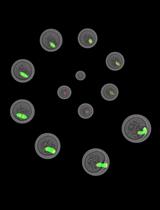
Isolation and in vitro Culture of Mouse Oocytes

小鼠卵母细胞的分离与体外培养

JG Jessica Greaney
GS Goutham Narayanan Subramanian
Yunan  Ye Yunan Ye
HH Hayden Homer
6054 Views
Aug 5, 2021
Females are endowed at birth with a fixed reserve of oocytes, which declines both in quantity and quality with advancing age. Understanding the molecular mechanisms regulating oocyte quality is crucial for improving the chances of pregnancy success in fertility clinics. In vitro culture systems enable researchers to analyse important molecular and genetic regulators of oocyte maturation and fertilisation. Here, we describe in detail a highly reproducible technique for the isolation and culture of fully grown mouse oocytes. We include the considerations and precautionary measures required for minimising the detrimental effects of in vitro culture conditions. This technique forms the starting point for a wide range of experimental approaches such as post-transcriptional gene silencing, immunocytochemistry, Western blotting, high-resolution 4D time-lapse imaging, and in vitro fertilization, which are instrumental in dissecting the molecular determinants of oocyte quality. Hence, this protocol serves as a useful, practical guide for any oocyte researcher beginning experiments aimed at investigating important oocyte molecular factors.Graphic abstract:A step-by-step protocol for the isolation and in vitro culture of oocytes from mice.
静脉畸形和视网膜血管瘤样血管畸形的基因工程小鼠模型

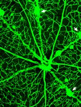
A Genetically Engineered Mouse Model of Venous Anomaly and Retinal Angioma-like Vascular Malformation

静脉畸形和视网膜血管瘤样血管畸形的基因工程小鼠模型

XC Xudong Cao
BX Beibei Xu
XL Xiao Li
TL Taotao Li
YH Yulong He
3456 Views
Aug 5, 2021
Characterization of key regulators in vein development will advance our understanding of mechanisms underlying venous anomalies and provide therapeutic targets for the treatment of vascular malformations. Here, we provide a detailed protocol for the generation of genetically engineered mouse models targeting the Tek gene for the analysis of vein formation and vein-associated vascular diseases at the embryonic and postnatal stages. It includes steps involved in the whole-mount processing of mouse skin, mesentery, and retina for the examination of vascular malformation during embryonic and postnatal development.

医学

基于游泳的测定成年斑马鱼心肌病模型运动能力的方法

A Swimming-based Assay to Determine the Exercise Capacity of Adult Zebrafish Cardiomyopathy Models

基于游泳的测定成年斑马鱼心肌病模型运动能力的方法

XM Xiao Ma
XX Xiaolei Xu
2279 Views
Aug 5, 2021
Exercise capacity, measured by treadmill in humans and other mammals, is an important diagnostic and prognostic index for patients with cardiomyopathy and heart failure. The adult zebrafish is increasingly used as a vertebrate model to study human cardiomyopathy due to its conserved cardiovascular physiology, convenience for genetic manipulation, and amenability to high-throughput genetic and compound screening. Owing to the small size of its body and heart, new phenotyping assays are needed to unveil phenotypic traits of cardiomyopathy in adult zebrafish. Here, we describe a swimming-based functional assay that measures exercise capacity in an adult zebrafish doxorubicin-induced cardiomyopathy model. This protocol can be applied to any adult zebrafish model of acquired or inherited cardiomyopathy and potentially to other cardiovascular diseases.Graphic abstract:Clinical relevance of the swimming-based phenotyping assay in adult zebrafish cardiomyopathy models.

微生物学

大肠杆菌表达的微生物紫红质离子转运活性测定

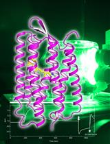
Ion Transport Activity Assay for Microbial Rhodopsin Expressed in Escherichia coli Cells

大肠杆菌表达的微生物紫红质离子转运活性测定

MK Masae Konno
KI Keiichi Inoue
HK Hideki Kandori
2481 Views
Aug 5, 2021
Microbial rhodopsins have diverse functions, including roles as light-driven ion pumps, light-gated ion channels, photosensors, and light-regulated enzymes. As the number of rhodopsin-like genes identified has increased in recent years, so has the requirement for rapid identification of their functions. The patch-clamp method is often used to investigate the ion transport mechanism of microbial rhodopsins in mammalian cells; however, this requires a dedicated system and advanced techniques. The ion transport assay using the Escherichia coli expression system described here evaluates the ion transport capacity by monitoring the pH change in E. coli suspensions; if the target rhodopsin has a light-dependent ion transport activity, a light-dependent pH change is observed. The pH increase or decrease corresponds to proton release from the cell or proton uptake into the cell, respectively. This method can be used to evaluate ion transport capacity in a high-throughput manner using a combination of general-purpose equipment and common techniques.Graphic abstract:Schematic diagram of the ion transport assay in rhodopsin-expressing E. coli cells.

分子生物学

小鼠背根神经节细胞核的分离及其单核基因组学研究

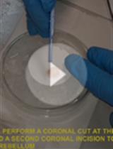
Isolation of Nuclei from Mouse Dorsal Root Ganglia for Single-nucleus Genomics

小鼠背根神经节细胞核的分离及其单核基因组学研究

LY Lite Yang
IT Ivan Tochitsky
CW Clifford J. Woolf
WR William Renthal
3953 Views
Aug 5, 2021
Primary somatosensory neurons, whose cell bodies reside in the dorsal root ganglion (DRG) and trigeminal ganglion, are specialized to transmit sensory information from the periphery to the central nervous system. Our molecular understanding of peripheral sensory neurons has been limited by both their heterogeneity and low abundance compared with non-neuronal cell types in sensory ganglia. We describe a protocol to isolate nuclei from mouse DRGs using iodixanol density gradient centrifugation, which enriches for neuronal nuclei while still sampling non-neuronal cells such as satellite glia and Schwann cells. This protocol is compatible with a range of downstream applications such as single-nucleus transcriptional and epigenomic assays.
Fe-NTA微柱纯化免疫沉淀(IP)洗脱物中磷酸肽的质谱分析

Fe-NTA Microcolumn Purification of Phosphopeptides from Immunoprecipitation (IP) Eluates for Mass Spectrometry Analysis

Fe-NTA微柱纯化免疫沉淀(IP)洗脱物中磷酸肽的质谱分析

ES Ethan J. Sanford
MS Marcus B. Smolka
2996 Views
Aug 5, 2021
Protein phosphorylation is a nearly universal signaling mechanism. To date, a number of proteomics tools have been developed to analyze phosphorylation. Phosphoproteome-wide analyses using whole cell extracts suffer from incomplete coverage, often missing phosphorylation events from low-abundance proteins. In order to increase coverage of phosphorylation sites on individual proteins of interest (“phospho-mapping”), immunoprecipitation (IP) followed by phosphoenrichment is necessary. Unfortunately, most commercially available phosphoenrichment kits are not readily scalable to the low-microgram quantities of protein present in IP eluates. Here, we describe a simple method specifically optimized for the enrichment of phosphopeptides from IP samples using an Fe-NTA based method. This method can be added downstream of any standard immunoprecipitation protocol and upstream of any MS analysis pipeline. The protocol described herein is cost effective, uses commonly available laboratory reagents, and can be used to obtain deep coverage of individual protein phosphorylation patterns, supplementary to phosphoproteomics data.Graphical abstract:Phospho-mapping workflow for a hypothetical protein of interest

神经科学

小鼠和人胰腺的光学清除和三维分析优化

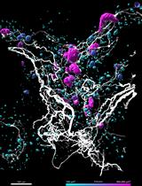
Optical Clearing and 3D Analysis Optimized for Mouse and Human Pancreata

小鼠和人胰腺的光学清除和三维分析优化

AA Alexandra Alvarsson
MJ Maria Jimenez-Gonzalez
RL Rosemary Li
CR Carolina Rosselot
NT Nikolaos Tzavaras
ZW Zhuhao Wu
SS Sarah A. Stanley
4447 Views
Aug 5, 2021
The pancreas is a heavily innervated organ, but pancreatic innervation can be challenging to comprehensively assess using conventional histological methods. However, recent advances in whole-mount tissue clearing and 3D rendering techniques have allowed detailed reconstructions of pancreatic innervation. Optical clearing is used to enhance tissue transparency and reduce light scattering, thus eliminating the need to section the tissue. Here, we describe a modified version of the optical tissue clearing protocol iDISCO+ (immunolabeling-enabled three-dimensional imaging of solvent-cleared organs) optimized for pancreatic innervation and endocrine markers. The protocol takes 13-19 days, depending on tissue size. In addition, we include protocols for imaging using light sheet and confocal microscopes and for 3D segmentation of pancreatic innervation and endocrine cells using Imaris.
小鼠的一个简单的空间独立的联想和逆转学习任务

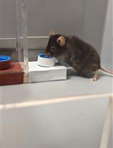
A Simple Spatial-independent Associative and Reversal Learning Task in Mice

小鼠的一个简单的空间独立的联想和逆转学习任务

FM Fulvio Magara
BB Benjamin Boury-Jamot
HH Hanna Hörnberg
2595 Views
Aug 5, 2021
The ability to adapt one's behavior in response to changing circumstances, or cognitive flexibility, is often altered in neuropsychiatric and neurodevelopmental conditions. In rodents, cognitive flexibility is frequently assessed using associative learning paradigms with a reversal component. The majority of existing protocols rely on unrestrictive exploration with no discouragement of wrong responses and are often influenced by spatial cues, at least during the test's learning phase. Here, we present a rewarded contingency discrimination learning test that minimizes the task's spatial component and contains an element that actively discourages pure exploratory responses. The method described herein is a manual version that can be performed using home-made equipment, but the test setup is amenable to automatization and can be adapted to address more complex cognitive demands, including conditional associative learning, attentional set formation, and attention shifting.

植物科学

利用本生烟叶片基因瞬时表达或沉默研究青枯菌毒力的快速简便方法

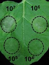
A Fast and Easy Method to Study Ralstonia solanacearum Virulence upon Transient Gene Expression or Gene Silencing in Nicotiana benthamiana Leaves

利用本生烟叶片基因瞬时表达或沉默研究青枯菌毒力的快速简便方法

WY Wenjia Yu
AM Alberto P. Macho
3734 Views
Aug 5, 2021
Ralstonia solanacearum is a devastating soil-borne bacterial pathogen that causes disease in multiple host plants worldwide. Typical assays to measure virulence of R. solanacearum in laboratory conditions rely on soil-drenching inoculation followed by observation and scoring of disease symptoms. Here, we describe a novel inoculation protocol to analyze the replication of R. solanacearum upon infiltration into the leaves of Nicotiana benthamiana, in which gene expression has been altered using Agrobacterium tumefaciens. The protocol includes five major steps: 1) growth of N. benthamiana plants; 2) infiltration of A. tumefaciens; 3) R. solanacearum inoculation; 4) sample collection and bacterial quantitation; 5) data analysis and representation. The transient gene expression or gene silencing prior to R. solanacearum inoculation provides a straightforward way to perform genetic analysis of plant functions involved in the interaction between pathogen and host, using the appropriate combination of A. tumefaciens and R. solanacearum strains, with high sensitivity and accuracy provided by the quantitation of bacterial numbers in plant tissues.

干细胞

小鼠骨膜细胞培养、体外分化及胫骨骨折的体内移植

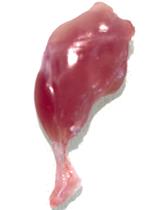
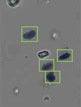
Mouse Periosteal Cell Culture, in vitro Differentiation, and in vivo Transplantation in Tibial Fractures

小鼠骨膜细胞培养、体外分化及胫骨骨折的体内移植

SP Simon Perrin
AJ Anais Julien
OD Oriane Duchamp de Lageneste
RA Rana Abou-Khalil
CC Céline Colnot
3588 Views
Aug 5, 2021
The periosteum covering the outer surface of bone contains skeletal stem/progenitor cells that can efficiently form cartilage and bone during bone repair. Several methods have been described to isolate periosteal cells based on bone scraping and/or enzymatic digestion. Here, we describe an explant culture method to isolate periosteum-derived stem/progenitor cells for subsequent in vitro and in vivo analyses. Periosteal cells (PCs) isolated using this protocol express mesenchymal markers, can be expanded in vitro, and exhibit high regenerative potential after in vivo transplantation at a fracture site, suggesting that this protocol can be employed for PC production to use in new cell-based therapies.