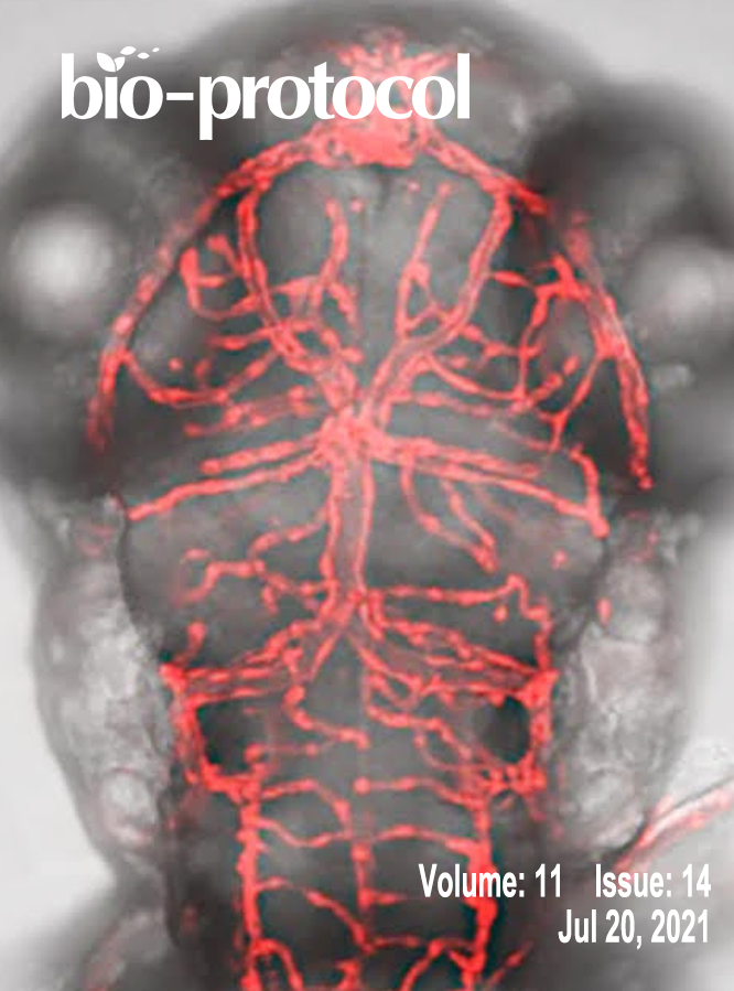
往期刊物2021

卷册: 11, 期号: 14

left

Dec

20

Dec

5

Nov

20

Nov

5

Oct

20

Oct

5

Sep

20

Sep

5

Aug

20

Aug

5

Jul

20

Jul

5

Jun

20

Jun

5

May

20

May

5

Apr

20

Apr

5

Mar

20

Mar

5

Feb

20

Feb

5

Jan

20

Jan

5

right

生物化学

一种基于凝胶的检测dsDNA蛋白易位的方法

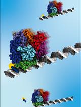
A Gel-Based Assay for Probing Protein Translocation on dsDNA

一种基于凝胶的检测dsDNA蛋白易位的方法

CB Christiane Brugger
AD Alexandra M. Deaconescu
3539 Views
Jul 20, 2021
Protein translocation on DNA represents the key biochemical activity of ssDNA translocases (aka helicases) and dsDNA translocases such as chromatin remodelers. Translocation depends on DNA binding but is a distinct process as it typically involves multiple DNA binding states, which are usually dependent on nucleotide binding/hydrolysis and are characterized by different affinities for the DNA. Several translocation assays have been described to distinguish between these two modes of action, simple binding as opposed to directional movement on dsDNA. Perhaps the most widely used is the triplex-forming oligonucleotide displacement assay. Traditionally, this assay relies on the formation of a DNA triplex from a dsDNA segment and a short radioactively labeled oligonucleotide. Upon translocation of the protein of interest along the DNA substrate, the third DNA strand is destabilized and eventually released off the DNA duplex. This process can be visualized and quantitated by polyacrylamide electrophoresis. Here, we present an effective, sensitive, and convenient variation of this assay that utilizes a fluorescently labeled oligonucleotide, eliminating the need to radioactively label DNA. In short, our protocol provides a safe and user-friendly alternative.Graphical abstract:Figure 1. Schematic of the triplex-forming oligonucleotide displacement assay.
一个GFP慢折叠突变体的自发和伴侣蛋白辅助的体外重折叠方案

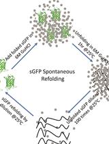
Protocol for Spontaneous and Chaperonin-assisted in vitro Refolding of a Slow-folding Mutant of GFP, sGFP

一个GFP慢折叠突变体的自发和伴侣蛋白辅助的体外重折叠方案

AS Anwar Sadat
ST Satyam Tiwari
KM Koyeli Mapa
2918 Views
Jul 20, 2021
Understanding the folding pathway of any protein is of utmost importance for deciphering the folding problems under adverse conditions. We can obtain important information about the folding pathway by monitoring the folding of any protein from its unfolded state. It is usually very difficult to monitor the folding process in real time as the process is generally very fast, and we need a suitable read out. In this protocol, we have solved this issue by using a protein that is non-fluorescent in its unfolded state but fluoresces in its native state after folding. The kinetics of refolding can be monitored by following the increase in fluorescence in real time. Previously, this was generally achieved by either monitoring a protein’s enzymatic activity or measuring the tryptophan fluorescence, where the signal output depends on well-described enzymatic activity or the frequency of tryptophan residues present in the proteins, respectively. Here, we describe a simple and real-time assay to monitor the refolding of sGFP, a recently described slow-folding mutant of yeGFP (yeast enhanced GFP). We unfold this protein using chemical denaturant and refold in a suitable buffer, monitoring the increase in fluorescence over time. GFP is fluorescent only when correctly folded; thus, using this technique, we can measure the true rate of protein refolding by following the increase in fluorescence over time. Therefore, sGFP can be used as an ideal model to study the in vitro protein folding process. Accordingly, the effects of different conditions and molecules on the protein folding pathway can be efficiently studied using sGFP as a model protein.Graphical abstract:Schematic of the steps involved in the sGFP refolding pathway. Native sGFP is unfolded by chemical denaturation using 6 M GuHCl at 25°C for 1 hour and then refolded in refolding buffer by 100-fold dilution.

生物物理学

一种多色双顺反子生物传感器比较活细胞中不同开放阅读框在单分子分辨率下的翻译动力学

A Multi-color Bicistronic Biosensor to Compare the Translation Dynamics of Different Open Reading Frames at Single-molecule Resolution in Live Cells

一种多色双顺反子生物传感器比较活细胞中不同开放阅读框在单分子分辨率下的翻译动力学

AK Amanda L. Koch
TM Tatsuya Morisaki
TS Timothy J. Stasevich
3065 Views
Jul 20, 2021
Here, we describe how to image and quantitate the translation dynamics of a bicistronic biosensor that we recently created to fairly compare cap-dependent and IRES-mediated translation at single-molecule resolution in live human cells. This technique employs a pair of complementary intrabodies loaded into living cells that co-translationally bind complementary epitopes in the two separate ORFs of the bicistronic biosensor. This causes the biosensor to fluoresce in different colors depending on which ORF/epitopes are translated. Using the biosensor together with high-resolution fluorescence microscopy and single-molecule tracking analysis allows for the quantitative comparison of translation dynamics between the two ORFs at a resolution of tens-of-nanometers in space and sub-seconds in time, which is not possible with more traditional GFP or luciferase reporters. Since both ORFs are on the same biosensor, they experience the same microenvironment, allowing a fair comparison of their relative translational activities. In this protocol, we describe how to get this assay up and running in cultured human cells so that translation dynamics can be studied under both normal and stressful cellular conditions. We also provide a number of useful tips and notes to help express components at appropriate levels inside cells for optimal live cell imaging.Graphical abstract:Steps required for 3-color single-molecule translation imaging and analysis.
用NAMD和PTools/Heligeom模拟蛋白质丝的微尺度和中尺度扰动

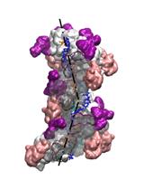
Modeling Perturbations in Protein Filaments at the Micro and Meso Scale Using NAMD and PTools/Heligeom

用NAMD和PTools/Heligeom模拟蛋白质丝的微尺度和中尺度扰动

BB Benjamin Boyer
BL Benoist Laurent
CR Charles H. Robert
CP Chantal Prévost
2921 Views
Jul 20, 2021
Protein filaments are dynamic entities that respond to external stimuli by slightly or substantially modifying the internal binding geometries between successive protomers. This results in overall changes in the filament architecture, which are difficult to model due to the helical character of the system. Here, we describe how distortions in RecA nucleofilaments and their consequences on the filament-DNA and bound DNA-DNA interactions at different stages of the homologous recombination process can be modeled using the PTools/Heligeom software and subsequent molecular dynamics simulation with NAMD. Modeling methods dealing with helical macromolecular objects typically rely on symmetric assemblies and take advantage of known symmetry descriptors. Other methods dealing with single objects, such as MMTK or VMD, do not integrate the specificities of regular assemblies. By basing the model building on binding geometries at the protomer-protomer level, PTools/Heligeom frees the building process from a priori knowledge of the system topology and enables irregular architectures and symmetry disruption to be accounted for.Graphical abstract:Model of ATP hydrolysis-induced distortions in the recombinant nucleoprotein, obtained by combining RecA-DNA and two RecA-RecA binding geometries.

癌症生物学

细胞内信号网络的非线性动力学建模

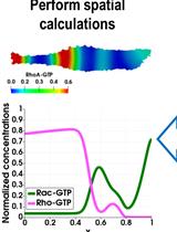
Modeling the Nonlinear Dynamics of Intracellular Signaling Networks

细胞内信号网络的非线性动力学建模

OR Oleksii S. Rukhlenko
BK Boris N. Kholodenko
3361 Views
Jul 20, 2021
This protocol illustrates a pipeline for modeling the nonlinear behavior of intracellular signaling pathways. At fixed spatial points, nonlinear signaling dynamics are described by ordinary differential equations (ODEs). At constant parameters, these ODEs may have multiple attractors, such as multiple steady states or limit cycles. Standard optimization procedures fine-tune the parameters for the system trajectories localized within the basin of attraction of only one attractor, usually a stable steady state. The suggested protocol samples the parameter space and captures the overall dynamic behavior by analyzing the number and stability of steady states and the shapes of the assembly of nullclines, which are determined as projections of quasi-steady-state trajectories into different 2D spaces of system variables. Our pipeline allows identifying main qualitative features of the model behavior, perform bifurcation analysis, and determine the borders separating the different dynamical regimes within the assembly of 2D parametric planes. Partial differential equation (PDE) systems describing the nonlinear spatiotemporal behavior are derived by coupling fixed point dynamics with species diffusion.

细胞生物学

用高选择性肽HK2pep分析己糖激酶2脱离线粒体相关膜的作用

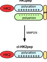
Analysis of the Effects of Hexokinase 2 Detachment From Mitochondria-Associated Membranes with the Highly Selective Peptide HK2pep

用高选择性肽HK2pep分析己糖激酶2脱离线粒体相关膜的作用

FC Francesco Ciscato
FC Federica Chiara
RF Riccardo Filadi
AR Andrea Rasola
3631 Views
Jul 20, 2021
The crucial role of hexokinase 2 (HK2) in the metabolic rewiring of tumors is now well established, which makes it a suitable target for the design of novel therapies. However, hexokinase activity is central to glucose utilization in all tissues; thus, enzymatic inhibition of HK2 can induce severe adverse effects. In an effort to find a selective anti-neoplastic strategy, we exploited an alternative approach based on HK2 detachment from its location on the outer mitochondrial membrane. We designed a HK2-targeting peptide named HK2pep, corresponding to the N-terminal hydrophobic domain of HK2 and armed with a metalloprotease cleavage sequence and a polycation stretch shielded by a polyanion sequence. In the tumor microenvironment, metalloproteases unleash polycations to allow selective plasma membrane permeation in neoplastic cells. HK2pep delivery induces the detachment of HK2 from mitochondria-associated membranes (MAMs) and mitochondrial Ca2+ overload caused by the opening of inositol-3-phosphate receptors on the endoplasmic reticulum (ER) and Ca2+ entry through the plasma membrane leading to Ca2+-mediated calpain activation and mitochondrial depolarization. As a result, HK2pep rapidly elicits death of diverse tumor cell types and dramatically reduces in vivo tumor mass. HK2pep does not affect hexokinase enzymatic activity, avoiding any noxious effect on non-transformed cells. Here, we make available a detailed protocol for the use of HK2pep and to investigate its biological effects, providing a comprehensive panel of assays to quantitate both HK2 enzymatic activity and changes in mitochondrial functions, Ca2+ flux, and cell viability elicited by HK2pep treatment of tumor cells.Graphical abstract:Flowchart for the analysis of the effects of HK2 detachment from MAMs.
用飞行时间(cyTOF flight)流式细胞术制备人软骨细胞用于分析研究

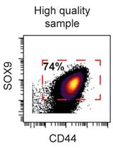
Preparation of Human Chondrocytes for Profiling Using Cytometry by Time-of-flight (cyTOF)

用飞行时间(cyTOF flight)流式细胞术制备人软骨细胞用于分析研究

FG Fiorella Carla Grandi
NB Nidhi Bhutani
3398 Views
Jul 20, 2021
Single-cell technologies have allowed high-resolution profiling of tissues and thus a deeper understanding of tissue homeostasis and disease heterogeneity. Understanding this heterogeneity can be especially important for tailoring treatments in a patient-specific manner. Here, we detail methods for preparing human cartilage tissue for profiling via cytometry by time-of-flight (cyTOF). We have previously utilized this method to characterize several rare cell populations in cartilage, including cartilage-progenitor cells, inflammation-amplifying cells (Inf-A), and inflammation-dampening cells (Inf-D). Previous bio-protocols have focused on cyTOF staining of PBMCs. Therefore, here we detail the steps unique to the processing of human cartilage and chondrocytes. Briefly, cartilage tissue is digested to release individual chondrocytes, which can be expanded and manipulated in culture. These cells are then collected and fixed in preparation for cyTOF, followed by standard staining and analysis protocols.

发育生物学

GeneWeld:斑马鱼短同源性诱导的高效靶向整合

GeneWeld: Efficient Targeted Integration Directed by Short Homology in Zebrafish

GeneWeld:斑马鱼短同源性诱导的高效靶向整合

JW Jordan M. Welker
WW Wesley A. Wierson
MA Maira P. Almeida
CM Carla M. Mann
MT Melanie E. Torrie
ZM Zhitao Ming
SE Stephen C. Ekker
KC Karl J. Clark
DD Drena L. Dobbs
JE Jeffrey J. Essner
MM Maura McGrail
10589 Views
Jul 20, 2021
Efficient precision genome engineering requires high frequency and specificity of integration at the genomic target site. Multiple design strategies for zebrafish gene targeting have previously been reported with widely varying frequencies for germline recovery of integration alleles. The GeneWeld protocol and pGTag (plasmids for Gene Tagging) vector series provide a set of resources to streamline precision gene targeting in zebrafish. Our approach uses short homology of 24-48 bp to drive targeted integration of DNA reporter cassettes by homology-mediated end joining (HMEJ) at a CRISPR/Cas induced DNA double-strand break. The pGTag vectors contain reporters flanked by a universal CRISPR sgRNA sequence to liberate the targeting cassette in vivo and expose homology arms for homology-driven integration. Germline transmission rates for precision-targeted integration alleles range 22-100%. Our system provides a streamlined, straightforward, and cost-effective approach for high-efficiency gene targeting applications in zebrafish.Graphic Abstract:GeneWeld method for CRISPR/Cas9 targeted integration.

免疫学

小胶质细胞的分离及蛋白表达的流式细胞仪分析:避免小胶质细胞背景荧光的陷阱

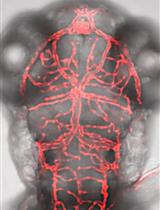
Isolation of Microglia and Analysis of Protein Expression by Flow Cytometry: Avoiding the Pitfall of Microglia Background Autofluorescence

小胶质细胞的分离及蛋白表达的流式细胞仪分析:避免小胶质细胞背景荧光的陷阱

JB Jeremy C. Burns
RR Richard M. Ransohoff
MM Michaël Mingueneau
6132 Views
Jul 20, 2021
Microglia are a unique type of tissue-resident innate immune cell found within the brain, spinal cord, and retina. In the healthy nervous system, their main functions are to defend the tissue against infectious microbes, support neuronal networks through synapse remodeling, and clear extracellular debris and dying cells through phagocytosis. Many existing microglia isolation protocols require the use of enzymatic tissue digestion or magnetic bead-based isolation steps, which increase both the time and cost of these procedures and introduce variability to the experiment. Here, we report a protocol to generate single-cell suspensions from freshly harvested murine brains or spinal cords, which efficiently dissociates tissue and removes myelin debris through simple mechanical dissociation and density centrifugation and can be applied to rat and non-human primate tissues. We further describe the importance of including empty channels in downstream flow cytometry analyses of microglia single-cell suspensions to accurately assess the expression of protein targets in this highly autofluorescent cell type. This methodology ensures that observed fluorescence signals are not incorrectly attributed to the protein target of interest by appropriately taking into account the unique autofluorescence of this cell type, a phenomenon already present in young animals and that increases with aging to levels that are comparable to those observed with antibodies against highly abundant antigens.

微生物学

耻垢分枝杆菌粗提物硝酸还原酶活性的体外测定

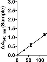
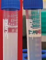
In vitro Nitrate Reductase Activity Assay of Mycolicibacterium smegmatis Crude Extract

耻垢分枝杆菌粗提物硝酸还原酶活性的体外测定

WT Wei Tan
ZS Zhi-Hui Shao
GZ Guo-Ping Zhao
2740 Views
Jul 20, 2021
Nitrate is one of the major inorganic nitrogen sources for microorganisms. Many bacterial and archaeal lineages can express cytoplasmic assimilatory nitrate reductase (NAS), which catalyzes the rate-limiting reduction of nitrate to nitrite in the nitrate assimilation pathway. Here, we present a detailed protocol for measuring in vitro nitrate reductase (NaR) activity of NAS enzymes from Mycolicibacterium smegmatis crude extract using both physiological and non-physiological electron donors.

分子生物学

用于下游分析的环状RNA的反义Oligo Pulldown

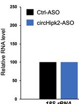
Antisense Oligo Pulldown of Circular RNA for Downstream Analysis

用于下游分析的环状RNA的反义Oligo Pulldown

DD Debojyoti Das
AD Aniruddha Das
AP Amaresh C. Panda
4252 Views
Jul 20, 2021
Circular RNAs (circRNAs) are a large family of noncoding RNA molecules that have emerged as novel regulators of gene expression by sequestering microRNAs (miRNAs) and RNA-binding proteins (RBPs). Several computational tools have been developed to predict circRNA interaction with target miRNAs and RBPs with a view to studying their potential effect on downstream target genes and cellular physiology. Biochemical assays, including reporter assays, AGO2 pulldown, ribonucleoprotein pulldown, and biotin-labeled RNA pulldown, are used to capture the association of miRNAs and RBPs with circRNAs. Only a few studies have used circRNA pulldown assays to capture the associated miRNAs and RBPs under physiological conditions. In this detailed protocol, the circRNA of interest (e.g., circHipk2) was captured using a biotin-labeled antisense oligo (ASO) targeting the circHipk2 backsplice junction sequence followed by pulldown with streptavidin-conjugated magnetic beads. The specific enrichment of circRNA was analyzed using reverse transcription quantitative PCR (RT-qPCR). Furthermore, the ASO pulldown assay can be coupled to miRNA RT-qPCR and western blotting analysis to confirm the association of miRNAs and RBPs predicted to interact with the target circRNA. In summary, the specific pulldown of circRNA using this quick and easy method makes it a useful tool for identifying and validating circRNA interaction with specific miRNAs and RBPs.
小胶质细胞的分离及蛋白表达的流式细胞仪分析:避免小胶质细胞背景荧光的陷阱

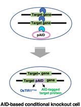
A Simple Method to Generate Super-sensitive AID (ssAID)-based Conditional Knockouts using CRISPR-based Gene Knockout in Various Vertebrate Cell Lines

小胶质细胞的分离及蛋白表达的流式细胞仪分析:避免小胶质细胞背景荧光的陷阱

KN Kohei Nishimura
TF Tatsuo Fukagawa
3423 Views
Jul 20, 2021
Inducing loss of function of a target protein using methods such as gene knockout is a powerful and useful strategy for analyzing protein function in cells. In recent years, the CRISPR/Cas-9-based gene knockout technology has been widely used across a variety of eukaryotes; however, this type of simple gene knockout strategy is not applicable to essential genes, which require a conditional knockout system. The auxin-inducible degron (AID) system enables rapid depletion of the target protein in an auxin-dependent manner and has been used to generate conditional mutants in various eukaryotic cell lines. One problem with the AID system is the use of high auxin concentrations for protein degradation, which can cause cytotoxicity. Recently, we established a super-sensitive AID (ssAID) system that allowed a reduction in the amount of auxin required by more than 1,000-fold. We also utilized a single-step method to generate AID-based conditional knockout cells with a ssAID system in various cell lines. In this protocol, we introduce our improved method, which provides a powerful tool for the investigation of the roles of essential genes.

神经科学

膜片钳移液管重复使用的快速酶清洗方法:通过消除膜片钳尝试之间的手动移液管更换来提高吞吐量

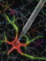
Method for Rapid Enzymatic Cleaning for Reuse of Patch Clamp Pipettes: Increasing Throughput by Eliminating Manual Pipette Replacement between Patch Clamp Attempts

膜片钳移液管重复使用的快速酶清洗方法:通过消除膜片钳尝试之间的手动移液管更换来提高吞吐量

CL Corey R. Landry
MY Mighten C. Yip
IK Ilya Kolb
WS William A. Stoy
MG Mercedes M. Gonzalez
CF Craig R. Forest
3512 Views
Jul 20, 2021
The whole-cell patch-clamp method is a gold standard for single-cell analysis of electrical activity, cellular morphology, and gene expression. Prior to our discovery that patch-clamp pipettes could be cleaned and reused, experimental throughput and automation were limited by the need to replace pipettes manually after each experiment. This article presents an optimized protocol for pipette cleaning, which enables it to be performed quickly (< 30 s), resulting in a high yield of whole-cell recording success rate (> 90%) for over 100 reuses of a single pipette. For most patch-clamp experiments (< 30 whole-cell recordings per day), this method enables a single pipette to be used for an entire day of experiments. In addition, we describe easily implementable hardware and software as well as troubleshooting tips to help other labs implement this method in their own experiments. Pipette cleaning enables patch-clamp experiments to be performed with higher throughput, whether manually or in an automated fashion, by eliminating the tedious and skillful task of replacing pipettes. From our experience with numerous electrophysiology laboratories, pipette cleaning can be integrated into existing patch-clamp setups in approximately one day using the hardware and software described in this article.Graphic abstract:Rapid enzymatic cleaning for reuse of patch-clamp pipettes

植物科学

拟南芥花粉管胞质pH的时空定量研究

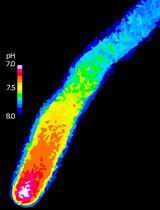
Spatiotemporal Quantification of Cytosolic pH in Arabidopsis Pollen Tubes

拟南芥花粉管胞质pH的时空定量研究

MP Maria Teresa Portes
DD Daniel S.C. Damineli
JF José A. Feijó
2935 Views
Jul 20, 2021
Ion-specific probes and fluorescent indicators have been key in establishing the role of ion signaling, namely calcium, protons, and anions, in plant development, providing a robust approach for monitoring spatiotemporal changes in intracellular ion dynamics. The integration of protons/pH in signaling mechanisms is especially important as reports of their biological functions continue to expand; however, attaining quantitative estimates with high spatiotemporal resolution in single cells poses a major research challenge. Here, we detail the use of the genetically encoded pH-sensitive pHluorin reporter expressed in Arabidopsis thaliana pollen tubes to assess cytosolic measurements with calibration to provide actual pH values. This technique enabled us to identify critical phenotypes and establish the importance of tip-focused pH gradient for pollen tube growth, although it can be adapted to other experimental systems.

更新

更正:用于活性氧生成和快速细胞蛋白质标准化的简单微孔板检测

Update Notice: A Simple Microplate Assay for Reactive Oxygen Species Generation and Rapid Cellular Protein Normalization

更正:用于活性氧生成和快速细胞蛋白质标准化的简单微孔板检测

NN Neville S. Ng
LO Lezanne Ooi
963 Views
Jul 20, 2021