往期刊物2021

卷册: 11, 期号: 12

left

Dec

20

Dec

5

Nov

20

Nov

5

Oct

20

Oct

5

Sep

20

Sep

5

Aug

20

Aug

5

Jul

20

Jul

5

Jun

20

Jun

5

May

20

May

5

Apr

20

Apr

5

Mar

20

Mar

5

Feb

20

Feb

5

Jan

20

Jan

5

right

生物化学

从人细胞中分离的线粒体蛋白质输入测定

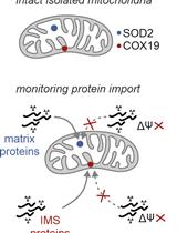
Protein Import Assay into Mitochondria Isolated from Human Cells

从人细胞中分离的线粒体蛋白质输入测定

LM Lena M. Murschall
EP Esra Peker
TM Thomas MacVicar
TL Thomas Langer
JR Jan Riemer
5142 Views
Jun 20, 2021
Mitochondria are essential organelles containing approximately 1,500 proteins. Only approximately 1% of these proteins are synthesized inside mitochondria, whereas the remaining 99% are synthesized as precursors on cytosolic ribosomes and imported into the organelle. Various tools and techniques to analyze the import process have been developed. Among them, in vitro reconstituted import systems are of importance to study these processes in detail. These experiments monitor the import reaction of mitochondrial precursors that were previously radiolabeled in a cell-free environment. However, the methods described have been mostly performed in mitochondria isolated from S. cerevisiae. Here, we describe the adaptation of this powerful assay to import proteins into crude mitochondria isolated from human tissue culture cells.Graphic abstract:Overview of the assay to monitor protein import into mitochondria isolated from human cells
用于内皮细胞分析小鼠胚胎脑血管片段富集

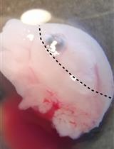
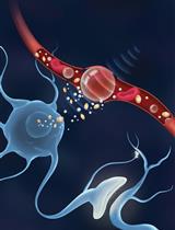
Enrichment of Vascular Fragments from Mouse Embryonic Brains for Endothelial Cell Analysis

用于内皮细胞分析小鼠胚胎脑血管片段富集

NS Nicolas Santander
TA Thomas D. Arnold
2668 Views
Jun 20, 2021
Endothelial cells in the brain interact with other cell types, forming the blood-brain barrier. This barrier controls the movement of solutes into and out of the brain, regulating pathophysiological processes and drug delivery to the brain. Common isolation methods used to study these cells during embryonic development involve enzymatic treatment and cell sorting using specific markers. This process modifies the cell state and produces minute amounts of sample. Here, we describe a protocol for the enrichment of vascular cells from embryonic brains based on dextran separation. In this method, the brain is lightly disrupted with a pestle and then resuspended in a dextran solution. Low-speed centrifugation permits the separation of the parenchymal and vascular fractions. Further centrifugation steps improve fractionation. This method is simple and fast and produces enough sample for biochemical assays.Graphic abstract:Purification of vascular fragments from an embryonic brain

生物工程

一种制备毫米大小的允许在内部有效的真核细胞游离翻译的超巨大脂质体的详细方案

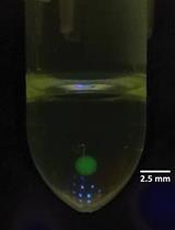
A Detailed Protocol for Preparing Millimeter-sized Supergiant Liposomes that Permit Efficient Eukaryotic Cell-free Translation in the Interior

一种制备毫米大小的允许在内部有效的真核细胞游离翻译的超巨大脂质体的详细方案

HT Hajime Takahashi
AO Atsushi Ogawa
2445 Views
Jun 20, 2021
Liposomes have been used as a pseudo cell membrane for encapsulating biomolecules and creating an artificial cell in the interior where biochemical reactions can occur. Among the several methods used to prepare biomolecule-encapsulating liposomes, the spontaneous emulsion transfer method is superior to others in that it allows us to readily prepare relatively large liposomes whose sizes are controlled (from micrometer- to millimeter-sized liposomes) without special equipment. However, conventional protocols for this method require liposomes to contain a considerably high concentration of sucrose (high-density solute), which severely inhibits gene expression, one of the most important biochemical reactions. Thus, we optimized the preparation conditions to develop a wheat germ extract (WGE)-based protocol that requires a much lower concentration of sucrose and has almost no effect on eukaryotic cell-free translation. Our protocol allows us to successfully prepare millimeter-sized, moderately stable, WGE-encapsulating liposomes in which WGE translation takes place efficiently. Since a broad range of genes derived from various types of organisms can be efficiently translated in a WGE-based translation system, liposomes prepared using our protocol would be useful as a versatile research tool for artificial cells.

细胞生物学

神经元的电生理特性:小鼠脑切片中的电流钳记录和放电模式分析

Electrophysiological Properties of Neurons: Current-Clamp Recordings in Mouse Brain Slices and Firing-Pattern Analysis

神经元的电生理特性:小鼠脑切片中的电流钳记录和放电模式分析

EN Elina Nagaeva
IZ Ivan Zubarev
EK Esa R. Korpi
5884 Views
Jun 20, 2021
Characterization of an electrically active cell, such as a neuron, demands measurement of its electrical properties. Due to differences in gene activation, location, innervation patterns, and functions, the millions of neurons in the mammalian brain are tremendously diverse in their membrane characteristics and abilities to generate action potentials. These features can be measured with a patch-clamp technique in whole-cell current-clamp configuration followed by detailed post-hoc analysis of firing patterns. This analysis can be time-consuming, and different laboratories have their own methods to perform it, either manually or with custom-written scripts. Here, we describe in detail a protocol for firing-pattern registration in neurons of the ventral tegmental area (VTA) as an example and introduce a software for its fast and convenient analysis. With the help of this article, other research groups can easily apply this method and generate unified types of data that are comparable between brain regions and various studies.Graphic abstract:Workflow of the Protocol
基于荧光的全染色单细胞分析和清除的微组织和类器官高通量筛选

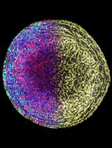
Fluorescence-based Single-cell Analysis of Whole-mount-stained and Cleared Microtissues and Organoids for High Throughput Screening

基于荧光的全染色单细胞分析和清除的微组织和类器官高通量筛选

HR Henrik Renner
MO Mandy Otto
MG Martha Grabos
HS Hans R. Schöler
JB Jan M. Bruder
4928 Views
Jun 20, 2021
Three-dimensional (3D) cell culture, especially in the form of organ-like microtissues (“organoids”), has emerged as a novel tool potentially mimicking human tissue biology more closely than standard two-dimensional culture. Typically, tissue sectioning is the standard method for immunohistochemical analysis. However, it removes cells from their native niche and can result in the loss of 3D context during analyses. Automated workflows require parallel processing and analysis of hundreds to thousands of samples, and sectioning is mechanically complex, time-intensive, and thus less suited for automated workflows. Here, we present a simple protocol for combined whole-mount immunostaining, tissue-clearing, and optical analysis of large-scale (approx. 1 mm) 3D tissues with single-cell level resolution. While the protocol can be performed manually, it was specifically designed to be compatible with high-throughput applications and automated liquid handling systems. This approach is freely scalable and allows parallel automated processing of large sample numbers in standard labware. We have successfully applied the protocol to human mid- and forebrain organoids, but, in principle, the workflow is suitable for a variety of 3D tissue samples to facilitate the phenotypic discovery of cellular behaviors in 3D cell culture-based high-throughput screens.Graphic abstract:Automatable organoid clearing and high-content analysis workflow and timeline

免疫学

用脂质组学方法测定培养细胞亚细胞膜组分中磷脂酸的种类

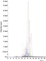
A Lipidomics Approach to Measure Phosphatidic Acid Species in Subcellular Membrane Fractions Obtained from Cultured Cells

用脂质组学方法测定培养细胞亚细胞膜组分中磷脂酸的种类

NK Nawal Kassas
LF Laetitia Fouillen
SG Stéphane Gasman
NV Nicolas Vitale
3057 Views
Jun 20, 2021
Over the last decade, lipids have emerged as possessing an ever-increasing number of key functions, especially in membrane trafficking. For instance, phosphatidic acid (PA) has been proposed to play a critical role in different steps along the secretory pathway or during phagocytosis. To further investigate in detail the precise nature of PA activities, we need to identify the organelles in which PA is synthesized and the PA subspecies involved in these biological functions. Indeed, PA, like all phospholipids, has a large variety based on its fatty acid composition. The recent development of PA sensors has helped us to follow intracellular PA dynamics but has failed to provide information on individual PA species. Here, we describe a method for the subcellular fractionation of RAW264.7 macrophages that allows us to obtain membrane fractions enriched in specific organelles based on their density. Lipids from these membrane fractions are precipitated and subsequently processed by advanced mass spectrometry-based lipidomics analysis to measure the levels of different PA species based on their fatty acyl chain composition. This approach revealed the presence of up to 50 different species of PA in cellular membranes, opening up the possibility that a single class of phospholipid could play multiple functions in any given organelle. This protocol can be adapted or modified and used for the evaluation of other intracellular membrane compartments or cell types of interest.

微生物学

基于荧光的弓形虫血红素定量

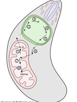
Fluorescence-based Heme Quantitation in Toxoplasma Gondii

基于荧光的弓形虫血红素定量

AB Amy Bergmann
ZD Zhicheng Dou
2968 Views
Jun 20, 2021
Toxoplasma gondii is a highly prevalent protozoan pathogen throughout the world. As a eukaryotic intracellular pathogen, Toxoplasma ingests nutrients from host cells to support its intracellular growth. The parasites also encode full or partial metabolic pathways for the biosynthesis of certain nutrients, such as heme. Heme is an essential nutrient in virtually all living organisms, acting as a co-factor for mitochondrial respiration complexes. Free heme is toxic to cells; therefore, it gets conjugated to proteins or other metabolites to form a “labile heme pool,” which is readily available for the biosynthesis of hemoproteins. Previous literature has shown that Toxoplasma gondii carries a fully functional de novo heme biosynthesis pathway and principally depends on this pathway for intracellular survival. Our recent findings also showed that the parasite’s intracellular replication is proportional to the total abundance of heme within the cells. Moreover, heme abundance is linked to mitochondrial oxygen consumption for ATP production in these parasites; thus, they may need to regulate their cellular heme levels for optimal infection when present in different environments. Therefore, quantitative measurement of heme abundance within Toxoplasma will help us to understand the roles of heme in subcellular activities such as mitochondrial respiration and other events related to energy metabolism.
连缀转录:测定酵母特定基因组位点的新生转录

Transcriptional Run-on: Measuring Nascent Transcription at Specific Genomic Sites in Yeast

连缀转录:测定酵母特定基因组位点的新生转录

VB Victoria Begley
Ld Lola de Miguel-Jiménez
SC Sebastián Chávez
2505 Views
Jun 20, 2021
DNA transcription by RNA polymerases has always interested the scientific community as it is one of the most important processes involved in genome expression. This has led scientists to come up with different protocols allowing analysis of this process in specific locations across the genome by quantitating the amount of RNA polymerases transcribing that genomic site in a cell population. This can be achieved by either detecting the total number of polymerases in contact with that region (i.e., by chromatin immunoprecipitation (ChIP) with anti-RNA polymerase antibodies) or by measuring the number of polymerases that are effectively engaged in transcription in that position. This latter strategy is followed using transcription run-on (TRO), also known as nuclear run-on (NRO), which was first developed in mammalian cells over 40 years ago and has since been adapted to many other different organisms and high-throughput methods. Here, we detail the procedure for performing TRO in Saccharomyces cerevisiae for single genomic regions to study active transcription on a single gene scale. To do so, we wash the cells in the detergent sarkosyl, which prevents new initiations at the promoter level, and then perform an in situ reaction, leading to the radiolabeling of transcripts by RNA polymerases that were already engaged in transcription at the moment of harvesting. By subsequently quantitating the signal of these transcripts, we can determine the level of active transcription in a single gene. This presents a major advantage over other forms of transcription quantitation such as RNA polymerase ChIP, since in the latter, both active and inactive polymerases are measured. By combining both ChIP and TRO, the amount of inactive or paused polymerases on a particular gene can be estimated.Graphic abstract:Transcriptional run-on scheme

神经科学

MRI引导下聚焦超声对小鼠基底前脑静脉及无创给药影响的研究

Intravenous and Non-invasive Drug Delivery to the Mouse Basal Forebrain Using MRI-guided Focused Ultrasound

MRI引导下聚焦超声对小鼠基底前脑静脉及无创给药影响的研究

KX Kristiana Xhima
DM Dallan McMahon
EN Edward Ntiri
MG Maged Goubran
KH Kullervo Hynynen
IA Isabelle Aubert
4204 Views
Jun 20, 2021
Basal forebrain cholinergic neurons (BFCNs) regulate circuit dynamics underlying cognitive processing, including attention, memory, and cognitive flexibility. In Alzheimer’s disease and related neurodegenerative conditions, the degeneration of BFCNs has long been considered a key player in cognitive decline. The cholinergic system thus represents a key therapeutic target. A long-standing obstacle for the development of effective cholinergic-based therapies is not only the production of biologically active compounds but also a platform for safe and efficient drug delivery to the basal forebrain. The blood-brain barrier (BBB) presents a significant challenge for drug delivery to the brain, excluding approximately 98% of small-molecule biologics and nearly 100% of large-molecule therapeutic agents from entry into the brain parenchyma. Current modalities to achieve effective drug delivery to deep brain structures, such as the basal forebrain, are particularly limited. Direct intracranial injection via a needle or catheter carries risks associated with invasive neurosurgery. Intra-arterial injection of hyperosmotic solutions or therapeutics modified to penetrate the BBB using endogenous transport systems lack regional specificity, which may not always be desirable. Intranasal, intrathecal, and intraventricular administration have limited drug distribution beyond the brain surface. Here, we present a protocol for non-invasively, locally, and transiently increasing BBB permeability using MRI-guided focused ultrasound (MRIgFUS) in the murine basal forebrain for delivery of therapeutic agents targeting the cholinergic system. Ongoing work in preclinical models and clinical trials supports the safety and feasibility of MRIgFUS-mediated BBB modulation as a promising drug delivery modality for the treatment of debilitating neurological diseases.
小鼠嗅上皮嗅觉感觉神经元的体内光学通路研究

In vivo Optical Access to Olfactory Sensory Neurons in the Mouse Olfactory Epithelium

小鼠嗅上皮嗅觉感觉神经元的体内光学通路研究

SI Shigenori Inagaki
RI Ryo Iwata
TI Takeshi Imai
3223 Views
Jun 20, 2021
In neuroscience, it is fundamental to understand how sensory stimuli are translated into neural activity at the entry point of sensory systems. In the olfactory system, odorants inhaled into the nasal cavity are detected by ~1,000 types of odorant receptors (ORs) that are expressed by olfactory sensory neurons (OSNs). Since each OSN expresses only one type of odorant receptor, the odor-evoked responses reflect the interaction between odorants and the expressed OR. The responses of OSN somata are often measured by calcium imaging and electrophysiological techniques; however, previous techniques require tissue dissection or cell dissociation, rendering it difficult to investigate physiological responses. Here, we describe a protocol that allows us to observe odor-evoked responses of individual OSN somata in the mouse olfactory epithelium in vivo. Two-photon excitation through the thinned skull enables highly-sensitive calcium imaging using a genetically encoded calcium indicator, GCaMP. Recording of odor-evoked responses in OSN somata in freely breathing mice will be fundamental to understanding how odor information is processed at the periphery and higher circuits in the brain.
培养感觉神经元轴突囊泡运输的双色实时成像

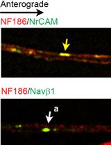
Dual Color, Live Imaging of Vesicular Transport in Axons of Cultured Sensory Neurons

培养感觉神经元轴突囊泡运输的双色实时成像

YB Yoko Bekku
JS James L. Salzer
2922 Views
Jun 20, 2021
The function of neurons in afferent reception, integration, and generation of electrical activity relies on their strikingly polarized organization, characterized by distinct membrane domains. These domains have different compositions resulting from a combination of selective targeting and retention of membrane proteins. In neurons, most proteins are delivered from their site of synthesis in the soma to the axon via anterograde vesicular transport and undergo retrograde transport for redistribution and/or lysosomal degradation. A key question is whether proteins destined for the same domain are transported in separate vesicles for local assembly or whether these proteins are pre-assembled and co-transported in the same vesicles for delivery to their cognate domains. To assess the content of transport vesicles, one strategy relies on staining of sciatic nerves after ligation, which drives the accumulation of anterogradely and retrogradely transported vesicles on the proximal and distal side of the ligature, respectively. This approach may not permit confident assessment of the nature of the intracellular vesicles identified by staining, and analysis is limited to the availability of suitable antibodies. Here, we use dual color live imaging of proteins labeled with different fluorescent tags, visualizing anterograde and retrograde axonal transport of several proteins simultaneously. These proteins were expressed in rat dorsal root ganglion (DRG) neurons cultured alone or with Schwann cells under myelinating conditions to assess whether glial cells modify the patterns of axonal transport. Advantages of this protocol are the dynamic identification of transport vesicles and characterization of their content for various proteins that is not limited by available antibodies.

植物科学

利用硅芯片进行拟南芥微嫁接

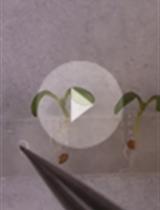
Micrografting in Arabidopsis Using a Silicone Chip

利用硅芯片进行拟南芥微嫁接

HT Hiroki Tsutsui
YK Yaichi Kawakatsu
MN Michitaka Notaguchi
6550 Views
Jun 20, 2021
The micrografting technique in the model plant Arabidopsis has been widely used in the field of plant science. Grafting experiments have demonstrated that signal transductions are systematically regulated in many plant characteristics, including defense mechanisms and responses to surrounding environments such as soil and light conditions. Hypocotyl micrografting is a powerful tool for the analysis of signal transduction between shoots and roots; however, the requirement for a high level of skill for micrografting, during which small seedlings are microdissected and micromanipulated, has limited its use. Here, we developed a silicone-made microdevice, called a micrografting chip, to perform Arabidopsis micrografting easily and uniformly. The micrografting chip has tandemly arrayed units, each of which consists of a seed pocket for seed germination and a micro-path to hold hypocotyl. All micrografting procedures are performed on the chip. This method using a micrografting chip will avoid the need for training and promote studies of systemic signaling in plants.Graphic abstract:A silicone chip for easy grafting
多肽介导的化学物质纳米粒对拟南芥叶绿体的靶向作用

Peptide-mediated Targeting of Nanoparticles with Chemical Cargoes to Chloroplasts in Arabidopsis Plants

多肽介导的化学物质纳米粒对拟南芥叶绿体的靶向作用

IS Israel Santana
PH Peiguang Hu
SJ Su-Ji Jeon
CC Chris Castillo
HT Hann Tu
JG Juan Pablo Giraldo
3747 Views
Jun 20, 2021
Plant nanobiotechnology is a flourishing field that uses nanomaterials to study and engineer plant function. Applications of nanotechnology in plants have great potential as tools for improving crop yield, tolerance to disease and environmental stress, agrochemical delivery of pesticides and fertilizers, and genetic modification and transformation of crop plants. Previous studies have used nanomaterials functionalized with chemicals, including biocompatible polymers with charged, neutral, or hydrophobic functional groups, to improve nanomaterial uptake and localization in plant cells. Recently, the use of biorecognition motifs such as peptides has been demonstrated to enable the targeted delivery of nanoparticles in plants (Santana et al., 2020). Herein, we describe a bio-protocol to target nanoparticles with chemical cargoes to chloroplasts in plant leaves and assess targeting efficiency using advanced analytical tools, including confocal microscopy and elemental analysis. We also describe the use of isothermal titration calorimetry to determine the affinity of nanomaterials for their chemical cargoes. Nanotechnology-based methods for targeted delivery guided by conserved plant molecular recognition mechanisms will provide more robust plant bioengineering tools across diverse plant species.Graphic abstract:Targeted delivery of nanomaterials with chemical cargoes to chloroplasts enabled by plant biorecognition

干细胞

CRISPR/Cas9介导的人iPSC细胞系SNP精确编辑

CRISPR/Cas9-mediated Precise SNP Editing in Human iPSC Lines

CRISPR/Cas9介导的人iPSC细胞系SNP精确编辑

HZ Hanwen Zhang
SZ Siwei Zhang
5096 Views
Jun 20, 2021
Human induced pluripotent stem cells (hiPSCs) have been extensively used in the fields of developmental biology and disease modeling. CRISPR/Cas9 gene editing in iPSC lines often has a low frequency, which hampers its application in precise allele editing of disease-associated single nucleotide polymorphisms (SNPs), especially those in the noncoding parts of the genome. Here, we present a unique workflow to engineer isogenic iPSC lines by SNP editing from heterozygous to homozygous for disease risk alleles or non-risk alleles using a transient and straightforward transfection-based protocol. This protocol enables us to simultaneously obtain pure and clonal isogenic lines of all three possible genotypes of a SNP site within about 4 to 5 weeks.

系统生物学

用于研究人类疾病中蛋白质N-糖基化失调的N-糖肽丰度和N-糖基化位点的差异分析

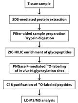
Differential Analysis of N-glycopeptide Abundance and N-glycosylation Site Occupancy for Studying Protein N-glycosylation Dysregulation in Human Disease

用于研究人类疾病中蛋白质N-糖基化失调的N-糖肽丰度和N-糖基化位点的差异分析

QZ Qi Zhang
CM Cheng Ma
LL Lian Li
LC Lih-Shen Chin
3198 Views
Jun 20, 2021
Protein N-glycosylation plays a vital role in diverse cellular processes, and dysregulated N-glycosylation is implicated in a variety of human diseases including neurodegenerative disorders and cancer. With recent advances in high-resolution mass spectrometry-based glycoproteomics technologies enabling large-scale N-glycoproteome profiling of disease and control samples, analysis of the large datasets has become a challenge. Here, we provide a protocol for the systems-level analysis of in vivo N-glycosylation sites on N-glycosylated proteins and their changes in human disease, such as Alzheimer's disease. The protocol includes quantitation and differential analysis of N-glycopeptide abundance, in addition to integrative N-glycoproteome and proteome data analyses, to determine disease-associated changes in N-glycosylation site occupancy and identify differentially N-glycosylated proteins in human disease versus control samples. This protocol can be modified and applied to study proteome-wide N-glycosylation alterations in response to different cellular stresses or pathophysiological states in other organisms or model systems.