往期刊物2021

卷册: 11, 期号: 1

left

Dec

20

Dec

5

Nov

20

Nov

5

Oct

20

Oct

5

Sep

20

Sep

5

Aug

20

Aug

5

Jul

20

Jul

5

Jun

20

Jun

5

May

20

May

5

Apr

20

Apr

5

Mar

20

Mar

5

Feb

20

Feb

5

Jan

20

Jan

5

right

癌症生物学

基于瞬时转染的293T细胞粘附实验

A Transient Transfection-based Cell Adhesion Assay with 293T Cells

基于瞬时转染的293T细胞粘附实验

RS Rohit Singh
BC Beom K. Choi
6113 Views
Jan 5, 2021
The in vitro cell adhesion assay is a quantitative method for measuring selective cell adhesion to specific proteins. Traditionally, cell adhesion assays employ purified protein immobilized on a solid glass or plastic surface. Here, we describe a transient 293T cell transfection-based cell adhesion assay to study selective cell adhesion of a specific cell type to a protein of interest. In this protocol, 293T cells are transfected with a mammalian expression plasmid containing mSiglec1 cDNA or an empty plasmid as a mock control and are then cultured to form a monolayer. Subsequently, these Siglec1-expressing and mock-transfected 293T cell monolayers are used for cell adhesion assays with GFP-expressing B16F10 cells. The number of GFP+ cancer cells adhering to each 293T monolayer is a quantitative mean to compare the selective adhesiveness of cancer cells to Siglec1. This method eliminates the need to express and purify the protein of interest to perform in vitro cell adhesion assays and can easily be performed with difficult-to-purify proteins while maintaining their native in situ structure.
吞噬试验的严格定量分析方法

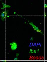
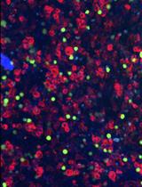
A Rigorous Quantitative Approach to Analyzing Phagocytosis Assays

吞噬试验的严格定量分析方法

MC Michael D. Caponegro
KT Kaitlyn Koenig Thompson
MT Maryam Tayyab
ST Stella E. Tsirka
4908 Views
Jan 5, 2021
Studying monocytic cells in isolated systems in vitro contributes significantly to the understanding of innate immune physiology. Functional assays produce read outs which can be used to measure responses to selected stimuli, such as pathogen exposure, antigen loading, and cytokine stimulation. Integration of these results with high quality in vivo models allows for the development of therapeutics which target these cell populations. Current methodologies to quantify phagocytic function of monocytic cells in vitro either measure phagocytic activity of individual cells (average number of beads or particles/cell), or a population outcome (% cells that contain phagocytosed material). Here we address technical challenges and shortcomings of these methods and present a protocol for collecting and analyzing data derived from a functional assay which measures phagocytic activity of macrophage and macrophage-like cells. We apply this method to two different experimental conditions, and compare to existing work flows. We also provide an online tool for users to upload and analyze data using this method.
CDK1体外磷酸化CENP-C的研究

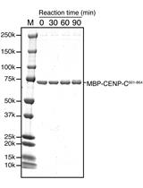
CENP-C Phosphorylation by CDK1 in vitro

CDK1体外磷酸化CENP-C的研究

RW Reito Watanabe
MH Masatoshi Hara
MA Mariko Ariyoshi
TF Tatsuo Fukagawa
4252 Views
Jan 5, 2021
Accurate chromosome segregation during mitosis requires the kinetochore, a large protein complex, which makes a linkage between chromosomes and spindle microtubes. An essential kinetochore component, CENP-C, is phosphorylated by Cyclin-B-Cyclin dependent kinase 1 (CDK1) that is a master kinase for mitotic progression, promoting proper kinetochore assembly during mitosis. Here, we describe an in vitro CDK1 kinase assay to detect CENP-C phosphorylation using Phos-tag SDS-PAGE without radiolabeled ATP. Our protocol has advantages in ease and safety over conventional phosphorylation assays using [γ-32P]-ATP, which has potential hazards despite their better sensitivity. The protocol described here can be applicable to other kinases and be also useful for analysis of phospho-sites in substrates in vitro.

细胞生物学

用于活性氧生成和快速细胞蛋白质标准化的简单微孔板检测

A Simple Microplate Assay for Reactive Oxygen Species Generation and Rapid Cellular Protein Normalization

用于活性氧生成和快速细胞蛋白质标准化的简单微孔板检测

NN Neville S. Ng
LO Lezanne Ooi
7760 Views
Jan 5, 2021
2',7'-dichlorofluorescein (DCF) and derivatives are commonly used as fluorescent indicators of a broad spectrum of reactive oxygen species (ROS) generation in cell-based assays. However, there are numerous challenges inherent to the utilization of DCF probes for intracellular microscopic analysis, including bleaching and probe efflux. Plate spectroscopy is comparatively simple and scalable compared to microscopy or flow cytometry-based acquisition, however is often subject to artefacts, including those introduced by thermal gradients and normalization methods. In this protocol we demonstrate a simple and sensitive plate spectrometry-based protocol utilizing the probes H2DCFDA and sulforhodamine B. The truncated sulforhodamine B assay (SRB) for cellular protein allows for a stable endpoint measurement of total cell population while also preserving morphology, can be combined or run in parallel with any other assay for normalization of readout to cell mass, and complemented by microscopic scoring of cell number and nuclear count. The oxidative stress and normalisation methods may enhance fields of research investigating cell differentiation, stress, or toxicity.Graphical abstract
LUHMES细胞中与帕金森氏病相关的线粒体和神经元形态学高通量筛选分析

A Parkinson’s Disease-relevant Mitochondrial and Neuronal Morphology High-throughput Screening Assay in LUHMES Cells

LUHMES细胞中与帕金森氏病相关的线粒体和神经元形态学高通量筛选分析

Tom  Leah Tom Leah
IV Irina Vazquez-Villaseñor
LF Laura Ferraiuolo
SW Stephen B Wharton
HM Heather Mortiboys
4834 Views
Jan 5, 2021
Parkinson’s disease is a devastating neurodegenerative disorder affecting 2-3% of the population over 65 years of age. There is currently no disease-modifying treatment. One of the predominant pathological features of Parkinson’s disease is mitochondrial dysfunction, and much work has aimed to identify therapeutic compounds which can restore the disrupted mitochondrial physiology. However, modelling mitochondrial dysfunction in a disease-relevant model, suitable for screening large compound libraries for ameliorative effects, represents a considerable challenge. Primary patient derived cells, SHSY-5Y cells and in vivo models of Parkinson’s disease have been utilized extensively to study the contribution of mitochondrial dysfunction in Parkinson’s. Indeed many studies have utilized LUHMES cells to study Parkinson’s disease, however LUHMES cells have not been used as a compound screening model for PD-associated mitochondrial dysfunction previously, despite possessing several advantages compared to other frequently used models, such as rapid differentiation and high uniformity (e.g., in contrast to iPSC-derived neurons), and relevant physiology as human mesencephalic tissue capable of differentiating into dopaminergic-like neurons that highly express characteristic markers. After previously generating GFP+-LUHMES cells to model metabolic dysfunction, we report this protocol using GFP+-LUHMES cells for high-throughput compound screening in a restoration model of PD-associated mitochondrial dysfunction. This protocol describes the use of a robust and reproducible toxin-induced GFP+-LUHMES cell model for high throughput compound screening by assessing a range of mitochondrial and neuronal morphological parameters. We also provide detailed instructions for data and statistical analysis, including example calculations of Z’-score to assess statistical effect size across independent experiments.

发育生物学

显微ct测量多重密度骨体积

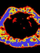
Measuring Bone Volume at Multiple Densities by Micro-computed Tomography

显微ct测量多重密度骨体积

EW Emma C. Walker
NM Narelle E. McGregor
AC Audrey S. M. Chan
NS Natalie A. Sims
3825 Views
Jan 5, 2021
Bone strength is controlled by both bone mass, and the organization and quality of the bone material. The current standard method for measuring bone mass in mouse and rat studies is micro-computed tomography. This method typically uses a single threshold to identify bone material in the cortical and trabecular regions. However, this single threshold method obscures information about the mineral content of the bone material and depends on normal morphology to separately analyze cortical and trabecular structures. To extend this method to identify bone mass at multiple density levels, we have established a protocol for unbiased selection and application of multiple thresholds using a standard laboratory-based micro-computed tomography instrument. This non-invasive method can be applied to longitudinal studies and archived samples and provides additional information about bone structure and strength.

微生物学

使用薄层和离子交换色谱评估二腺苷酸环化酶和c-di-AMP-磷酸二酯酶的活性

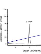
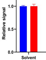
Assessment of Diadenylate Cyclase and c-di-AMP-phosphodiesterase Activities Using Thin-layer and Ion Exchange Chromatography

使用薄层和离子交换色谱评估二腺苷酸环化酶和c-di-AMP-磷酸二酯酶的活性

AL Andreas Latoscha
DD David Jan Drexler
GW Gregor Witte
NT Natalia Tschowri
3153 Views
Jan 5, 2021
All living cells use cyclic nucleotides as second messengers for signal sensing and transduction. Cyclic di-3′,5′-adenosine monophosphate (c-di-AMP) is primarily involved in the control of bacterial and euryarcheal osmoadaptation and is produced by diadenylate cyclases from two molecules of ATP. Specific phosphodiesterases hydrolyze c-di-AMP to the linear phosphoadenylate adenosine 5′-pApA or to AMP. Different methods including high-performance liquid chromatography (HPLC), thin-layer chromatography (TLC) and ion exchange chromatography (IEX) can be used to determine activities of c-di-AMP-synthesizing and degrading enzymes. Here, we describe in detail the TLC and IEX methods adapted for characterization of the diadenylate cyclase DisA and the phosphodiesterase AtaC from Streptomyces venezuelae. TLC allows quick and easy separation of radioactive-labeled substrates and products, while IEX avoids utilization of potentially hazardous radioactive substrates and can be used as a good substitute if an HPLC system is not available. Unlike in TLC assays, samples cannot be analyzed in parallel by using the IEX assay, thus it is more time consuming.

神经科学

胚胎和产后早期小鼠脑切片的优化免疫染色

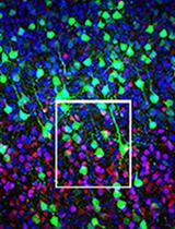
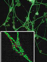
Optimized Immunostaining of Embryonic and Early Postnatal Mouse Brain Sections

胚胎和产后早期小鼠脑切片的优化免疫染色

KH Kawssar Harb
MB Michele Bertacchi
MS Michèle Studer
5186 Views
Jan 5, 2021
The mammalian neocortex, the outer layer of the cerebrum and most recently evolved brain region, is characterized by its unique areal and laminar organization. Distinct cortical layers and areas can be identified by the protein expression of graded transcription factors and molecular determinants that define the identity of different projection neurons. Thus, specific detection and visualization of protein expression is crucial for assessing the identity of neocortical neurons and, more broadly, for understanding early and late developmental mechanisms and function of this complex system. Several immunostaining/immunofluorescence methods exist to detect protein expression. Published protocols vary with regard to subtle details, which may impact the final outcome of the immunofluorescence. Here, we provide a detailed protocol, suitable for both thin cryostat sections and thick vibratome sections, which has successfully worked for a wide range of antibodies directed against key molecular players of neocortical development. Ranging from early technical steps of brains collection down to image analysis and statistics, we include every detail concerning sample inclusion and sectioning, slide storage and optimal antibody dilutions aimed at reducing non-specific background. Routinely used in the lab, our background-optimized immunostaining protocol allows efficient detection of area- and layer- specific molecular determinants of distinct neocortical projection neurons.Graphic abstractWorkflow chart for the optimized immunostaining protocol of mouse brain sections. A. A flow chart for different steps of the optimized immunostaining protocol on both thin cryostat and thick vibratome sections. B. Example for immunostaining against Satb2 and Ctip2 on a thin coronal section (20 μm) at the level of the somatosensory cortex. The first column to the left shows the binning system where 6 bins can be overlaid on the image. On the bottom, an example of counting analysis showing the percentage of marker-positive cells normalized to the total number of DAPI or Hoechst-positive cells. C. Example for immunostaining against Satb2 and Ctip2 on a GFP+ thick vibratome section (200 μm). Images are taken at low magnification (10x, left) and high magnification (40x, right). The graph shows a counting of the percentage of Ctip2-positive neurons normalized to the total number of GFP-electroporated neurons on high-magnification images. Images on B and C are modified from Harb et al. (2016).
用于追踪和离体神经发生药理学操作的成年小鼠脑器官型海马切片培养系统的建立

Establishing an Adult Mouse Brain Hippocampal Organotypic Slice Culture System that Allows for Tracing and Pharmacological Manipulation of ex vivo Neurogenesis

用于追踪和离体神经发生药理学操作的成年小鼠脑器官型海马切片培养系统的建立

SM Steffen Mayerl
Cf Charles ffrench-Constant
4813 Views
Jan 5, 2021
The function of the hippocampus depends on the process of adult hippocampal neurogenesis which underpins the exceptional neural plasticity of this structure, and is also frequently affected in CNS pathologies. Thus, manipulation of this process represents an important therapeutic goal. To identify potential strategies, organotypic adult brain slices are emerging as a valuable tool. Over the recent years, this methodology has been refined and here we present a combined protocol that brings together these refinements to enable long-term culture of adult hippocampal slices. We employ a sectioning technique that retains essential afferent inputs onto the hippocampus as well as serum-free culture conditions, so allowing an extended culture period. To sustain the neurogenic potential in the slices, we utilize the gliogenesis-inhibitor Indomethacin. Using EdU retention analysis enables us to assess the effects of pharmacological intervention on neurogenesis. With these improvements, we have established an easy and reliable method to study the effects of small molecules/drugs on proliferation and neuron formation ex vivo which will facilitate future discovery driven drug screenings.
渐变气味刺激下秀丽隐杆线虫神经元活动的钙成像

Calcium Imaging of Neuronal Activity under Gradually Changing Odor Stimulation in Caenorhabditis elegans

渐变气味刺激下秀丽隐杆线虫神经元活动的钙成像

YT Yuki Tanimoto
KK Koutarou D. Kimura
3403 Views
Jan 5, 2021
Olfactory behavior is among the most fundamental animal behaviors both in the wild and in the laboratory. To elucidate the neural mechanisms underlying olfactory behavior, it is critical to measure neural responses to odorant concentration changes resembling those that animals actually sense during olfactory behavior. However, reproducing the dynamically changing olfactory stimuli to an animal during such measurements of neural activity is technically challenging. Here, we describe technical details and protocols for odor stimulation during calcium imaging of the sensory neurons of the nematode Caenorhabditis elegans. In this system, the neuronal activity of C. elegans is measured using ratiometric calcium imaging during exposure to quantitatively controlled olfactory stimuli over time. Temporal changes in odor concentrations around the animal are precisely controlled according to a predesigned temporal odor gradient to reproduce a realistic odor concentration change during olfactory behavior in a behavioral arena. By monitoring neural activity in response to the realistic olfactory stimulus, it is possible to elucidate the mechanisms by which olfactory input is processed by neural activities and reflected in behavioral output.
小鼠巩膜脉络膜/视网膜色素上皮/视网膜不分离染色及用于视网膜下层整体观察的全组织染色法

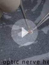
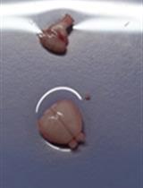
Non-separate Mouse Sclerochoroid/RPE/Retina Staining and Whole Mount for the Integral Observation of Subretinal Layer

小鼠巩膜脉络膜/视网膜色素上皮/视网膜不分离染色及用于视网膜下层整体观察的全组织染色法

SL Sung-Jin Lee
Soo-Young  Kim Soo-Young Kim
3335 Views
Jan 5, 2021
The subretinal layer between retinal pigment epithelium (RPE) and photoreceptors is a region involved in inflammation and angiogenesis during the procession of diseases such as age-related macular degeneration. The current protocols of whole mounts (retina and RPE) are unable to address the intact view of the subretinal layer because the separation between retina and RPE is required, and each separate tissue is then stained. Non-separate Sclerochoroid/RPE/Retina whole mount staining was recently developed and reported. The method can be further combined and optimized with melanin bleaching and tissue clearing. Here, we describe steps of both non-pigmented and pigmented mouse Sclerochoroid/RPE/Retina whole mount including eyeball preparation, staining, mounting and confocal scanning. In addition, we present the confocal images of RPE, subretinal microglia and the neighboring photoreceptors in Sclerochoroid/RPE/Retina whole mounts.

植物科学

大麻种子快速高效发芽规程的建立与标准化

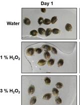
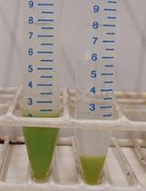
Development and Standardization of Rapid and Efficient Seed Germination Protocol for Cannabis sativa

大麻种子快速高效发芽规程的建立与标准化

Aleksei   Sorokin Aleksei Sorokin
Narendra Singh  Yadav Narendra Singh Yadav
dg Daniel Gaudet
IK Igor Kovalchuk
5783 Views
Jan 5, 2021
Cannabis seed germination is an important process for growers and researchers alike. Many biotechnological applications require a reliable sterile method for seed germination. This protocol outlines a seed germination procedure for Cannabis sativa using a hydrogen peroxide (H2O2) solution as liquid germination media. In this protocol, all three steps including seed sterilization, germination, and seedlings development were carried out in an H2O2 solution of different concentrations; 1% H2O2 solution showed the fastest and the most efficient germination. This protocol also exhibited high germination efficiency for very old cannabis seeds with lower viability. Overall, this protocol demonstrates superior germination compared to water control and reduces the risk of contamination, making it suitable for tissue culture and other sensitive applications.
应用碳纳米载体对烟草基因进行高效瞬时敲减

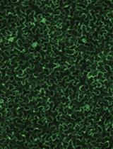
Efficient Transient Gene Knock-down in Tobacco Plants Using Carbon Nanocarriers

应用碳纳米载体对烟草基因进行高效瞬时敲减

Gozde S. Demirer Gozde S. Demirer
ML Markita P. Landry
5279 Views
Jan 5, 2021
Gene knock-down in plants is a useful approach to study genotype-phenotype relationships, render disease resistance to crops, and enable efficient biosynthesis of molecules in plants. Small interfering RNA (siRNA)-mediated gene silencing is one of the most common ways to achieve gene knock-down in plants. Traditionally, siRNA is delivered into intact plant cells by coding the siRNA sequences into DNA vectors, which are then delivered through viral and/or bacterial methods. In this protocol, we provide an alternative direct delivery method of siRNA molecules into intact plant cells for efficient transient gene knock-down in model tobacco plant, Nicotiana benthamiana, leaves. Our approach uses one dimensional carbon-based nanomaterials, single-walled carbon nanotubes (SWNTs), to deliver siRNA, and does not rely on viral/bacterial delivery. The distinct advantages of our method are i) there is no need for DNA coding of siRNA sequences, ii) this abiotic method could work in a broader range of plant species than biotic methods, and iii) there are fewer regulatory complications when using abiotic delivery methods, whereby gene silencing is transient without permanent modification of the plant genome.Graphic abstract
拟南芥细胞悬浮培养的稳定转化:COI1受体过度表达的案例研究

Stable Transformation of Arabidopsis thaliana Cell Suspension Cultures: A Case Study for the Overexpression of The COI1 Receptor

拟南芥细胞悬浮培养的稳定转化:COI1受体过度表达的案例研究

ED Eva K. Díaz-Sánchez
JO José A. O’Brien
IP Imma Pérez-Salamó
JK Jovaras Krasauskas
MB Moritz Bömer
AD Alessandra Devoto
4884 Views
Jan 5, 2021
Cell suspension cultures have been studied for decades to produce natural molecules. However, the difficulty in generating stably transformed cell lines has limited their use to produce high value chemicals reproducibly and in elevated quantities.In this protocol, a method to stably transform and maintain Arabidopsis cell suspension cultures is devised and presented in detail. Arabidopsis cell cultures were directly transformed with A. tumefaciens for the overexpression of the CORONATINE INSENSITIVE 1 (COI1) jasmonate receptor. Cell cultures were established after transformation and continuously maintained and tested for the overexpression of COI1. The protocol was also previously used to silence Arabidopsis peroxidases and allows for long term maintenance of transformed cells. Details on culture maintenance, both in liquid and solid media are provided, alongside with evidence of protein expression to confirm transformation.The system described provides a powerful tool for synthetic biology to study signaling independent of developmental control and to obtain metabolites of interest for the biotechnological and medical sectors.
脉冲追踪EdU法检测拟南芥和笄石菖叶原基细胞分裂方向

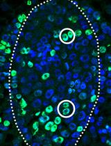
A Pulse–chase EdU Method for Detection of Cell Division Orientation in Arabidopsis and Juncus prismatocarpus Leaf Primordia

脉冲追踪EdU法检测拟南芥和笄石菖叶原基细胞分裂方向

Xiaofeng  Yin Xiaofeng Yin
Hirokazu Tsukaya Hirokazu Tsukaya
3617 Views
Jan 5, 2021
In plants, the morphological diversity of leaves is largely determined by cell division, especially cell division orientation. Whereas cell division itself is easily monitored, the detection and quantification of cell division orientation are difficult. The few existing methods for detection and quantification of cell division orientation are either inefficient or laborious. Here, we describe a pulse-chase strategy using a 5-ethynyl-2’-deoxyuridine (EdU) labeling assay. Plant tissues are first incubated with EdU for a short period (pulse), followed by a long incubation without EdU (chase). Using this method, the positions of daughter cells are easily detected and can be used to quantify cell division orientation. Our protocol is rapid and very efficient for quantitative analysis of cell division orientation, and can be applied to both model and non-model plant species.Graphic abstract Plant cell division pairs clearly visualized by a pulse-chase EdU method
用于确定短链异戊烯基二磷酸合酶产物谱的非放射性分析

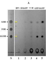
Non-radioactive Assay to Determine Product Profile of Short-chain Isoprenyl Diphosphate Synthases

用于确定短链异戊烯基二磷酸合酶产物谱的非放射性分析

AR Avanish Rai
DN Dinesh A. Nagegowda
2972 Views
Jan 5, 2021
Isoprenoids represent the largest class of metabolites with amazing diversities in structure and function. They are involved in protecting plants against pathogens or herbivores or involved in attracting pollinators. Isoprenoids are derived from geranyl diphosphate (GPP; C10), farnesyl diphosphate (FPP; C15), geranylgeranyl diphosphate (GGPP; C20), and geranylfarnesyl diphosphate (GFPP; C25) that are in turn formed by sequential condensations of isopentenyl diphosphate (IPP; C5) with an allylic acceptor such as dimethylallyl diphosphate (DMAPP; C5), GPP, FPP, or GGPP in a reaction catalyzed by isoprenyl diphosphate synthases (IDSs). IDS enzyme assay for determination of prenyl diphosphate products is generally performed using radiolabelled substrates, and the products formed are identified by employing expensive instruments such as phosphor imager, radio-GC, or radioHPLC. Though a non-radioactive assay for measuring IDS activity in crude plant extract has been reported, it requires a complex methodology utilizing chromatography coupled with tandem mass spectrometry (LC/MS-MS). Here, we describe a non-radioactive and simple inexpensive assay for determining the IDS assay products using non-radiolabeled IPP and its co-allylic substrates DMAPP,GPP, and FPP. The detection of prenyl diphosphate products generated in the assay was highly efficient and spots corresponding to prenyl alcohols were visible at >40 µM concentrations of IPP and DMAPP/GPP/FPP substrates. The protocol described here is sensitive, reliable, and technically simple, which could be used for functional characterization of IDS candidates.