往期刊物2020

卷册: 10, 期号: 23

left

Dec

20

Dec

5

Nov

20

Nov

5

Oct

20

Oct

5

Sep

20

Sep

5

Aug

20

Aug

5

Jul

20

Jul

5

Jun

20

Jun

5

May

20

May

5

Apr

20

Apr

5

Mar

20

Mar

5

Feb

20

Feb

5

Jan

20

Jan

5

right

生物化学

天然共免疫沉淀法鉴定哺乳动物细胞中染色质相关蛋白的“伙伴蛋白质”

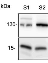
Native Co-immunoprecipitation Assay to Identify Interacting Partners of Chromatin-associated Proteins in Mammalian Cells

天然共免疫沉淀法鉴定哺乳动物细胞中染色质相关蛋白的“伙伴蛋白质”

AH Afzal Husain
NB Nasim A. Begum
MK Maki Kobayashi
TH Tasuku Honjo
6714 Views
Dec 5, 2020
Protein-protein interactions play key roles in nuclear processes including transcription, replication, DNA damage repair, and recombination. Co-immunoprecipitation (Co-IP) followed by western blot or mass spectrometry is an invaluable approach to identify protein-protein interactions. One of the challenges in the Co-IP of a protein localized to nucleus is the extraction of nuclear proteins from sub-nuclear fractions without losing physiologically relevant protein interactions. Here we describe a protocol for native Co-IP, which was originally used to successfully identify previously known as well novel topoisomerase 1 (TOP1) interacting proteins. In this protocol, we first extracted nuclear proteins by sequentially increasing detergent and salt concentrations, the extracted fractions were then diluted, pooled, and used for Co-IP. This protocol can be used to identify protein-interactome of other chromatin-associated proteins in a variety of mammalian cells.

生物物理学

使用微压痕仪对腺泡进行机械表征

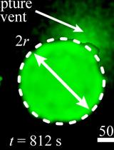
Mechanical Characterization of Glandular Acini Using a Micro-indentation Instrument

使用微压痕仪对腺泡进行机械表征

CO Christopher S. O’Bryan
QZ Qiao Zhang
TL Tanmay P. Lele
TA Thomas E. Angelini
3556 Views
Dec 5, 2020
The linker of nucleoskeleton and cytoskeleton (LINC) complex is responsible for tethering the nucleus to the cytoskeleton, providing a pathway for the cell’s nucleus to sense mechanical signals from the environment. Recently, we explored the role of the LINC complex in the development of glandular epithelial acini, such as those found in kidneys, breasts, and other organs. Acini developed with disrupted LINC complexes exhibited a loss of structural integrity, including filling of the lumen structures. As part of our investigation, we performed a mechanical indentation assay of LINC disrupted and undisrupted MDCK II cells using a micro-indentation instrument mounted above a laser-scanning confocal microscope. Through a combination of force measurements acquired from the micro-indentation instrument and contact area measurements taken from fluorescence images, we determined the average contact pressure at which the acini structure ruptured. Here, we provide a detailed description of the design of the micro-indentation instrument, as well as the experimental steps developed to perform these bio-indentation measurements. Furthermore, we discuss the data analysis steps necessary to determine the rupture pressure of the acini structures. While this protocol is focused on the indentation of individual glandular acini, the methods presented here can be adapted to perform a variety of mechanical indentation experiments for both 2D and 3D biological systems.

癌症生物学

基于球体的三维皮肤黑色素瘤模型评价肿瘤免疫细胞相互作用

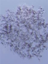
A 3D Skin Melanoma Spheroid-Based Model to Assess Tumor-Immune Cell Interactions

基于球体的三维皮肤黑色素瘤模型评价肿瘤免疫细胞相互作用

Marek Wagner Marek Wagner
Shigeo Koyasu Shigeo Koyasu
3689 Views
Dec 5, 2020
Three-dimensional (3D) tumor spheroids have the potential to bridge the gap between two-dimensional (2D) monolayer tumor cell cultures and solid tumors with which they share a significant degree of similarity. However, the progression of solid tumors is often influenced by the dynamic and reciprocal interactions between tumor and immune cells. Here we present a 3D tumor spheroid-based model that might shed new light on understanding the mechanisms of tumor and immune cell interactions. The model first utilizes the hanging drop assay, which serves as one of the simplest methods for generating 3D spheroids and requires no specialized equipment. Next, pre-established spheroids can be co-cultured either directly or indirectly with an immune cell population of interest. Using skin melanoma, we provide a detailed description of the model, which might hold a significant importance for the development of successful therapeutic strategies.

发育生物学

以跑步机运动小鼠为模型研究母体运动对后代的影响

Treadmill Running of Mouse as a Model for Studying Influence of Maternal Exercise on Offspring

以跑步机运动小鼠为模型研究母体运动对后代的影响

SC Song Ah Chae
JS Jun Seok Son
MZ Mei-Jun Zhu
Jd Jeanene M. de Avila
Min Du Min Du
3503 Views
Dec 5, 2020
Epidemiological studies robustly show the beneficial effects of maternal exercise in reducing maternal birth complications and improving neonatal outcomes, though underlying mechanisms remain poorly understood. To facilitate mechanistic exploration, a protocol for maternal exercise of mice is established, with the regimen following the exercise guidelines for pregnant women. Compared to volunteer wheel running, treadmill running allows precise control of exercise intensity and duration, dramatically reducing variations among individual mouse within treatments and facilitating translation into maternal exercise in humans. Based on the maximal oxygen consumption rate (VO2max) before pregnancy, the treadmill exercise protocol is separated into three stages: early stage (E1.5 to E7.5 at 40% VO2max), mid stage (E8.5 to E14.5 at 65% VO2max), and late stage of pregnancy (E15.5 to birth at 50% VO2max), which demonstrated persistent beneficial effects on maternal health and fetal development. This protocol can be useful for standardizing maternal treadmill exercise using mice as an experimental model.

免疫学

原代和iPSC来源的人类NK细胞的分离、刺激和功能分析方案

Protocol for Isolation, Stimulation and Functional Profiling of Primary and iPSC-derived Human NK Cells

原代和iPSC来源的人类NK细胞的分离、刺激和功能分析方案

JM Janine E. Melsen
MT Maria Themeli
Mv Monique M. van Ostaijen-ten Dam
Ev Els van Beelen
GL Gertjan Lugthart
RH Rob C. Hoeben
MS Marco W. Schilham
HM Harald M. Mikkers
6590 Views
Dec 5, 2020
Natural killer (NK) cells are innate immune cells, characterized by their cytotoxic capacity, and chemokine and cytokine secretion upon activation. Human NK cells are identified by CD56 expression. Circulating NK cells can be further subdivided into the CD56bright (~10%) and CD56dim NK cell subsets (~90%). NK cell-like cells can also be derived from human induced pluripotent stem cells (iPSC). To study the chemokine and cytokine secretion profile of the distinct heterogenous NK cell subsets, intracellular flow cytometry staining can be performed. However, this assay is challenging when the starting material is limited. Alternatively, NK cell subsets can be enriched, sorted, stimulated, and functionally profiled by measuring secreted effector molecules in the supernatant by Luminex. Here, we provide a rapid and straightforward protocol for the isolation and stimulation of primary NK cells or iPSC-derived NK cell-like cells, and subsequent detection of secreted cytokines and chemokines, which is also applicable for a low number of cells.
B细胞迁移的活体成像仪观察分析

Analysis of B Cell Migration by Intravital Microscopy

B细胞迁移的活体成像仪观察分析

MS Michael Schnoor
LS Leopoldo Santos-Argumedo
Daniel Alberto Girón-Pérez Daniel Alberto Girón-Pérez
EV Eduardo Vadillo
3597 Views
Dec 5, 2020
During immune responses, B cells home to lymph nodes (LN), where they encounter antigens. Homing starts with capture and L-selectin-dependent rolling on the activated endothelium of high endothelial venules (HEV). After recognition of chemokines presented on HEV, activation of B cell integrins occurs mediating firm arrest. Subsequently, B cells crawl to the spot of extravasation to enter the LN. Extravasation can be visualized and quantified in vivo by intravital microscopy (IVM) of the inguinal LN. Here, we describe an established protocol that permits detailed in vivo analysis of B cell recruitment to LN under sterile inflammatory conditions. We describe data acquisition, exportation, quantification, and statistical analysis using specialized software. IVM of LN is a powerful technique that can provide a better understanding of B cell migratory behavior during inflammation in vivo.

微生物学

重组Skd3(人ClpB)蛋白和烟草蚀刻病毒(TEV)蛋白酶在大肠杆菌中的表达与纯化

Expression and Purification of Recombinant Skd3 (Human ClpB) Protein and Tobacco Etch Virus (TEV) Protease from Escherichia coli

重组Skd3(人ClpB)蛋白和烟草蚀刻病毒(TEV)蛋白酶在大肠杆菌中的表达与纯化

RC Ryan R. Cupo
JS James Shorter
4343 Views
Dec 5, 2020
Skd3 (encoded by human CLPB) is a mitochondrial AAA+ protein comprised of an N-terminal ankyrin-repeat domain and a C-terminal HCLR-clade nucleotide-binding domain. The function of Skd3 has long remained unknown due to challenges in purifying the protein to high quality and near homogeneity. Recently we described Skd3 as a human mitochondrial protein disaggregase that solubilizes proteins in the mitochondrial intermembrane space. This protocol overcomes the challenges associated with purifying Skd3 and allows for in depth in vitro study of Skd3 activity. Tobacco etch virus (TEV) protease is required in the purification of Skd3. Thus, we also describe how to purify high quality TEV protease for use in the purification of Skd3, other purification protocols, and in vitro assays requiring TEV protease.
结合凝胶阻滞和足印法测定特异性和/或不稳定复合物的蛋白质-DNA相互作用

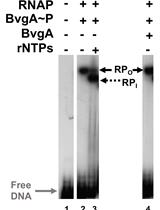
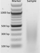
Combining Gel Retardation and Footprinting to Determine Protein-DNA Interactions of Specific and/or Less Stable Complexes

结合凝胶阻滞和足印法测定特异性和/或不稳定复合物的蛋白质-DNA相互作用

MH Meng-Lun Hsieh
AB Alice Boulanger
LK Leslie G. Knipling
DH Deborah M. Hinton
2738 Views
Dec 5, 2020
DNA footprinting is a classic technique to investigate protein-DNA interactions. However, traditional footprinting protocols can be unsuccessful or difficult to interpret if the binding of the protein to the DNA is weak, the protein has a fast off-rate, or if several different protein-DNA complexes are formed. Our protocol differs from traditional footprinting protocols, because it provides a method to isolate the protein-DNA complex from a native gel after treatment with the footprinting agent, thus removing the bound DNA from the free DNA or other protein-DNA complexes. The DNA is then extracted from the isolated complex before electrophoresis on a sequencing gel to determine the footprinting pattern. This analysis provides a possible solution for those who have been unable to use traditional footprinting methods to determine protein-DNA contacts.
α-变形杆菌核糖体的纯化及Northern印迹分析

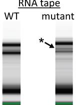
Ribosome Purification from an α-proteobacterium and rRNA Analysis by Northern Blot

α-变形杆菌核糖体的纯化及Northern印迹分析

LY Liang Yin
Caroline S. Harwood Caroline S. Harwood
2627 Views
Dec 5, 2020
Ribosomes are an integral part of cellular life. They are complex molecular machines consisting of multiple ribosomal proteins and RNAs. To study different aspects of ribosome composition, many methods have been developed over the decades. Here, we describe how to purify ribosomes from the α-proteobacterium Rhodopseudomonas palustris. Following this protocol, RNA can be extracted from either purified ribosomes or directly from cell cultures, and ribosomal RNAs quantified using Northern blot. This protocol gives an example of studying ribosomes in a bacterium other than the commonly used E. coli. The challenge of performing Northern blots with rRNA is also addressed in detail.

分子生物学

α-变形杆菌转运RNA的装载状态分析

Charging State Analysis of Transfer RNA from an α-proteobacterium

α-变形杆菌转运RNA的装载状态分析

LY Liang Yin
Caroline S. Harwood Caroline S. Harwood
2566 Views
Dec 5, 2020
Transfer RNA (tRNA) is an essential link between the genetic code and proteins. During the process of translation, tRNA is charged with its cognate amino acid and delivers it to the ribosome, thus serving as a substrate of protein synthesis. To analyze the charging state of a particular tRNA, total RNA is purified and analyzed on an acid-urea gel. Separated RNA is then transferred to a membrane and detected with a probe for the tRNA of interest. Here, we present an improved protocol to analyze the tRNA charging state in the α-proteobacterium Rhodopseudomonas palustris. Compared to the classical method, the RNA isolation step is optimized to suit this organism. Additionally, a non-radioactive platform is used for electrophoresis and Northern blots. This significantly reduces the time and the effort required for this protocol.

神经科学

利用光遗传学淀粉样蛋白-β监测黑腹果蝇、斑马鱼和秀丽隐杆线虫体内蛋白质聚集

Use of Optogenetic Amyloid-β to Monitor Protein Aggregation in Drosophila melanogaster, Danio rerio and Caenorhabditis elegans

利用光遗传学淀粉样蛋白-β监测黑腹果蝇、斑马鱼和秀丽隐杆线虫体内蛋白质聚集

PK Prameet Kaur
CK Caroline Kibat
ET Emelyne Teo
JG Jan Gruber
AM Ajay Mathuru
NT Nicholas S. Tolwinski
4438 Views
Dec 5, 2020
Alzheimer’s Disease (AD) has long been associated with accumulation of extracellular amyloid plaques (Aβ) originating from the Amyloid Precursor Protein. Plaques have, however, been discovered in healthy individuals and not all AD brains show plaques, suggesting that extracellular Aβ aggregates may play a smaller role than anticipated. One limitation to studying Aβ peptide in vivo during disease progression is the inability to induce aggregation in a controlled manner. We developed an optogenetic method to induce Aβ aggregation and tested its biological influence in three model organisms–D. melanogaster, C. elegans and D. rerio. We generated a fluorescently labeled, optogenetic Aβ peptide that oligomerizes rapidly in vivo in the presence of blue light in all organisms. Here, we detail the procedures for expressing this fusion protein in animal models, investigating the effects on the nervous system using time lapse light-sheet microscopy, and performing metabolic assays to measure changes due to intracellular Aβ aggregation. This method, employing optogenetics to study the pathology of AD, allows spatial and temporal control in vivo that cannot be achieved by any other method at present.
用于运动过程中小鼠下丘体内电生理记录的头部手术

Headpost Surgery for in vivo Electrophysiological Recording in the Mouse Inferior Colliculus during Locomotion

用于运动过程中小鼠下丘体内电生理记录的头部手术

YY Yoonsun Yang
GK Gunsoo Kim
3030 Views
Dec 5, 2020
The inferior colliculus (IC) is a critical midbrain integration center for auditory and non-auditory information. Although much is known about the response properties of the IC neurons to auditory stimuli, how the IC neural circuits function during movement such as locomotion remains poorly understood. Mice offer a valuable model in this respect, but previous studies of the mouse IC were performed in anesthetized or restrained preparations, making it difficult to study the IC function during behavior. Here we describe a neural recording protocol for the mouse IC in which mice are head-fixed, but can run on a passive treadmill. Mice first receive a headpost surgery, and become habituated to head-fixing while being on a treadmill. Following a few days of habituation, neural recordings of the IC neuron activity are performed. The neural activity can be compared across different behavioral conditions, such as standing still versus running on a treadmill. We describe how to overcome the challenges of headpost surgery for awake IC recording, presented by the location and overlying bones. This protocol allows investigations of the IC function in behaving mice, while allowing precise stimulus control and the use of recording methods similar to those for anesthetized preparations.
 应用膜辅助显微切割(PAM)对啮齿类伏隔核进行采样

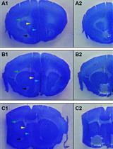
Using the Parafilm-assisted Microdissection (PAM) Method to Sample Rodent Nucleus Accumbens

应用膜辅助显微切割(PAM)对啮齿类伏隔核进行采样

MP Marco Pagliusi Jr.
AB Arthur F. Brandão
GZ Gabriel G. Zanetti
IB Ivan J. M. Bonet
CS Cesar R. Sartori
AV Andre S. Vieira
2713 Views
Dec 5, 2020
Microdissection techniques are very important for anatomical and functional studies focused on neuroscience, where it is often necessary microdissect specific brain areas to perform molecular or anatomical analyses. The parafilm®-assisted microdissection (PAM) was previously described and involves the microdissection of tissue sections mounted on parafilm-covered glass slides. In this work, we describe the use of the PAM method to microdissect rodent nucleus accumbens (NAc). (1) We first describe the best way to perform the mouse euthanasia and how to remove the brain. (2) Next, we describe how to prepare the slides with parafilm® that will be used to receive the brain slices. (3) Following, we describe how to handle the brain in the cryostat, how to align the hemispheres and how to identify the NAc antero-posterior limits. (4) We also describe how to perform the staining and dehydration of the slices, a critical step to facilitate the microdissection and preserve macromolecules. (5) In the final step, we describe how to identify the dorso-ventral and latero-medial limits of the NAc and, finally, how to perform the manual microdissection of the area. This is a low-cost technique that allows the researcher to specifically microdissect any brain region, from which intact RNA and proteins can be extracted to perform several molecular analyses (e.g., real-time PCR, Western blot, and RNA-seq).

植物科学

染色质免疫沉淀法(ChIP)检测生长素处理拟南芥花序组织中的组蛋白标记

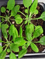
Chromatin Immunoprecipitation (ChIP) to Assess Histone Marks in Auxin-treated Arabidopsis thaliana Inflorescence Tissue

染色质免疫沉淀法(ChIP)检测生长素处理拟南芥花序组织中的组蛋白标记

AK André Kuhn
Lars Østergaard
4215 Views
Dec 5, 2020
Chromatin immunoprecipitation coupled with quantitative PCR (ChIP-qPCR) or high-throughput sequencing (ChIP-seq) has become the gold standard for the identification of binding sites of DNA binding proteins and the localization of histone modification on a locus-specific or genome-wide scale, respectively. ChIP experiments can be divided into seven critical steps: (A) sample collection, (B) crosslinking of proteins to DNA, (C) nuclear extraction, (D) chromatin isolation and fragmentation by sonication, (E) immunoprecipitation of histone marks by appropriate antibodies, (F) DNA recovery, and (G) identification of precipitated protein-associated DNA by qPCR or high-throughput sequencing. Here, we describe a time-efficient protocol that can be used for ChIP-qPCR experiments to study the localization of histone modifications in young inflorescences of the model plants Arabidopsis thaliana.
哌啶酸的气相色谱-质谱定量分析

Pipecolic Acid Quantification Using Gas Chromatography-coupled Mass Spectrometry

哌啶酸的气相色谱-质谱定量分析

KY Keshun Yu
HL Huazhen Liu
PK Pradeep Kachroo
3313 Views
Dec 5, 2020
Pipecolic acid (Pip), a non-proteinacious product of lysine catabolism, is an important regulator of immunity in plants and humans alike. For instance, Pip accumulation is associated with the genetic disorder Zellweger syndrome, chronic liver diseases, and pyridoxine-dependent epilepsy in humans. In plants, Pip accumulates upon pathogen infection and is required for plant defense. The aminotransferase ALD1 catalyzes biosynthesis of Pip precursor piperideine-2-carboxylic acid, which is converted to Pip via ornithine cyclodeaminase. A variety of methods are used to quantify Pip, and some of these involve use of expensive amino acid analysis kits. Here, we describe a simplified procedure for quantitative analysis of Pip from plant tissues. Pipecolic acid was extracted from leaf tissues along with an internal standard norvaline, derivatized with propyl chloroformate and analyzed by gas chromatography-coupled mass spectrometry using selective ion mode. This procedure is simple, economical, and efficient and does not involve isotopic internal standards or multiple-step derivatizations.

更正

 更正:利用胞嘧啶脱氨酶表达进行氟尿嘧啶标记RNA标记和分离

Correction Notice: Labeling and Isolation of Fluorouracil Tagged RNA by Cytosine Deaminase Expression

更正:利用胞嘧啶脱氨酶表达进行氟尿嘧啶标记RNA标记和分离

HB Harihar Basnet
JM Joan Massague
1180 Views
Dec 5, 2020