

往期刊物2020
卷册: 10, 期号: 22
生物化学
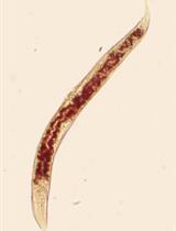
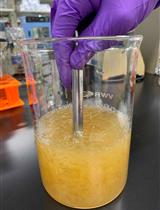
Total Triglyceride Quantification in Caenorhabditis elegans
秀丽隐杆线虫总甘油三酯的定量研究


Cleavable Affinity Purification (Cl-AP): A One-step Procedure to Affinity Purify Protein Complexes
CI-AP:一种亲和纯化蛋白质复合物的一步法
生物物理学
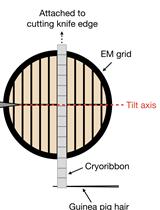
Serial Cryomicrotomy of Saccharomyces cerevisiae for Serial Electron Cryotomography
用于连续电子低温断层扫描的酿酒酵母连续冷冻切片
癌症生物学
Dissecting the Rat Mammary Gland: Isolation, Characterization, and Culture of Purified Mammary Epithelial Cells and Fibroblasts
解剖大鼠乳腺:纯化的乳腺上皮细胞和成纤维细胞的分离,鉴定和培养


Magnet-assisted Flow Cytometry of in vivo Tumors to Quantitate Cell-specific Responses to Magnetic Iron Oxide Nanoparticles
磁辅助流式细胞术检测体内肿瘤细胞以量化对磁性氧化铁纳米粒子的特异性反应

免疫学
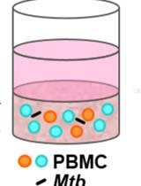
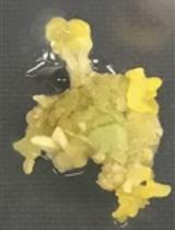
Generating Three-dimensional Human Granulomas in vitro to Study Mycobacterium tuberculosis-host Interaction
通过体外生成三维人体肉芽肿研究结核分枝杆菌与宿主的相互作用

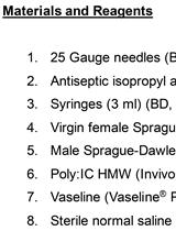
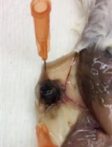
Maternal Immune Activation with the Viral Mimetic Poly:IC in Pregnant Rats
Poly:IC模拟病毒感染对孕鼠的母体免疫激活
微生物学
Rapid Isolation and Purification of Secreted Bacteriocins from Streptococcus mutans and Other Lactic Acid Bacteria
变形链球菌及其他乳酸菌分泌细菌素的快速分离纯化


A Quick Method for Screening Biocontrol Efficacy of Bacterial Isolates against Bacterial Wilt Pathogen Ralstonia solanacearum in Tomato
细菌分离物对番茄青枯病病原菌青枯雷尔氏菌生防效果的快速筛选方法


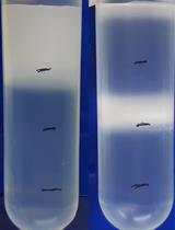
Giant Mimiviridae CsCl Purification Protocol
拟菌病毒科巨型病毒的CsCl纯化方案
神经科学
In vitro Time-lapse Imaging of Primary Cilium in Migrating Neuroblasts
迁移神经母细胞中初级纤毛的体外延时成像
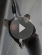
Confocal Microscopy of Reovirus Transport in Living Dorsal Root Ganglion Neurons
活体背根节神经元中呼肠孤病毒转运的共聚焦显微镜观察
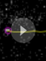
Visual-looming Shadow Task with in-vivo Calcium Activity Monitoring to Assess Defensive Behaviors in Mice
视觉变化阴影任务结合体内钙离子活性监测评估小鼠防御行为
植物科学
Agrobacterium-mediated Transformation of Sweet Basil (Ocimum basilicum)
农杆菌介导的罗勒(Ocimum basilicum)转化