往期刊物2020

卷册: 10, 期号: 21

left

Dec

20

Dec

5

Nov

20

Nov

5

Oct

20

Oct

5

Sep

20

Sep

5

Aug

20

Aug

5

Jul

20

Jul

5

Jun

20

Jun

5

May

20

May

5

Apr

20

Apr

5

Mar

20

Mar

5

Feb

20

Feb

5

Jan

20

Jan

5

right

生物化学

使用聚丙烯酰胺凝胶生成巨型磷脂囊泡(GUVs)

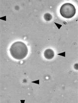
Generation of Giant Unilamellar Vesicles (GUVs) Using Polyacrylamide Gels

使用聚丙烯酰胺凝胶生成巨型磷脂囊泡(GUVs)

Eric Parigoris Eric Parigoris
Daniel L. Dunkelmann Daniel L. Dunkelmann
US Unai Silvan
4069 Views
Nov 5, 2020
Giant unilamellar vesicles (GUVs) are a widely used model system for a range of applications including membrane biophysics, drug delivery, and the study of actin dynamics. While several protocols have been developed for their generation in recent years, the use of these techniques involving charged lipid types and buffers of physiological ionic strength has not been widely adopted. This protocol describes the generation of large numbers of free-floating GUVs, even for charged lipid types and buffers of higher ionic strength, using a simple approach involving soft polyacrylamide (PAA) gels. This method entails glass cover slip functionalization with (3-Aminopropyl)trimethoxysilane (APTES) and glutaraldehyde to allow for covalent bonding of PAA onto the glass surface. After polymerization of the PAA, the gels are dried in vacuo. Subsequently, a lipid of choice is evenly dispersed on the dried gel surface, and buffers of varying ionic strength can be used to rehydrate the gels and form GUVs. This protocol is robust for the production of large numbers of free-floating GUVs composed of different lipid compositions under physiological conditions. It can conveniently be performed with commonly utilized laboratory reagents.

发育生物学

PLISH技术的研究进展

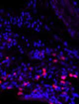
Advances in Proximity Ligation in situ Hybridization (PLISH)

PLISH技术的研究进展

MN Monica Nagendran
AA Adam M Andruska
PH Pehr B Harbury
TD Tushar J Desai
6013 Views
Nov 5, 2020
Understanding tissues in the context of development, maintenance and disease requires determining the molecular profiles of individual cells within their native in vivo spatial context. We developed a Proximity Ligation in situ Hybridization technology (PLISH) that enables quantitative measurement of single cell gene expression in intact tissues, which we have now updated. By recording spatial information for every profiled cell, PLISH enables retrospective mapping of distinct cell classes and inference of their in vivo interactions. PLISH has high sensitivity, specificity and signal to noise ratio. It is also rapid, scalable, and does not require expertise in molecular biology so it can be easily adopted by basic and clinical researchers.
DigiTAG–一种分析果蝇稀有细胞群转录组的RNA测序方法

DigiTAG–a RNA Sequencing Approach to Analyze Transcriptomes of Rare Cell Populations in Drosophila melanogaster

DigiTAG–一种分析果蝇稀有细胞群转录组的RNA测序方法

LL Lisa Landskron
FB Francois Bonnay
TB Thomas R. Burkard
JK Jürgen A. Knoblich
3139 Views
Nov 5, 2020
Cell-type specific transcriptional programs underlie the development and maintenance of organs. Not only distinct cell types within a tissue, even cells with supposedly identical cell fates show a high degree of transcriptional heterogeneity. Inevitable, low cell numbers are a major hurdle to study transcriptomes of pure cell populations. Here we describe DigiTAG, a high-throughput method that combines transposase fragmentation and molecular barcoding to retrieve high quality transcriptome data of rare cell types in Drosophila melanogaster. The protocol showcases how DigiTAG can be used to analyse the transcriptome of rare neural stem cells (type II neuroblasts) of Drosophila larval brains, but can also be utilized for other cell types or model systems.

免疫学

利用受激发射损耗显微镜对T细胞中央超分子信号簇的超分辨成像

Super-resolution Imaging of the T cell Central Supramolecular Signaling Cluster Using Stimulated Emission Depletion Microscopy

利用受激发射损耗显微镜对T细胞中央超分子信号簇的超分辨成像

ST Sin Lih Tan
DA Dominic Alibhai
SC Stephen J. Cross
HT Harry Thompson
CW Christoph Wülfing
3045 Views
Nov 5, 2020
Supramolecular signaling assemblies are of interest for their unique signaling properties. A µm scale signaling assembly, the central supramolecular signaling cluster (cSMAC), forms at the center interface of T cells activated by antigen presenting cells (APC). The adaptor protein linker for activation of T cells (LAT) is a key cSMAC component. The cSMAC has widely been studied using total internal reflection fluorescence microscopy of CD4+ T cells activated by planar APC substitutes. Here we provide a protocol to image the cSMAC in its cellular context at the interface between a T cell and an APC. Super resolution stimulated emission depletion microscopy (STED) was utilized to determine the localization of LAT, that of its active, phosphorylated form and its entire pool. Agonist peptide-loaded APCs were incubated with TCR transgenic CD4+ T cells for 4.5 min before fixation and antibody staining. Fixed cell couples were imaged using a 100x 1.4 NA objective on a Leica SP8 AOBS confocal laser scanning microscope. LAT clustered in multiple supramolecular complexes and their number and size distributions were determined. Using this protocol, cSMAC properties in its cellular context at the interface between a T cell and an APC could be quantified.

微生物学

严重急性呼吸综合征冠状病毒2(SARS-CoV-2)的RT-LAMP比色法检测

Colorimetric RT-LAMP Methods to Detect Severe Acute Respiratory Syndrome Coronavirus 2 (SARS-CoV-2)

严重急性呼吸综合征冠状病毒2(SARS-CoV-2)的RT-LAMP比色法检测

GP Gun-Soo Park
SB Seung-Hwa Baek
KK Keunbon Ku
SK Seong Jun Kim
SK Seung Il Kim
BK Bum-Tae Kim
JM Jin-Soo Maeng
4943 Views
Nov 5, 2020
Standard diagnostic methods of Coronavirus Disease 2019 (COVID-19) rely on RT-qPCR technique which have limited point-of-care test (POCT) potential due to necessity of dedicated equipment and specialized personnel. LAMP, an isothermal nucleic acid amplification test (NAAT), is a promising technique that may substitute RT-qPCR for POCT of genomic materials. Here, we provide a protocol to perform reverse transcription LAMP targeting SARS-CoV-2. We adopted both real-time fluorescence detection and end-point colorimetric detection approaches. Our protocol would be useful for screening diagnosis of COVID-19 and be a baseline for development of improved POCT NAAT.
白念珠菌培养、细胞收获和总RNA提取

Candida albicans Culture, Cell Harvesting, and Total RNA Extraction

白念珠菌培养、细胞收获和总RNA提取

MC Max V. Cravener
Aaron P. Mitchell Aaron P. Mitchell
4047 Views
Nov 5, 2020
Transcriptional analysis has become a cornerstone of biological research, and with the advent of cheaper and more efficient sequencing technology over the last decade, there exists a need for high-yield and efficient RNA extraction techniques. Fungi such as the human pathogen Candida albicans present a unique obstacle to RNA purification in the form of the tough cell wall made up of many different components such as chitin that are resistant to many common mammalian or bacterial cell lysis methods. Typical in vitro C. albicans cell harvesting methods can be time consuming and expensive if many samples are being processed with multiple opportunities for product loss or sample variation. Harvesting cells via vacuum filtration rather than centrifugation cuts down on time before the cells are frozen and therefore the available time for the RNA expression profile to change. Vacuum filtration is preferred for C. albicans for two main reasons: cell lysis is faster on non-pelleted cells due to increased exposed surface area, and filamentous cells are difficult to pellet in the first place unlike yeast or bacterial cells. Using mechanical cell lysis, by way of zirconia/silica beads, cuts down on time for processing as well as overall cost compared to enzymatic treatments. Overall, this method is a fast, efficient, and high-yield way to extract total RNA from in vitro cultures of C. albicans.
铜绿假单胞菌肺部感染的小鼠急性肺炎模型

Murine Acute Pneumonia Model of Pseudomonas aeruginosa Lung Infection

铜绿假单胞菌肺部感染的小鼠急性肺炎模型

XP Xiaolei Pan
WW Weihui Wu
3695 Views
Nov 5, 2020
Animal infection models play significant roles in studying bacterial pathogenic mechanisms, host pathogen interaction as well as evaluating drug and vaccine efficacies. We have been utilizing an acute pneumonia model to study bacterial colonization in lungs and assess virulence to the host by determination of bacterial loads and survival assays, as well as examine the bacterial gene expression in vivo. Additionally, the host's immune response to the pathogen can be explored through this infection model.
感染葡萄酒腐败细菌的噬菌体分离与冷冻电镜观察

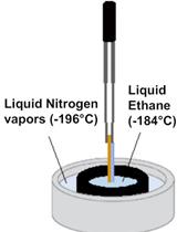
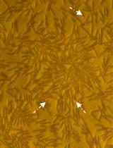
Isolation and CryoTEM of Phages Infecting Bacterial Wine Spoilers

感染葡萄酒腐败细菌的噬菌体分离与冷冻电镜观察

AC Amel Chaïb
MD Marion Decossas
CP Cécile Philippe
OC Olivier Claisse
OL Olivier Lambert
Claire Le Marrec Claire Le Marrec
3606 Views
Nov 5, 2020
With the objective to isolate phages infecting wine bacterial spoilers, we designed a method for the isolation and purification of phages infecting grape-associated bacteria. The method proved successful to isolate GC1 tectivirus infecting the acetic acid bacterium Gluconobacter cerinus. The isolated phage represents a new genus within the Tectiviridae, named "Gammatectivirus". Using a traditional technique for the concentration of phage particles involving several steps of centrifugation, further insights in the ultrastructure of GC1 could be observed by cryo electron microscopy, saving time and effort. The simple workflow presented may be applied to other viruses infecting bacteria inhabiting other vegetal niches.Graphic abstractFlowchart illustrating the protocol to isolate, concentrate and observe GC1 under cryo-EM
低成本、小规模、复合微生物连续培养系统的构建与验证

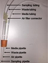
Construction and Validation of A Low-cost, Small-scale, Multiplex Continuous Culturing System for Microorganisms

低成本、小规模、复合微生物连续培养系统的构建与验证

Lilit Tonoyan Lilit Tonoyan
Freddy Guihéneuf Freddy Guihéneuf
RF Ruari Friel
VO Vincent O’Flaherty
3178 Views
Nov 5, 2020
This protocol details the construction of a simple, low-cost, small-scale, multiplex chemostat system designed for the continuous cultivation of microorganisms in suspension (i.e., bacteria, yeast, microalgae). The continuous culture device can operate at a working volume of 25 ml and allows the run of 8 chemostats in parallel by a single person. It provides a platform for parallel, long-term studies of evolution and adaptation of microorganisms under the stress of antimicrobial agents and/or toxic pollutants. The system complies with the varied needs of researchers for an accessible, highly-throughput and reliable tool that is nevertheless easy to construct, use and operate, and not demanding of space, materials, medium supply and workload. Here, we also validate the use of this system to generate de novo resistance towards a novel antimicrobial and a commonly used antibiotic in an antimicrobial-sensitive model organism. We believe that this "Do It Yourself" (DIY) system may constitute a useful tool to address the global problem of antibiotic resistance and to develop non-antibiotic based therapies.
嗜肺军团菌介导的泛素修饰和磷酸核糖泛素连接的体外谷氨酰化抑制

In vitro Glutamylation Inhibition of Ubiquitin Modification and Phosphoribosyl-Ubiquitin Ligation Mediated by Legionella pneumophila Effectors

嗜肺军团菌介导的泛素修饰和磷酸核糖泛素连接的体外谷氨酰化抑制

AS Alan G. Sulpizio
MM Marena E. Minelli
YM Yuxin Mao
2874 Views
Nov 5, 2020
Glutamylation is a posttranslational modification where the amino group of a free glutamate amino acid is conjugated to the carboxyl group of a glutamate side chain within a target protein. SidJ is a Legionella kinase-like protein that has recently been identified to perform protein polyglutamylation of the Legionella SdeA Phosphoribosyl-Ubiquitin (PR-Ub) ligase to inhibit SdeA’s activity. The attachment of multiple glutamate amino acids to the catalytic glutamate residue of SdeA by SidJ inhibits SdeA’s modification of ubiquitin (Ub) and ligation activity. In this protocol, we will discuss a SidJ non-radioactive, in vitro glutamylation assay using its substrate SdeA. This will also include a second reaction to assay the inhibition of SdeA by using both modification of free Ub and ligation of ADP-ribosylated Ubiquitin (ADPR-Ub) to SdeA’s substrate Rab33b. Prior to the identification and publication of SdeA’s activity, no SdeA inhibition assays existed. Our group and others have demonstrated various methods to display inhibition of SdeA’s activity. The alternatives include measurement of ADP-ribosylation of Ub using radioactive NAD, NAD hydrolysis, and Western blot analysis of HA-Ub ligation by SdeA. This protocol will describe the inhibition of both ubiquitin modification and the PR-Ub ligation by SdeA using inexpensive standard gels and Coomassie staining.

分子生物学

使用TubuleJ从冷冻电子显微镜图像确定微管晶格参数

Determination of Microtubule Lattice Parameters from Cryo-electron Microscope Images Using TubuleJ

使用TubuleJ从冷冻电子显微镜图像确定微管晶格参数

SK Siou Ku
CM Cédric Messaoudi
CG Charlotte Guyomar
CK Charles Kervrann
DC Denis Chrétien
3413 Views
Nov 5, 2020
The α-β tubulin heterodimer undergoes subtle conformational changes during microtubule assembly. These can be modulated by external factors, whose effects on microtubule structure can be characterized on 2D views obtained by cryo-electron microscopy. Analysis of microtubule images is facilitated if they are straight enough to interpret and filter their image Fourier transform, which provide useful information concerning the arrangement of tubulin molecules inside the microtubule lattice. Here, we describe the use of the TubuleJ software to straighten microtubules and determine their lattice parameters. Basic 3D reconstructions can be performed to evaluate the relevance of these parameters. This approach can be used to analyze the effects of nucleotide analogues, drugs or MAPs on microtubule structure, or to select microtubule images prior to high-resolution 3D reconstructions.
通过单纳米粒子追踪揭示细菌集群的长距离运输

Long-distance Transport in Bacterial Swarms Revealed by Single Nanoparticle Tracking

通过单纳米粒子追踪揭示细菌集群的长距离运输

JF Jingjing Feng
Yan He Yan He
2414 Views
Nov 5, 2020
During swarming, high density flagella-driven bacteria migrate collectively in a swirling pattern on wet agar surfaces, immersed in a thin viscous fluid layer called “swarm fluid”. Though the fluid environment has essential role in the emergence of swarming behavior, the microscopic mechanisms of it in mediating the cooperation of bacteria populations are not fully understood. Here, instead of micro-sized tracers used in previous research, we use gold nanorods as single particle tracers to probe the dynamics of the swarm fluid. This protocol includes five major parts: (1) the culture of swarming bacterial colony; (2) the preparations of gold nanorod tracers and the micro-spraying technique which are used to put the nanotracers into the upper fluid of bacterial swarms; (3) imaging and tracking; (4) other necessary control experiments; (5) data analysis and fitting of physical models. With this method, the nano-sized tracers could move long distances above motile cells without direct collisions with the bacteria bodies. In this way, the microscopic dynamics of the swarm fluid could be tracked with high spatiotemporal resolution. Moreover, the comprehensive analysis of multi-particle trajectories provides systematic visualization of the fluid dynamics. The method is promising to probe the fluid dynamics of other natural or artificial active matter systems.

植物科学

卵母细胞K+释放量及K+水平测定

K+ Release Assay and K+ Measurement in Oocyte Assay

卵母细胞K+释放量及K+水平测定

HL Hong Li
3113 Views
Nov 5, 2020
The Xenopus oocyte is a powerful system for the exogenous expression and functional characterization of plant membrane transport proteins. Until now, a number of potassium transporters and channels have been identified in oocytes expression system by the two-electrode voltage clamp technology. It is difficult to characterize K+/H+ anti-transporters, especially, electroneutral transporter. The K+ efflux assay system enables easy, fast, large-scale measurement of the transporters activity without two-electrode voltage clamp technology. This protocol describes a technique to measure the efflux activity of potassium transporter in oocytes expressing system.

系统生物学

利用k-mers进行测序读取的无参考关联映射

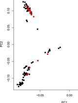
Reference-free Association Mapping from Sequencing Reads Using k-mers

利用k-mers进行测序读取的无参考关联映射

ZM Zakaria Mehrab
JM Jaiaid Mobin
IT Ibrahim Asadullah Tahmid
LP Lior Pachter
AR Atif Rahman
4742 Views
Nov 5, 2020
Association mapping is the process of linking phenotypes with genotypes. In genome wide association studies (GWAS), individuals are first genotyped using microarrays or by aligning sequenced reads to reference genomes. However, both these approaches rely on reference genomes which limits their application to organisms with no or incomplete reference genomes. To address this, reference free association mapping methods have been developed. Here we present the protocol of an alignment free method for association studies which is based on counting k-mers in sequenced reads, testing for associations between k-mers and the phenotype of interest, and local assembly of the k-mers of statistical significance. The method can map associations of categorical phenotypes to sequence and structural variations without requiring prior sequencing of reference genomes.