往期刊物2020

卷册: 10, 期号: 19

left

Dec

20

Dec

5

Nov

20

Nov

5

Oct

20

Oct

5

Sep

20

Sep

5

Aug

20

Aug

5

Jul

20

Jul

5

Jun

20

Jun

5

May

20

May

5

Apr

20

Apr

5

Mar

20

Mar

5

Feb

20

Feb

5

Jan

20

Jan

5

right

生物物理学

PIE-Scope的组装和成像设置

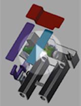
Assembly and Imaging Set up of PIE-Scope

PIE-Scope的组装和成像设置

SG Sergey Gorelick
DD David A. Dierickx
GB Genevieve Buckley
JW James C. Whisstock
AM Alex de Marco
4138 Views
Oct 5, 2020
Cryo-Electron Tomography (cryo-ET) is a method that enables resolving the structure of macromolecular complexes directly in the cellular environment. However, sample preparation for in situ Cryo-ET is labour-intensive and can require both cryo-lamella preparation through cryo-Focused Ion Beam (FIB) milling and correlative light microscopy to ensure that the event of interest is present in the lamella. Here, we present an integrated cryo-FIB and light microscope setup called the Photon Ion Electron microscope (PIE-scope) that enables direct and rapid isolation of cellular regions containing protein complexes of interest. The PIE-scope can be retrofitted on existing microscopes, although the drawings we provide are meant to work on ThermoFisher DualBeams with small mechanical modifications those can be adapted on other brands.

癌症生物学

一种新颖、可靠的数字微流控单细胞捕获方法

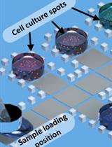
A Novel and Robust Single-cell Trapping Method on Digital Microfluidics

一种新颖、可靠的数字微流控单细胞捕获方法

JZ Jiao Zhai
HL Haoran Li
AW Ada Hang-Heng Wong
Cheng Dong Cheng Dong
SY Shuhong Yi
Yanwei  Jia Yanwei Jia
PM Pui-In Mak
CD Chuxia Deng
RM Rui Martins
3269 Views
Oct 5, 2020
Due to cell heterogeneity, the differences among individual cells are averaged out in bulk analysis methods, especially in the analysis of primary tumor biopsy samples from patients. To deeply understand the cell-to-cell variation in a primary tumor, single-cell culture and analysis with limited amount of cells are in high demand. Microfluidics has been an optimum platform to address the issue given its small reaction volume requirements. Digital microfluidics, which utilizes an electric signal to manipulate individual droplets has shown promise in cell-culture with easy controls. In this work, we realize single cell trapping on digital microfluidic platform by fabricating 3D microstructures on-chip to form semi-closed micro-wells. With this design, 20% of 30 x 30 array can be occupied by isolated single cells. We also use a low evaporation silicon oil and a fluorinated surfactant to lower the droplet actuation voltage and prevent the drop from evaporation, while allowing cell respiration during the long term of culture (24 h). The main steps for single cell trapping on digital microfluidics, as illustrated in this protocol, include 3D microstructures design, 3D microstructures construction on chip and oil film with surfactant for single cell trapping on chip.

细胞生物学

用于定量比率荧光分析的大肠杆菌GO-Matryoshka生物传感器亲和纯化

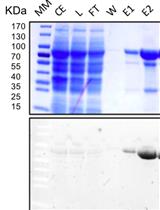
Affinity Purification of GO-Matryoshka Biosensors from E. coli for Quantitative Ratiometric Fluorescence Analyses

用于定量比率荧光分析的大肠杆菌GO-Matryoshka生物传感器亲和纯化

Mayuri Sadoine Mayuri Sadoine
VC Vanessa Castro-Rodríguez
TP Tobias Poloczek
HJ Helene Javot
ES Erdem Sunal
MW Michael M. Wudick
WF Wolf B. Frommer
3832 Views
Oct 5, 2020
Genetically encoded biosensors are powerful tools for quantitative visualization of ions and metabolites in vivo. Design and optimization of such biosensors typically require analyses of large numbers of variants. Sensor properties determined in vitro such as substrate specificity, affinity, response range, dynamic range, and signal-to-noise ratio are important for evaluating in vivo data. This protocol provides a robust methodology for in vitro binding assays of newly designed sensors. Here we present a detailed protocol for purification and in vitro characterization of genetically encoded sensors, exemplified for the His affinity-tagged GO-(Green-Orange) MatryoshCaMP6s calcium sensor. GO-Matryoshka sensors are based on single-step insertion of a cassette containing two nested fluorescent proteins, circularly permutated fluorescent green FP (cpGFP) and Large Stoke Shift LSSmOrange, within the binding protein of interest, producing ratiometric sensors that exploit the analyte-triggered change in fluorescence of a cpGFP.
双标记PDGFR-β和α-SMA在猪急性肾损伤模型中检测周细胞-肌成纤维细胞转化作为早期纤维化标志物的研究

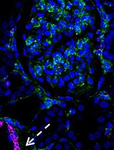
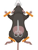
Double Labeling of PDGFR-β and α-SMA in Swine Models of Acute Kidney Injury to Detect Pericyte-to-Myofibroblast Transdifferentation as Early Marker of Fibrosis

双标记PDGFR-β和α-SMA在猪急性肾损伤模型中检测周细胞-肌成纤维细胞转化作为早期纤维化标志物的研究

AS Alessandra Stasi
Rossana  Franzin Rossana Franzin
CD Chiara Divella
LG Loreto Gesualdo
GS Giovanni Stallone
GC Giuseppe Castellano
3360 Views
Oct 5, 2020
Growing evidences suggest that peritubular capillaries pericytes are the main source of scar-forming myofibroblasts during chronic kidney disease (CKD), as well as early phases of acute kidney injury (AKI). In a swine model of sepsis and I/R (Ischemia Reperfusion) injury-induced AKI we demonstrated that renal pericytes are able to transdifferentiate toward α-SMA+ myofibroblasts leading to interstitial fibrosis. Even if precise pericytes identification requires transmission electron microscopy and the co-immunostaining of several markers (i.e., Gli, NG2 chondroitin sulphate proteoglycan, CD146, desmin or CD73) and emerging new markers (CD248 or TEM1, endosialin), previous studies suggested that PDGFR-β could be used as marker for renal pericytes characterization. Recently, double immunofluorescence staining of PDGFR-β and α-SMA was performed to identify the damage activated pericytes (PDGFR-β+/α-SMA+ cells) in the early phase of fibrosis development. Our data highlighted the crucial role of renal pericytes in the physiopathology of sepsis and I/R associated AKI. In this protocol, we describe the procedure for double immunofluorescence staining of PDGFR-β and α-SMA in swine Formalin-Fixed Paraffin-Embedded (FFPE) kidney biopsies and the method for image analysis and quantification.

发育生物学

海星卵母细胞内pH值的荧光测定与校准

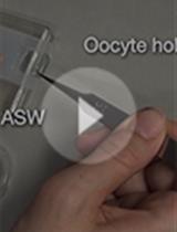
Fluorescence Measurement and Calibration of Intracellular pH in Starfish Oocytes

海星卵母细胞内pH值的荧光测定与校准

Enako  Hosoda Enako Hosoda
KC Kazuyoshi Chiba
3568 Views
Oct 5, 2020
Oocyte maturation is a process wherein an oocyte arrested at prophase of meiosis I resumes meiosis to become a fertilizable egg. In starfish ovaries, a hormone released from follicle cells activates the oocytes, resulting in an increase in their intracellular pH (pHi), which is required for spindle assembly. Herein, we describe a protocol for pHi measurement in living oocytes microinjected with the pH-sensitive dye BCECF. For in vivo BCECF calibration, we treated oocytes with artificial seawater containing CH3COONH4 to clamp pHi, injected pH-standard solutions, and converted the BCECF fluorescence intensity ratios to pHi values. Of note, if the actual pHi is higher or lower than the known pH of injected standard solutions, the BCECF fluorescence intensity ratio will decrease or increase, respectively. On the other hand, the pH of the injected solution displaying no change in fluorescence intensity should be considered the actual pHi. These methods for pHi calibration and clamping are simple and reproducible.

微生物学

革兰阴性菌肽聚糖的超高效液相色谱法分析

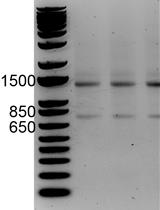
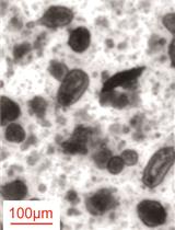
Analysis of Gram-negative Bacteria Peptidoglycan by Ultra-performance Liquid Chromatography

革兰阴性菌肽聚糖的超高效液相色谱法分析

LA Laura Alvarez
BC Baptiste Cordier
Sv Sven van Teeffelen
FC Felipe Cava
4456 Views
Oct 5, 2020
Bacteria are surrounded by a protective peptidoglycan cell wall. Provided that this structure and the enzymes involved are the preferred target for our most successful antibiotics, determining its structural and chemical complexity is of the highest interest. Traditionally, high-performance liquid chromatography (HPLC) analyses have been performed, but these methods are very time consuming in terms of sample preparation and chromatographic separation. Here we describe an optimized method for preparation of Gram-negative bacteria peptidoglycan and its subsequent analysis by ultra-performance liquid chromatography (UPLC). The use of UPLC in peptidoglycan analyses provides a dramatic reduction of the sample volume and hands-on time required and, furthermore, permits in-line mass spectrometry (MS) of the UPLC resolved muropeptides, thus facilitating their identification. This method improves our capability to perform high throughput analysis to better understand the cell-wall biology.
嗜肺军团菌效应蛋白SidJ体外谷氨酰化活性的放射性测定

Radioactive Assay of in vitro Glutamylation Activity of the Legionella pneumophila Effector Protein SidJ

嗜肺军团菌效应蛋白SidJ体外谷氨酰化活性的放射性测定

AS Alan G. Sulpizio
JS Jung-Ho Shin
MM Marena E. Minelli
YM Yuxin Mao
3109 Views
Oct 5, 2020
The Legionella effector protein SidJ has recently been identified to perform polyglutamylation on another Legionella effector, SdeA, ablating SdeA’s activity. SidJ is a kinase-like protein that requires the small eukaryotic protein calmodulin to perform glutamylation. Glutamylation is a relatively uncommon type of post-translational modification, where the amino group of a free glutamate amino acid is covalently linked to the γ-carboxyl group of a glutamate sidechain in a substrate protein. This protocol describes the SidJ glutamylation reaction using radioactive [U-14C] glutamate and its substrate SdeA, the separation of proteins by gel electrophoresis, preparation of gels for radioactive exposure, and relative quantification of glutamylation activity. This procedure is useful for the identification of substrates for glutamylation, characterization of substrate and glutamylase activities due to mutations, and identification of proteins with glutamylation activity. Some studies have assayed glutamylation with the use of [3H] glutamate (Regnard et al., 1998) and the use of the GT335 antibody (Wolff et al., 1992). However, the use of [U-14C] glutamate requires a shorter radioactive exposure time with no dependence on antibody specificity.
以小鼠霍乱弧菌感染为模型系统的TetR调控体内抑制技术鉴定基因工程菌的条件基因沉默

TetR Regulated in vivo Repression Technology to Identify Conditional Gene Silencing in Genetically Engineerable Bacteria Using Vibrio cholerae Murine Infections as Model System

以小鼠霍乱弧菌感染为模型系统的TetR调控体内抑制技术鉴定基因工程菌的条件基因沉默

FZ Franz G. Zingl
FM Fabian Mitterer
HT Himadri B. Thapa
SS Stefan Schild
2746 Views
Oct 5, 2020
Investigation of bacterial gene regulation upon environmental changes is still a challenging task. For example, Vibrio cholerae, a pathogen of the human gastrointestinal tract, faces diverse transient conditions in different compartments upon oral ingestion. Genetic reporter systems have been demonstrated to be extremely powerful tools to unravel gene regulation events in complex conditions, but so far focused mainly on gene induction. Herein, we describe the TetR-controlled recombination-based in vivo expression technology TRIVET, which allows detection of gene silencing events. TRIVET resembles a modified variant of the in vivo expression technology (IVET) as well as recombination-based in vivo expression technology (RIVET), which were used to identify conditional gene induction in several bacteria during host colonization. Like its predecessors, TRIVET is a single cell based reporter system, which allows the analysis of bacterial gene repression in a spatiotemporal manner via phenotypical changes in the resistance profile. Briefly, a promoterless tetR (encoding the transcriptional repressor TetR) can be integrated randomly into the bacterial genome via transposon mutagenesis or site-specific downstream of a promoter of interest via homologous recombination. Reduction of transcriptional expression of TetR results in a de-repression of the TetR-controlled resolvase TnpR, which in turn leads to excision of an antibiotic resistance cassette (also known as res-cassette) and altered resistance profile observable via streaking on ampicillin and kanamycin plates. This alteration can then be quantified as the ratio between resistant and non-resistant isolates. Furthermore, the newly introduced second reporter gene, a promoterless phoA (encoding the alkaline phosphatase PhoA) offers an additional validation step of the results via an independent colorimetric assay to measure enzyme activity. The protocol presented herein also offers an approach to identify the gene locus in case of the random screen for gene repression as well as a quantification of the conditional repression of a gene of interest. Although the current protocol is established for gene repression during host colonization, it can likely be adapted to study gene silencing under various conditions faced by a bacterium.
用于定量含氧光颗粒静水压力的简单时差成像

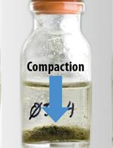
Simple Time-lapse Imaging for Quantifying the Hydrostatic Production of Oxygenic Photogranules

用于定量含氧光颗粒静水压力的简单时差成像

Esmee D. Joosten Esmee D. Joosten
Jérôme Hamelin Jérôme Hamelin
Kim Milferstedt Kim Milferstedt
2710 Views
Oct 5, 2020
Oxygenic photogranules (OPGs) are dense, three-dimensional aggregates containing a syntrophic, light-driven microbial community. Their temporal and spatial development interests microbial ecologists working at the bioprocess engineering interface, as this knowledge can be used to optimize biotechnological applications, such as wastewater treatment and biomass valorization. The method presented here enables the high-throughput quantification of photogranulation. OPGs are produced from a loose sludge-like microbial matrix in hydrostatic batch cultures exposed to light. This matrix transforms into a consolidated, roughly spherical aggregate over time. Photogranulation is quantified by time-lapse imaging coupled to automated image analysis. This allows studying the development of many OPGs simultaneously and in a fully automated way to systematically test what factors drive photogranulation. The protocol can also be used to quantify other types of (a)biotic aggregation.

分子生物学

应用RNA测序和RNAs峰值检测lncRNAs和mRNAs的细胞内丰度

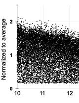
Using RNA Sequencing and Spike-in RNAs to Measure Intracellular Abundance of lncRNAs and mRNAs

应用RNA测序和RNAs峰值检测lncRNAs和mRNAs的细胞内丰度

Megan D. Schertzer Megan D. Schertzer
MM McKenzie M. Murvin
JC J. Mauro Calabrese
4413 Views
Oct 5, 2020
Long noncoding RNAs (lncRNAs) play essential roles in normal physiology and in disease but their mechanisms of action can be challenging to identify. For mechanistic studies, it is often useful to know a lncRNA’s intracellular abundance, i.e., approximately how many molecules of the lncRNA are present in a typical cell of a cell-type of interest. At least two approaches have been used to approximate lncRNA intracellular abundance: single-molecule sensitivity RNA fluorescence in situ hybridization (smFISH) and single-gene, calibrated reverse-transcription followed by quantitative PCR (RT-qPCR). However, like all experimental approaches, these methods have their limitations. smFISH, when analyzed using diffraction-limited microscopy, can underestimate intracellular abundance, especially for lncRNAs that accumulate in focused subcellular regions. Calibrated RT-qPCR may return inaccurate estimates of abundance because individual PCR amplicons spaced across the length of a transcript can vary in their efficiency of reverse transcription. Here, we describe a sequencing-based approach that is straightforward, orthogonal to smFISH and RT-qPCR, and can be used to approximate the intracellular abundance for most expressed long RNAs (lncRNAs and mRNAs) in a cell type of interest. Firstly, the average weight of total RNA per cell for the cell type of interest is estimated by replicate rounds of RNA purification from a known number of cells. Secondly, an rRNA-depletion RNA-Seq protocol is performed after adding spike-in control RNAs to a known quantity of total cellular RNA. Lastly, by comparing read counts per transcript to a standard curve derived from the spiked-in RNAs, the intracellular abundance for each transcript is estimated. The sequencing-based approach provides a powerful complement to existing methods, particularly in situations where it is desirable to quantify the abundance of multiple lncRNAs and/or mRNAs simultaneously.

神经科学

操作式条件反射模型结合化学遗传学方法研究小鼠食物成瘾的神经生物学机制

An Operant Conditioning Model Combined with a Chemogenetic Approach to Study the Neurobiology of Food Addiction in Mice

操作式条件反射模型结合化学遗传学方法研究小鼠食物成瘾的神经生物学机制

EM Elena Martín-García
LD Laura Domingo-Rodriguez
RM Rafael Maldonado
4848 Views
Oct 5, 2020
The study of food addiction comprises 3 hallmarks that include the persistence to response without an outcome, the strong motivation for palatable food, and the loss of inhibitory control over food intake that leads to compulsive behavior in addicted individuals. The complex multifactorial nature of this disorder and the unknown neurobiological mechanistic correlation explains the lack of effective treatments. Our operant conditioning model allows deciphering why some individuals are vulnerable and develop food addiction while others are resilient and do not. It is a translational approach since it is based on the Diagnostic and Statistical Manual of Mental Disorders 5th edition (DSM-5) and the Yale Food Addiction Scale (YFAS 2.0). This model allows to evaluate the addiction criteria in 2 time-points at an early and a late period by grouping them into 1) persistence to response during a period of non-availability of food, 2) motivation for food with a progressive ratio, and 3) compulsivity when the reward is associated with a punishment such as an electric foot-shock. The advantage of this model is that it allows us to measure 4 phenotypic traits suggested as predisposing factors related to vulnerability to addiction. Also, it is possible to evaluate the long food addiction mouse model with mice genetically modified. Importantly, the novelty of this protocol is the adaptation of this food addiction model to a short protocol to evaluate genetic manipulations targeting specific brain circuitries by using a chemogenetic approach that could promote the rapid development of this addictive behavior. These adaptations lead to a short food addiction mouse protocol, in which mice follow the same behavioral procedure of the early period in the long food addiction protocol with some variations due to the surgical viral vector injection. To our knowledge, there is no paradigm in mice allowing us to study the combination of such a robust behavioral approach that allows uncovering the neurobiology of food addiction at the brain circuit level. We can study using this protocol if modifying the excitability of a specific brain network confers resilience or vulnerability to developing food addiction. Understanding these neurobiological mechanisms is expected to help to find novel and efficient interventions to battle food addiction.
成年小鼠海马脑片的制备及其锐波波纹的研究

Preparing Viable Hippocampal Slices from Adult Mice for the Study of Sharp Wave-ripples

成年小鼠海马脑片的制备及其锐波波纹的研究

LL Linhua Liu
XZ Xiaojing Zhou
Jian-young Wu Jian-young Wu
4417 Views
Oct 5, 2020
We describe a protocol for preparing acute brain slices which can produce robust hippocampal sharp wave-ripples (SWRs) in vitro. The protocol is optimized for its simplicity and reliability for the preparation of solutions, slicing, and recovery incubation. Most slices in almost every mouse prepared though the protocol expressed vigorous spontaneous SWRs for ~24 h, compared to the 20-30% viability from "standard" low sodium slicing protocols. SWRs are spontaneous neuronal activity in the hippocampus and are essential for consolidation of episodic memory. Brain slices reliably expressing SWRs are useful for studying memory impairment and brain degeneration diseases in ex vivo experiments. Spontaneous expression of SWRs is sensitive to conditions of slicing and perfusion/oxygenation during recording. The amplitude and abundance of SWRs are often used as a biomarker for viable slices. Key improvements include fast circulation, a long recovery period (3-6 h) after slicing, and allowing tissue to recover at 32 °C in a well perfused incubation chamber. Slices in our custom-made apparatus can express spontaneous SWRs for many hours, suggesting a long period with balanced excitation and inhibition in the local networks. Slices from older mice (~postnatal 180 days) show similar viability to younger (postnatal 21-30) mice.

植物科学

植物细胞壁多糖共聚焦和超分辨成像的多靶点免疫组织化学研究

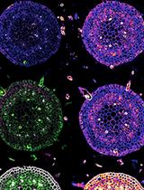
Multitarget Immunohistochemistry for Confocal and Super-resolution Imaging of Plant Cell Wall Polysaccharides

植物细胞壁多糖共聚焦和超分辨成像的多靶点免疫组织化学研究

KH Kalina T. Haas
MR Methieu Rivière
RW Raymond Wightman
AP Alexis Peaucelle
5981 Views
Oct 5, 2020
The plant cell wall (PCW) is a pecto-cellulosic extracellular matrix that envelopes the plant cell. By integrating extra-and intra-cellular cues, PCW mediates a plethora of essential physiological functions. Notably, it permits controlled and oriented tissue growth by tuning its local mechano-chemical properties. To refine our knowledge of these essential properties of PCW, we need an appropriate tool for the accurate observation of the native (in muro) structure of the cell wall components. The label-free techniques, such as AFM, EM, FTIR, and Raman microscopy, are used; however, they either do not have the chemical or spatial resolution. Immunolabeling with electron microscopy allows observation of the cell wall nanostructure, however, it is mostly limited to single and, less frequently, multiple labeling. Immunohistochemistry (IHC) is a versatile tool to analyze the distribution and localization of multiple biomolecules in the tissue. The subcellular resolution of chemical changes in the cell wall component can be observed with standard diffraction-limited optical microscopy. Furthermore, novel chemical imaging tools such as multicolor 3D dSTORM (Three-dimensional, direct Stochastic Optical Reconstruction Microscopy) nanoscopy makes it possible to resolve the native structure of the cell wall polymers with nanometer precision and in three dimensions.Here we present a protocol for preparing multi-target immunostaining of the PCW components taking as example Arabidopsis thaliana, Star fruit (Averrhoa carambola), and Maize thin tissue sections. This protocol is compatible with the standard confocal microscope, dSTORM nanoscope, and can also be implemented for other optical nanoscopy such as STED (Stimulated Emission Depletion Microscopy). The protocol can be adapted for any other subcellular compartments, plasma membrane, cytoplasmic, and intracellular organelles.
植物根尖中黄酮醇、山奈酚和槲皮素的染色方法

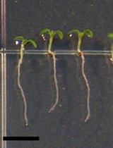
A Protocol for Flavonols, Kaempferol and Quercetin, Staining in Plant Root Tips

植物根尖中黄酮醇、山奈酚和槲皮素的染色方法

NN Nguyen Hoai Nguyen
5147 Views
Oct 5, 2020
Flavonols are a subclass of flavonoids of the group of plant secondary metabolites. In planta, flavonols play various functions such as antioxidant and natural regulator of auxin polar transport. Many lines of evidence have shown that flavonols also contribute to human health in anti-oxidation, anti-inflammation, and even prevention some types of cancer. Several methods have been utilized to measure flavonols such as high-performance liquid chromatography (HPLC), liquid chromatography-mass spectrometry (LC-MS), and diphenylboric acid-2-aminoethyl ester (DPBA) staining. While HPLC or LC-MS can quantitatively determine the level of flavonols, DPBA staining can provide an in-situ view of flavonols accumulation in the plants. In this protocol, a detailed procedure for staining the flavonols in Arabidopsis root tips is described. Five-day-old Arabidopsis seedlings are soaked in a solution containing DPBA and latterly the flavonols (kaempferol and quercetin) can be observed under a confocal microscope.

干细胞

基于Fluidigm的源于患者小肠类器官单细胞基因表达文库的制备

Fluidigm Based Single-cell Gene Expression Library Preparation from Patient-derived Small Intestinal Organoids

基于Fluidigm的源于患者小肠类器官单细胞基因表达文库的制备

KS Kohei Suzuki
RO Ryuichi Okamoto
3750 Views
Oct 5, 2020
In this protocol, we describe our methods to isolate crypts from patients' biopsy samples and to culture human intestinal stem cells as it’s called “organoid.” Beyond that, we describe how to dissociate organoids cells into single cells for single-cell analysis as a further application. This protocol should provide investigators sufficient tools to generate human organoids from biopsy samples and to accomplish a stable in-vitro assay system.