往期刊物2020

卷册: 10, 期号: 17

left

Dec

20

Dec

5

Nov

20

Nov

5

Oct

20

Oct

5

Sep

20

Sep

5

Aug

20

Aug

5

Jul

20

Jul

5

Jun

20

Jun

5

May

20

May

5

Apr

20

Apr

5

Mar

20

Mar

5

Feb

20

Feb

5

Jan

20

Jan

5

right

生物物理学

LOVTRAP对双层膜蛋白质结合的光遗传学调控

Optogenetic Tuning of Protein-protein Binding in Bilayers Using LOVTRAP

LOVTRAP对双层膜蛋白质结合的光遗传学调控

DT Doug Tischer
OW Orion D. Weiner
4819 Views
Sep 5, 2020
Modern microscopy methods are powerful tools for studying live cell signaling and biochemical reactions, enabling us to observe when and where these reactions take place from the level of a cell down to single molecules. With microscopy, each cell or molecule can be observed both before and after a given perturbation, facilitating better inference of cause and effect than is possible with destructive modes of signaling quantitation. As many inputs to cell signaling and biochemical systems originate as protein-protein interactions near the cell membrane, an outstanding challenge lies in controlling the timing, location and the magnitude of protein-protein interactions in these unique environments. Here, we detail our procedure for manipulating such spatial and temporal protein-protein interactions in a closed microscopy system using a LOVTRAP-based light-responsive protein-protein interaction system on a supported lipid bilayer. The system responds in seconds and can pattern details down to the one micron level. We used this technique to unlock fundamental aspects of T cell signaling, and this approach is generalizable to many other cell signaling and biochemical contexts.

细胞生物学

基于DNA传感器的真核细胞与病原体受体特异性粘附力的体外检测

An in vitro DNA Sensor-based Assay to Measure Receptor-specific Adhesion Forces of Eukaryotic Cells and Pathogens

基于DNA传感器的真核细胞与病原体受体特异性粘附力的体外检测

MW Maurizio Wack
TW Tina Wiegand
FF Friedrich Frischknecht
EC E. Ada Cavalcanti-Adam
4304 Views
Sep 5, 2020
Motility of eukaryotic cells or pathogens within tissues is mediated by the turnover of specific interactions with other cells or with the extracellular matrix. Biophysical characterization of these ligand-receptor adhesions helps to unravel the molecular mechanisms driving migration. Traction force microscopy or optical tweezers are typically used to measure the cellular forces exerted by cells on a substrate. However, the spatial resolution of traction force microscopy is limited to ~2 µm and performing experiments with optical traps is very time-consuming. Here we present the production of biomimetic surfaces that enable specific cell adhesion via synthetic ligands and at the same time monitor the transmitted forces by using molecular tension sensors. The ligands were coupled to double-stranded DNA probes with defined force thresholds for DNA unzipping. Receptor-mediated forces in the pN range are thereby semi-quantitatively converted into fluorescence signals, which can be detected by standard fluorescence microscopy at the resolution limit (~0.2 µm). The modular design of the assay allows to vary the presented ligands and the mechanical strength of the DNA probes, which provides a number of possibilities to probe the adhesion of different eukaryotic cell types and pathogens and is exemplified here with osteosarcoma cells and Plasmodium berghei Sporozoites.
组蛋白转换的定量动力学成像及流式细胞术研究

Quantitative Kinetic Analyses of Histone Turnover Using Imaging and Flow Cytometry

组蛋白转换的定量动力学成像及流式细胞术研究

HS Hanae Sato
RS Robert H. Singer
JG John M. Greally
4031 Views
Sep 5, 2020
Dynamic histone changes occur as a central part of chromatin regulation. Deposition of histone variants and post-translational modifications of histones are strongly associated with properties of chromatin status. Characterizing the kinetics of histone variants allows important insights into transcription regulation, chromatin maintenance and other chromatin properties. Here we provide a protocol of quantitative and sensitive approaches to test the timing of incorporation and dissociation of histones using a two-color SNAP-labeling system, labelling pre-existing and newly-incorporated histones distinctly. Together with cell cycle synchronization methods and cell cycle markers, this approach enables a pulse-chase analysis to determine the turnover of histone variants during the cell cycle, detected using imaging or flow cytometry methods at single cell resolution. As well as testing global histone turnover, cell cycle-dependent cellular localization of histone variants can be also addressed using imaging approaches.

发育生物学

秀丽隐杆线虫荧光多聚体分析

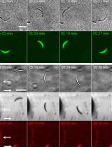
Fluorescent Polysome Profiling in Caenorhabditis elegans

秀丽隐杆线虫荧光多聚体分析

DS Dan Shaffer
Jarod A Rollins Jarod A Rollins
6643 Views
Sep 5, 2020
An important but often overlooked aspect of gene regulation occurs at the level of protein translation. Many genes are regulated not only by transcription but by their propensity to be recruited to actively translating ribosomes (polysomes). Polysome profiling allows for the separation of unbound 40S and 60S subunits, 80S monosomes, and actively translating mRNA bound by two or more ribosomes. Thus, this technique allows for actively translated mRNA to be isolated. Transcript abundance can then be compared between actively translated mRNA and all mRNA present in a sample to identify instances of post-transcriptional regulation. Additionally, polysome profiling can be used as a readout of global translation rates by quantifying the proportion of actively translating ribosomes within a sample. Previously established protocols for polysome profiling rely on the absorbance of RNA to visualize the presence of polysomes within the fractions. However, with the advent of flow cells capable of detecting fluorescence, the association of fluorescently tagged proteins with polysomes can be detected and quantified in addition to the absorbance of RNA. This protocol provides detailed instructions on how to perform fluorescent polysome profiling in C. elegans to collect actively translated mRNA, to quantify changes in global translation, and to detect ribosomal binding partners.

免疫学

细胞系和原代免疫细胞中的低水平活性氧的流式细胞仪检测

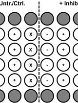
Flow-cytometric Detection of Low-level Reactive Oxygen Species in Cell Lines and Primary Immune Cells

细胞系和原代免疫细胞中的低水平活性氧的流式细胞仪检测

Kevin  Bode Kevin Bode
CL Corinna Link
PK Peter H. Krammer
HW Heiko Weyd
5897 Views
Sep 5, 2020
Depending on its concentration and cellular origin the production of reactive oxygen species (ROS) in the organism serves a variety of functions. While high concentrations during an oxidative burst are used to fight pathogens, low to moderate amounts of ROS act as signaling molecules important for several physiological processes such as regulation of immune responses. The ROS-sensitive dye 2',7'-dichlorodihydrofluorescein diacetate (H2DCFDA) is an inexpensive and well-established tool for measuring intracellular ROS levels. However, it needs to be carefully controlled to be able to draw firm conclusions on the nature of ROS species produced and the cellular source of ROS generation such as the enzyme complex NADPH-oxidase 2 (NOX-2). In this protocol, a robust method to determine low intracellular ROS production using H2DCFDA was validated by several ROS-specific as well as NOX-2-specific inhibitors. Cells were treated with inhibitors or control substances prior to treatment with the ROS-inducer of interest. H2DCFDA was added only for the last 30 min of the treatment schedule. To terminate its conversion, we used a ROS-specific inhibitor until analysis by flow cytometry within the FITC-channel (Ex: 488 nm/Em: 519 nm). In summary, this protocol allows the detection of signaling-relevant intracellular ROS production in cell lines and primary immune cells (e.g., Mono Mac 6 cells and Bone marrow-derived dendritic cells, respectively). Using this method in combination with specific inhibitors, we were able to validate even exceptionally low amounts of ROS produced by NOX-2 and relevant for immune-regulatory signaling.
人畜共患病疟原虫——诺氏疟原虫增殖和生长抑制活性测定

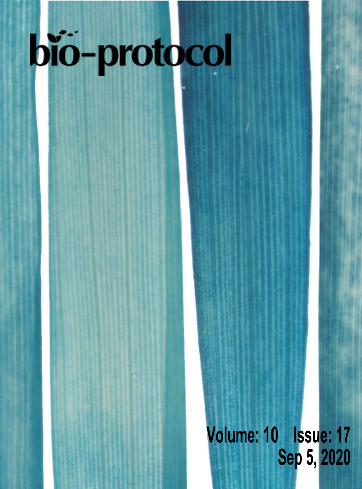
Multiplication and Growth Inhibition Activity Assays for the Zoonotic Malaria Parasite, Plasmodium knowlesi

人畜共患病疟原虫——诺氏疟原虫增殖和生长抑制活性测定

FM Franziska Mohring
TR Thomas A. Rawlinson
SD Simon J. Draper
RM Robert W. Moon
4817 Views
Sep 5, 2020
Malaria remains a major cause of morbidity and mortality globally. Clinical symptoms of the disease arise from the growth and multiplication of Plasmodium parasites within the blood of the host. Thus in vitro assays to determine how drug, antibody and genetic perturbations affect the growth rate of Plasmodium parasites are essential for the development of new therapeutics and improving our understanding of parasite biology. As both P. falciparum and P. knowlesi can be maintained in culture with human red blood cells, the effect of antimalarial drugs and inhibitory antibodies that target the invasion or growth capacity of Plasmodium parasites are routinely investigated by using multiplication assays or growth inhibition activity (GIA) assays against these two species. This protocol gives detailed step-by-step procedures to carry out flow cytometry-based multiplication assays and growth inhibition activity assays to test neutralizing antibodies based on the activity of the parasite enzyme lactate dehydrogenase of Plasmodium knowlesi adapted to human red blood cell culture. Whilst similar assays are well established for P. falciparum, P. knowlesi is more closely related to all other human infective species (Pacheco et al., 2018) and so can be used as a surrogate for testing drugs and vaccines for other malaria species such as P. vivax, which is the most widespread cause of malaria outside of Africa, but cannot yet be cultured under laboratory conditions.

微生物学

人单纯疱疹病毒CRISPR/Cas9抑制作用的筛选方法

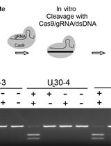
Screening Method for CRISPR/Cas9 Inhibition of a Human DNA Virus: Herpes Simplex Virus

人单纯疱疹病毒CRISPR/Cas9抑制作用的筛选方法

WN Werner M. Neuhausser
HO Hyung S. Oh
PE Pierce Eggan
MA Magdalena Angelova
RK Rory Kirchner
KE Kevin C. Eggan
DK David M. Knipe
5576 Views
Sep 5, 2020
The efficiency of cleavage of individual CRISPR/Cas9-sgRNAs remains difficult to predict based on the CRISPR target sequence alone. Different intracellular environments (dependent on cell type or cell cycle state for example) may affect sgRNA efficiency by altering accessibility of genomic DNA through DNA modifications such as epigenetic marks and DNA-binding proteins (e.g., histones) as well as alteration of the chromatin state of genomic DNA within the nucleus. We recently reported a multi-step screening method for the identification of efficient sgRNAs targeting the Herpes simplex virus (HSV-1) genome and reported a differential mechanism for viral inhibition by CRISPR-Cas9 in the latent versus lytic phase. The screening platform detailed in this protocol allows step-by-step testing of the efficiency of cleavage in a cell-free system and in the context of viral target cells such as human foreskin fibroblasts followed by functional testing of the effects of CRISPR/sgRNA on viral protein expression, replication, and reactivation. This strategy could be readily applied to other target cells such as pluripotent stem cell-derived human sensory neurons or other human DNA viruses.
超分辨率共聚焦显微镜活体细胞成像(SCLIM):高时空分辨率的三色、四维同步活体细胞成像

Live-cell Imaging by Super-resolution Confocal Live Imaging Microscopy (SCLIM): Simultaneous Three-color and Four-dimensional Live Cell Imaging with High Space and Time Resolution

超分辨率共聚焦显微镜活体细胞成像(SCLIM):高时空分辨率的三色、四维同步活体细胞成像

KK Kazuo Kurokawa
AN Akihiko Nakano
4813 Views
Sep 5, 2020
Many questions in cell biology can be solved by state-of-the-art technology of live cell imaging. One good example is the mechanism of membrane traffic, in which small membrane carriers are rapidly moving around in the cytoplasm to deliver cargo proteins between organelles. For directly visualizing the events in membrane trafficking system, researchers have long awaited the technology that enables simultaneous multi-color and four-dimensional observation at high space and time resolution. Super-resolution microscopy methods, for example STED, PALM/STORM, and SIM, provide greater spatial resolution, however, these methods are not enough in temporal resolution. The super-resolution confocal live imaging microscopy (SCLIM) that we developed has now achieved the performance required. By using SCLIM, we have conducted high spatiotemporal visualization of secretory cargo together with early and late Golgi resident proteins tagged with three different fluorescence proteins. We have demonstrated that secretory cargo is indeed delivered within the Golgi by cisternal maturation. In addition, we have visualized details of secretory cargo trafficking in the Golgi, including formation of zones within a maturing cisterna, in which Golgi resident proteins are segregated, and movement of cargo between these zones. This protocol can be used for simultaneous three-color and four-dimensional observation of various phenomena in living cells, from yeast to higher plants and animals, at high spatiotemporal resolution.
一个高通量细菌间竞争平台

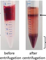
A High-throughput Interbacterial Competition Platform

一个高通量细菌间竞争平台

Hsiao-Han Lin Hsiao-Han Lin
Erh-Min  Lai Erh-Min Lai
4692 Views
Sep 5, 2020
Contact-dependent interbacterial competition is a common strategy used by bacteria to fight for their ecological niches. Interbacterial competition is monitored by a competition assay involving co-culturing the attacker and the recipient bacterial cells on agar, followed by recovery of the surviving recipient cells. Conventional interbacterial competition assays rely on serial dilution, plate spreading, and colony counting experiments for the readout. The high demand for time and labor in a competition assay limits its use for large-scale screening. However, a high-throughput interbacterial competition screening method is required to screen genetic factors involved in an interbacterial competition. Here, using Agrobacterium tumefaciens as an attacker and Escherichia coli as a recipient, we developed a robust, fast, efficient, and high-throughput type VI secretion system-dependent interbacterial competition screening platform. This system allows for 96 simultaneous competition assays without the need for serial dilution and plate spreading. Data analysis of this system relies on only direct and straightforward colony counting. This platform may be easily adapted to identify novel factors involved in any contact-dependent interbacterial competition systems.

分子生物学

BRIDGE:一个可重复的蛋白质配体模拟和结合自由能计算的开放平台

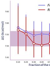
BRIDGE: An Open Platform for Reproducible Protein-Ligand Simulations and Free Energy of Binding Calculations

BRIDGE:一个可重复的蛋白质配体模拟和结合自由能计算的开放平台

TS Tharindu Senapathi
Christopher B. Barnett Christopher B. Barnett
Kevin J. Naidoo Kevin J. Naidoo
7744 Views
Sep 5, 2020
Protein-ligand binding prediction is central to the drug-discovery process. This often follows an analysis of genomics data for protein targets and then protein structure discovery. However, the complexity of performing reproducible protein conformational analysis and ligand binding calculations, using vetted methods and protocols can be a challenge. Here we show how Biomolecular Reaction and Interaction Dynamics Global Environment (BRIDGE), an open-source web-based compute and analytics platform for computational chemistry developed based on the Galaxy bioinformatics platform, makes protocol sharing seamless following genomics and proteomics. BRIDGE makes available tools and workflows to carry out protein molecular dynamics simulations and accurate free energy computations of protein-ligand binding. We illustrate the dynamics and simulation protocols for predicting protein-ligand binding affinities in silico on the T4 lysozyme system. This protocol is suitable for both novice and experienced practitioners. We show that with BRIDGE, protocols can be shared with collaborators or made publicly available, thus making simulation results and computations independently verifiable and reproducible.
N3'→P5'磷酰胺DNA的非酶RNA模板合成

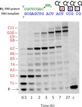
Nonenzymatic RNA-templated Synthesis of N3′→P5′ Phosphoramidate DNA

N3'→P5'磷酰胺DNA的非酶RNA模板合成

DO Derek K. O'Flaherty
Lijun Zhou Lijun Zhou
JS Jack W. Szostak
3680 Views
Sep 5, 2020
The RNA world hypothesis describes a scenario where early life forms relied on RNA to govern both inheritance and catalyze useful chemical reactions. Prior to the emergence of enzymes capable of replicating the RNA genome, a nonenzymatic replication process would have been necessary to initiate Darwinian Evolution. However, the one-pot nonenzymatic RNA chemical copying of templates with mixed-sequences is insufficient to generate strand products long enough to encode useful function. The use of alternate (RNA-like) genetic polymers may overcome hurdles associated with RNA copying, and further our understanding of nonenzymatic copying chemistry. This protocol describes the nonenzymatic copying of RNA templates into N3′→P5′ phosphoramidate DNA (3′-NP-DNA). We describe, in detail, the synthesis of 3′-amino-2′,3′-dideoxyribonucleotide monomers activated with 2-aminoimidazole (3′-NH2-2AIpddN), and their use in template-directed polymerization.

神经科学

社交激活神经元的识别

Identification of Socially-activated Neurons

社交激活神经元的识别

MP Mary L. Phillips
LP Lucas Pozzo-Miller
5832 Views
Sep 5, 2020
Determining the neuronal circuitry responsible for specific behaviors is a major focus in the field of neurobiology. Activity-dependent immediate early genes (IEGs), transcribed and translated shortly after neurons discharge action potentials, have been used extensively to either identify or gain genetic access to neurons and brain regions involved in such behaviors. By using immunohistochemistry for the protein product of the IEG c-Fos combined with retrograde labeling of specific neuronal populations, precise experimental timing, and identical data acquisition and processing, we present a method to quantitatively identify specific neuronal subpopulations that were active during social encounters. We have previously used this method to show a stronger recruitment of ventral hippocampal neurons that project to the medial prefrontal cortex, compared to those that project to the lateral hypothalamus, following social interactions. After optimization of surgeries for the injection of retrograde tracers, this method will be useful for the identification and mapping of neuronal populations engaged in many different behaviors.
通过全身体积描记法测量小鼠的呼吸模式

Measuring Breathing Patterns in Mice Using Whole-body Plethysmography

通过全身体积描记法测量小鼠的呼吸模式

PP Patricia Prada-Dacasa
AU Andrea Urpi
LS Laura Sánchez-Benito
PB Patrizia Bianchi
Albert Quintana Albert Quintana
5783 Views
Sep 5, 2020
Respiratory dysfunction is among the main cause of severe and fatal pathologies worldwide. The use of effective experimental models and methodologies for the study of the pulmonary pathophysiology is necessary to prevent, control and cure these diseases. Plethysmography, a technique for the assessment of lung function, has been widely applied in mice for the characterization of respiratory physiology. However, classical plethysmography methods present technical limitations such as the use of anesthesia and animal immobilization. Whole-body plethysmography (WBP) avoids these issues providing a non-invasive approach for the assessment of the respiratory function in conscious animals. WBP relies on the recording of pressure changes that are produced by the spontaneous breathing activity of an animal placed inside an airtight chamber. During normal respiration, pressure variation is directly proportional to the respiratory pattern of the animal allowing the measurement of the respiratory rate and tidal volume. These parameters are commonly used to evaluate pulmonary function in different physiological and disease models. In contrast to classical plethysmography methods, WBP technique allows reproducible serial measurements as it avoids animal restraint or the use of anesthesia. These key features rend WBP a suitable approach for longitudinal studies allowing the assessment of progressive respiratory alterations in physiological and pathological conditions. This protocol describes the procedures for the measurement of the breathing patterns in mice using the WBP method, the data analysis and results interpretation.
啮齿类动物的峰值间隔程序:研究人类疾病模型中间隔时间及其变化的神经生物学基础的工具

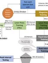
The Peak Interval Procedure in Rodents: A Tool for Studying the Neurobiological Basis of Interval Timing and Its Alterations in Models of Human Disease

啮齿类动物的峰值间隔程序:研究人类疾病模型中间隔时间及其变化的神经生物学基础的工具

FB Fuat Balcı
DF David Freestone
3315 Views
Sep 5, 2020
Animals keep track of time intervals in the seconds to minutes range with, on average, high accuracy but substantial trial-to-trial variability. The ability to detect the statistical signatures of such timing behavior is an indispensable feature of a good and theoretically-tractable testing procedure. A widely used interval timing procedure is the peak interval (PI) procedure, where animals learn to anticipate rewards that become available after a fixed delay. After learning, they cluster their responses around that reward-availability time. The in-depth analysis of such timed anticipatory responses leads to the understanding of an internal timing mechanism, that is, the processing dynamics and systematic biases of the brain’s clock. This protocol explains in detail how the PI procedure can be implemented in rodents, from training through testing to analysis. We showcase both trial-by-trial and trial-averaged analytical methods as a window into these internal processes. This protocol has the advantages of capturing timing behavior in its full-complexity in a fashion that allows for a theoretical treatment of the data.

植物科学

农杆菌介导的优良籼稻品种Komboka的高效转化

Efficient Agrobacterium-mediated Transformation of the Elite–Indica Rice Variety Komboka

农杆菌介导的优良籼稻品种Komboka的高效转化

VL Van T. Luu
MS Melissa Stiebner
PM Paula Emmerich Maldonado
SV Sandra Valdés
DM Didier Marín
GD Gerardo Delgado
VL Virginia Laluz
LW Lin-Bo Wu
PC Paul Chavarriaga
JT Joe Tohme
IS Inez H. Slamet-Loedin
WF Wolf B. Frommer
8650 Views
Sep 5, 2020
Genetic transformation is crucial for both investigating gene functions and for engineering of crops to introduce new traits. Rice (Oryza sativa L.) is an important model in plant research, since it is the staple food for more than half of the world’s population. As a result, numerous transformation methods have been developed for both indica and japonica rice. Since breeders continuously develop new rice varieties, transformation protocols have to be adapted for each new variety. Here we provide an optimized transformation protocol with detailed tips and tricks for a new African variety Komboka using immature embryos. In Komboka, we obtained an apparent transformation rate of up to 48% for GUS/GFP reporter gene constructs using this optimized protocol. This protocol is also applicable for use with other elite indica rice varieties.
转基因烟草瞬时表达后蛋白质丰度的比例计量测定

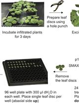
Ratiometric Measurement of Protein Abundance after Transient Expression of a Transgene in Nicotiana benthamiana

转基因烟草瞬时表达后蛋白质丰度的比例计量测定

Aashima  Khosla Aashima Khosla
DN David C. Nelson
4822 Views
Sep 5, 2020
Ratiometric reporters are tools to dynamically measure the relative abundance of a protein of interest. In these systems, a target protein fused to a fluorescent or bioluminescent reporter is expressed with fixed stoichiometry to a reference protein fused to a second reporter. Both fusion proteins are encoded on a single transcript but are separated during translation by a 2A “self-cleaving” peptide. This approach enables changes in the relative abundance of a target protein to be detected sensitively, reducing variability in expression of the ratiometric reporter transgene that may occur across different tissues or transformation events. We recently developed a set of Gateway-compatible plant transformation vectors termed pRATIO that combine a variety of promoters, fluorescent and bioluminescent reporters, and 2A peptides derived from foot-and-mouth disease virus. Here, we describe in detail how to use the dual-fluorescent ratiometric reporter pRATIO3212 to examine the relative abundance of a target protein after transient expression in Nicotiana benthamiana leaves. For this example, we analyze degradation of the SUPPRESSOR OF MAX2 1 (SMAX1) protein from Arabidopsis thaliana in response to treatments with karrikins and rac-GR24. This protocol provides a simple, rapid, and readily scalable method for in vivo analysis of relative protein abundance in Agrobacterium-infiltrated Nicotiana leaf tissues.
一种用于评估水稻白叶枯病泛菌致病性的高效接种技术

An Efficient Inoculation Technique to Assess the Pathogenicity of Pantoea Species Associated to Bacterial Blight of Rice

一种用于评估水稻白叶枯病泛菌致病性的高效接种技术

KK Kossi Kini
RA Raoul Agnimonhan
IW Issa Wonni
Drissa Silue Drissa Silue
4438 Views
Sep 5, 2020
Bacteria blight diseases of rice due to several genera of pathogenic bacteria are one of the major constraints worldwide for rice production. The disease can be best managed through host plant resistance sources. For most of these bacteria such as Xanthomonas oryzae pv. oryzae, X. oryzae pv. oryzicola, Pseudomonas fuscovaginae, Burkholderia glumae, Burkholderia gladioli and Acidovorax avenae subsp. avenae, specific diagnostic techniques that include molecular and pathogenicity tests have been developed. However, for Pantoea spp., information on pathogenicity assay is very limited and protocols used are not uniform. Most authors use the leaf clipping method. In this paper, we describe the protocol for mechanical inoculation of rice seedlings aged 35 days. The method consists of infiltrating bacterial suspensions at concentrations of 108 CFU/ml, with a needleless syringe into the intercellular and interveinal spaces of rice leaves underside at about 4-5 cm below the leaf tip. This method can be used for a standardized pathogenicity assessment, germplasm resistance evaluation for identifying and characterizing resistance sources.

干细胞

人诱导多能干细胞(hiPSC)源神经元与大鼠脑回路的整合

Integration of Human Induced Pluripotent Stem Cell (hiPSC)-Derived Neurons into Rat Brain

人诱导多能干细胞(hiPSC)源神经元与大鼠脑回路的整合

XY Xiling Yin
TD Ted M. Dawson
VD Valina L. Dawson
5239 Views
Sep 5, 2020
Human neuron transplantation offers novel opportunities for modeling human neurologic diseases and potentially replacement therapies. However, the complex structure of the human cerebral cortex, which is organized in six layers with tightly interconnected excitatory and inhibitory neuronal networks, presents significant challenges for in vivo transplantation techniques to obtain a balanced, functional and homeostatically stable neuronal network. Here, we present a protocol to introduce human induced pluripotent stem cell (hiPSC)-derived neural progenitors to rat brains. Using this approach, hiPSC-derived neurons structurally integrate into the rat forebrain, exhibit electrophysiological characteristics, including firing, excitatory and inhibitory synaptic activity, and establish neuronal connectivity with the host circuitry.