往期刊物2020

卷册: 10, 期号: 16

left

Dec

20

Dec

5

Nov

20

Nov

5

Oct

20

Oct

5

Sep

20

Sep

5

Aug

20

Aug

5

Jul

20

Jul

5

Jun

20

Jun

5

May

20

May

5

Apr

20

Apr

5

Mar

20

Mar

5

Feb

20

Feb

5

Jan

20

Jan

5

right

癌症生物学

使用电子细胞基质阻抗传感(ECIS)测量上皮细胞生长和连接发育

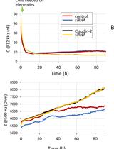
Measuring Cell Growth and Junction Development in Epithelial Cells Using Electric Cell-Substrate Impedance Sensing (ECIS)

使用电子细胞基质阻抗传感(ECIS)测量上皮细胞生长和连接发育

SA Shaista Anwer
KS Katalin Szaszi
4479 Views
Aug 20, 2020
Electric Cell-substrate Impedance Sensing (ECIS) is an automated method that can be used to quantify processes such as cell attachment, growth, migration and barrier functions (i.e., the properties of tight junctions). The method provides simultaneous information on cell number and tight junction function by detecting electric parameters of cells grown on electrodes. Samples are probed with small alternating current (AC) over a range of frequencies, and changes in capacitance and impedance are measured over time. Capacitance reflects the degree of electrode coverage by cells, that correlates with cell number, and can be used to assess cell proliferation or migration. Impedance values inform about barrier function. Obtaining real-time simultaneous information on these parameters is unique to this system and is of great value for addressing fundamental questions such as the role of tight junction proteins in cell growth and migration. This protocol describes the use of ECIS to follow cell growth and tight junction-dependent barrier generation in tubular epithelial cells. We used this method to explore how depleting claudin-2, a tight junction protein affects tubular cell growth and barrier function. During the process, cells are transfected with control or claudin-2-specific siRNA, and 24h later plated on electrodes. ECIS automatically collects information on cell growth and barrier as the monolayer develops. The data are initially analyzed using the ECIS software and exported into a graph software for further processing.

细胞生物学

STC-1细胞体外释放GLP-1的实验研究

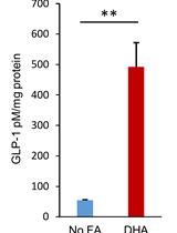
In-vitro GLP-1 Release Assay Using STC-1 Cells

STC-1细胞体外释放GLP-1的实验研究

LQ Liu Qi
TS Tan Shuai
JD Jia Da
BE Burstein Ezra
SL Sifuentes-Dominguez Luis
5244 Views
Aug 20, 2020
Enteroendocrine cells (EECs) are known chemosensors in the gastrointestinal (GI) epithelium. They release a diversity of gut hormones in response to various stimuli. Here, we report an in-vitro assay to measure GLP-1 release from cultured murine EEC’s under fatty acid stimulation.
红细胞甘油通透性的Stopped-flow 光散射分析

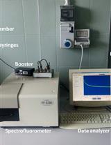
Stopped-flow Light Scattering Analysis of Red Blood Cell Glycerol Permeability

红细胞甘油通透性的Stopped-flow 光散射分析

PG Patrizia Gena
PP Piero Portincasa
Sabino Matera Sabino Matera
Yonathan Sonntag Yonathan Sonntag
MR Michael Rutzler
GC Giuseppe Calamita
3831 Views
Aug 20, 2020
Stopped-Flow Light Scattering (SFLS) is a method devised to analyze the kinetics of fast chemical reactions that result in a significant change of the average molecular weight and/or in the shape of the reaction substrates. Several modifications of the original stopped-flow system have been made leading to a significant extension of its technical applications. One of these modifications allows the biophysical characterization of the water and solute permeability of biological and artificial membranes. Here, we describe a protocol of SFLS to measure the glycerol permeability of isolated human red blood cells (RBCs) and evaluate the pharmacokinetics properties (selectivity and potency) of isoform-specific inhibitors of AQP3, AQP7 and AQP9, three mammalian aquaglyceroporins allowing transport of glycerol across membranes. Suspensions of RBCs (1% hematocrit) are exposed to an inwardly directed gradient of 100 mM glycerol in a SFLS apparatus at 20 °C and the resulting changes in scattered light intensity are recorded at a monochromatic wavelength of 530 nm for 120 s. The SFLS apparatus is set up to have a dead time of 1.6-ms and 99% mixing efficiency in less than 1 ms. Data are fitted to a single exponential function and the related time constant (, seconds) of the cell-swelling phase of light scattering corresponding to the osmotic movement of water that accompanies the entry of glycerol into erythrocytes is measured. The coefficient of glycerol permeability (Pgly, cm/s) of RBCs is calculated with the following equation: where (s) is the fitted exponential time constant and S/V is the surface-to-volume ratio (cm-1) of the analyzed RBC specimen. Pharmacokinetics of the isoform-specific inhibitors of AQP3, AQP7 and AQP9 are assessed by evaluating the extent of RBC Pgly values resulting after the exposure to serial concentrations of the blockers.

免疫学

LPS诱导NF-κB报告小鼠局部炎症及药物沉积的实时体内显像研究

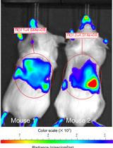
Real-time in vivo Imaging of LPS-induced Local Inflammation and Drug Deposition in NF-κB Reporter Mice

LPS诱导NF-κB报告小鼠局部炎症及药物沉积的实时体内显像研究

AS Artur Schmidtchen
MP Manoj Puthia
5071 Views
Aug 20, 2020
Wound, biomaterial, and surgical infections are all characterized by a localized and excessive inflammation, motivating the development of in vivo methods focused on the analysis of local immune events. However, current inflammation models, such as the commonly used in vivo models of endotoxin-induced inflammation are based on systemic, usually intraperitoneal, administration of lipopolysaccharide (LPS), causing endotoxin shock. Here, we describe a model of LPS-induced local inflammation in NF-κB-RE-Luc reporter mice. LPS, alone or with added therapeutic substances, is delivered locally via a hydrogel which is deposited subcutaneously, providing a spatially defined environment, enabling in vivo bioimaging analyses of local NF-κB activation. Evaluation of drug efficacy can be analyzed longitudinally in the same mouse, and using fluorescently labeled drugs, local drug deposition can be simultaneously analyzed, and correlated to the site of inflammation. Finally, the protocol can also be used to study retention and systemic release of the drug from locally deposited gels and other biomaterials.
白念珠菌中性粒细胞胞外诱杀试验

Neutrophil Extracellular Trap Killing Assay of Candida albicans

白念珠菌中性粒细胞胞外诱杀试验

Sheng-Yang Wu Sheng-Yang Wu
Betty A. Wu-Hsieh Betty A. Wu-Hsieh
5065 Views
Aug 20, 2020
Fungal pathogen Candida albicans is one of the top leading causes of overall healthcare-associated bloodstream infections worldwide. Neutrophil is the major effector cell to clear C. albicans infection. Our study showed that mouse neutrophils utilize two independent mechanisms to kill C. albicans: one is CR3 downstream NADPH oxidase-dependent mechanism that kills opsonized C. albicans; the other one is dectin-2-mediated NADPH oxidase-independent neutrophil extracellular trap (NET) that kills unopsonized C. albicans. Neutrophil killing of opsonized C. albicans requires phagocytosing the organism and production of reactive oxygen species production (ROS). Most existing protocols that assay for neutrophil killing of C. albicans requires a washing step after allowing neutrophils to phagocytose the organism. By definition, NET kills organisms extracellularly. Therefore, it is important to skip the washing step and add an optimal ratio of neutrophils and C. albicans to the wells. To demonstrate the effect of NET, it is necessary to compare killing ability of neutrophils treated with micrococcal nuclease (MNase), an enzyme that digests NET, to that treated with heat-inactivated MNase. MNase is also applied to release NET-bound fungal elements for counting. This protocol can be applied to assay NET killing of other biofilm-forming organisms.

微生物学

白念珠菌琼脂侵入实验

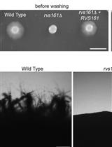
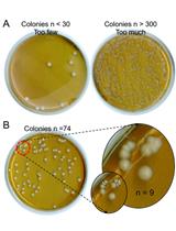
Candida albicans Agar Invasion Assays

白念珠菌琼脂侵入实验

SN Shamoon Naseem
LD Lois M. Douglas
James B. Konopka James B. Konopka
4582 Views
Aug 20, 2020
The ability of the human fungal pathogen Candida albicans to disseminate into tissues is promoted by a switch from budding to invasive hyphal growth. This morphological transition is stimulated by multiple environmental factors that can vary at different sites of infection. To identify genes that promote invasive growth, C. albicans mutants can be screened for defects in growing invasively into solid agar medium as a substitute for studying tissue invasion. This in vitro approach has advantages in that it permits the media conditions to be varied to mimic different host environments. In addition, the concentration of agar can be varied to determine the effects of altering the rigidity of the matrix into which the cells invade, as this provides a better indicator of invasive growth than the ability to form hyphae in a liquid culture. Testing under multiple conditions can be used to identify mutant cells with the strongest defects. Therefore, protocols and media for analyzing invasive growth of C. albicans under different conditions will be described that are appropriate for testing a single strain or high-throughput analysis of a collection of mutant C. albicans strains.
猴原代肝细胞体外培养及疟疾肝期观察

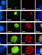
In vitro Cultivation and Visualization of Malaria Liver Stages in Primary Simian Hepatocytes

猴原代肝细胞体外培养及疟疾肝期观察

DG Devendra Kumar Gupta
TD Thierry Diagana
4209 Views
Aug 20, 2020
Human liver is the primary and obligatory site for malaria infection where sporozoites invade host hepatocytes. Malaria hepatic stages are asymptomatic and represent an attractive target for development of anti-malarial interventions and vaccines. However, owing to lack of robust and reproducible in vitro culture system, it is difficult to target and study this imperative malaria liver stage. Here, we describe a procedure that allow cultivation and visualization of malaria hepatic stages including dormant hypnozoites using primary simian hepatocytes. This method enables sensitive and quantitative assessment of different hepatic stages in vitro.
ICP-AES法测定酿酒酵母细胞离子浓度

Determination of the Cellular Ion Concentration in Saccharomyces cerevisiae Using ICP-AES

ICP-AES法测定酿酒酵母细胞离子浓度

LT Louise Thines
AI Anne Iserentant
PM Pierre Morsomme
2801 Views
Aug 20, 2020
The yeast Saccharomyces cerevisiae has been perceived over decades as a highly valuable model organism for the investigation of ion homeostasis. Indeed, many of the genes and biological systems that function in yeast ion homeostasis are conserved throughout unicellular eukaryotes to humans. In this context, measurement of the yeast cellular ionic content provides information regarding yeast response to gene deletion or exposure to chemicals for instance. We propose here a protocol that we tested for the analysis of 12 elements (Ba2+, Ca2+, Cd2+, Co2+, Cu2+, Fe2+, K+, Mg2+, Mn2+, Na+, Ni2+, Zn2+) in yeast using Inductively Coupled Plasma-Atomic Emission Spectrometry (ICP-AES). This technique enables determination of the cellular content of numerous ions from one biological sample.

分子生物学

生物流体中细胞外DNA的分离和定量

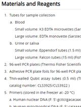
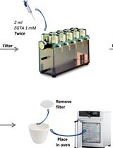
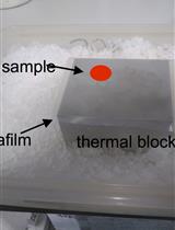
Isolation and Quantification of Extracellular DNA from Biofluids

生物流体中细胞外DNA的分离和定量

ĽJ Ľubica Janovičová
BK Barbora Konečná
BV Barbora Vlková
PC Peter Celec
4066 Views
Aug 20, 2020
Extracellular DNA is studied as a diagnostic biomarker, but also as a factor involved in the pathophysiology of several diseases due to its pro-inflammatory properties. Extracellular DNA can be extracted from plasma, urine, saliva or other biofluids using standard DNA isolation procedures and specialized commercial kits. Sample preparation for isolation is important, freezing and thawing may affect the amount of extracellular DNA extracted. Subsequent centrifugations remove cells and cell debris from the samples to obtain true extracellular DNA. Small volume of samples especially from animal experiments is often an issue and it affects the DNA yield. Very short fragments (˂ 100 bp) can be lost during isolation and are difficult to quantify using PCR. Fluorometric methods asses all stained DNA fragments. Selecting the method for quantification of extracellular DNA is crucial and combination of at least two methods is ideal. Standardization of procedures or at least their reporting in research papers is of utmost importance for comparison of results.
用逆转录酶终止位点和RNAseq定位翻译起始复合体内的mRNA-18S rRNA接触点

Mapping mRNA-18S rRNA Contacts Within Translation Initation Complex by Means of Reverse Transcriptase Termination Sites and RNAseq

用逆转录酶终止位点和RNAseq定位翻译起始复合体内的mRNA-18S rRNA接触点

ID Irene Díaz-López
RT René Toribio
IV Iván Ventoso
3986 Views
Aug 20, 2020
The nucleotides involved in RNA-RNA interaction can be tagged by chemical- or UV-induced crosslinking, and further identified by classical or modern high throughput techniques. The contacts of mRNA with 18S rRNA that occur along the mRNA channel of 40S subunit have been mapped by site-specific UV crosslinking followed by reverse transcriptase termination sites (RTTS) using radioactive or fluorescent oligonucleotides. However, the sensitivity of this technique is restricted to the detection of those fragments that resulted from the most frequent crosslinkings. Here, we combined RTTS with RNAseq to map the mRNA-18S rRNA contacts with a much deeper resolution. Although aimed to detect the interaction of mRNA with the ES6S region of 18S rRNA, this technique can also be applied to map the interaction of mRNA with other non-coding RNA molecules (e.g., snRNAs, microRNAs and lncRNAs) during transcription, splicing or RNA-mediated postranscriptional regulation.
存在不良成核核苷酸类似物的微管种晶组装

Microtubule Seeded-assembly in the Presence of Poorly Nucleating Nucleotide Analogues

存在不良成核核苷酸类似物的微管种晶组装

SK Siou Ku
CH Claire Heichette
LD Laurence Duchesne
DC Denis Chrétien
3080 Views
Aug 20, 2020
Microtubule dynamic instability is driven by the hydrolysis of the GTP bound to the β-subunit of the α-β tubulin heterodimer. Nucleotide analogues are commonly used to mimic the different steps of the tubulin GTPase cycle, but most of them are poor microtubule nucleators. Usually, microtubule assembly is seeded by guanylyl-(α, β)-methylene-diphosphonate (GMPCPP) or glycerol that can be limiting factors in monitoring the effect of other nucleotide analogs on their polymerization. Here, we describe a protocol that allows the assembly of microtubules in the presence of nucleotide analogues without the need of heterogeneous seeds and at a low final glycerol concentration. Microtubules are first assembled in the presence of the analogue of interest and glycerol to promote assembly. These microtubules are then sonicated to produce seeds that will be used to assemble microtubules in the absence of glycerol. This strategy produces homogeneous nucleotide-bound microtubules that can be further analyzed by biochemical or structural methods such as cryo-electron microscopy.

神经科学

一种可重复性测定成年斑马鱼临界游泳速度的方法

A Reproducible Protocol to Measure the Critical Swimming Speed of Adult Zebrafish

一种可重复性测定成年斑马鱼临界游泳速度的方法

YW Yuma Wakamatsu
MK Makoto Kashima
HH Hiromi Hirata
4877 Views
Aug 20, 2020
The quantitative measurement of water flow-induced swimming of fish species using a swimmill is a powerful method to evaluate motor ability of individual fish. Zebrafish is a commonly used vertebrate that enables the study of morphological, physiological and behavioral characteristics associated with genes. We here established a reproducible method that allows to measure the body length and the critical swimming speed of adult zebrafish using a swimmill.
使用IP-One评估Gαq/ 15信号:基于细胞高通量测定的单板转染和测定方案

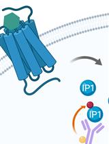
Assessing Gαq/15-signaling with IP-One: Single Plate Transfection and Assay Protocol for Cell-Based High-Throughput Assay

使用IP-One评估Gαq/ 15信号:基于细胞高通量测定的单板转染和测定方案

Élie Besserer-Offroy Élie Besserer-Offroy
RB Rebecca L Brouillette
JL Jean-Michel Longpré
PS Philippe Sarret
4498 Views
Aug 20, 2020
Cell-based functional assays are an important part of compound screening and drug lead optimization, and they can also play a crucial role in the determination of the residues involved in ligand binding and signaling for a particular G-protein-coupled receptor. Conventional methods used for Gαq/15-coupled receptors rely on the use of fluorescent probes for Ca++ sensing (such as Fura-2 and Fluo-4) or on the incorporation of [3H]-inositol into inositol 1,4,5- triphosphate (IP3). However, these methods are not suitable for screening large libraries of compounds or for screening several mutants of the same receptor. In contrast, the IP-One assay by Cisbio is a TR-FRET assay suitable for large compound library screening when using stable cell lines that express a specific 7TMR. However, when using transiently transfected mutants of a 7TMR, this assay is not ideal, as it requires a two-step protocol of cell culture. Therefore, we have optimized the IP-One assay protocol using the reverse transfection method in 384-well plates. This offers a time- and resource-efficient alternative to the two-step protocol previously used for the screening of several mutants of Gαq/15-coupled 7TMRs.
核转运蛋白-β2抑制和ALS / FTD相关蛋白FUS的逆转聚集和液-液相分离

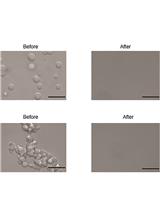
Karyopherin-β2 Inhibits and Reverses Aggregation and Liquid-liquid Phase Separation of the ALS/FTD-Associated Protein FUS

核转运蛋白-β2抑制和ALS / FTD相关蛋白FUS的逆转聚集和液-液相分离

Emma Robinson Emma Robinson
JS James Shorter
LG Lin Guo
4388 Views
Aug 20, 2020
The study of RNA-binding proteins (RBP) offers insight into the mechanisms of pathologic protein aggregation in neurodegenerative diseases. We developed a protocol for purifying an RBP FUS and a nuclear import receptor (NIR) Kapβ2 and testing the ability of Kapβ2 to mitigate FUS aggregation and liquid-liquid phase separation.
利用微流体装置共培养小鼠神经元研究Tau的错误折叠传播

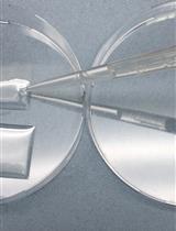
Co-culture of Murine Neurons Using a Microfluidic Device for The Study of Tau Misfolding Propagation

利用微流体装置共培养小鼠神经元研究Tau的错误折叠传播

GH Grace I Hallinan
DL Dianne M Lopez
Mariana Vargas-Caballero Mariana Vargas-Caballero
JW Jonathan West
Katrin Deinhardt Katrin Deinhardt
4238 Views
Aug 20, 2020
The deposition of misfolded, aggregated tau protein is a hallmark of several neurodegenerative diseases, collectively termed “tauopathies”. Tau pathology spreads throughout the brain along connected pathways in a prion-like manner. The process of tau pathology propagation across circuits is a focus of intense research and has been investigated in vivo in human post-mortem brain and in mouse models of the diseases, in vitro in diverse cellular systems including primary neurons, and in cell free assays using purified recombinant tau protein. Here we describe a protocol that takes advantage of a minimalistic neuronal circuit arrayed within a microfluidic device to follow the propagation of tau misfolding from a presynaptic to a postsynaptic neuron. This assay allows high-resolution imaging as well as individual manipulation of the releasing and receiving neuron, and is therefore beneficial for investigating the propagation of tau and other misfolded proteins in vitro.
猴子运动技能研究的连续性达标任务

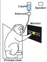
Sequential Reaching Task for the Study of Motor Skills in Monkeys

猴子运动技能研究的连续性达标任务

MO Machiko Ohbayashi
NP Nathalie Picard
2891 Views
Aug 20, 2020
The ability to perform a sequence of movements is a key component of motor skills, such as typing or playing a musical instrument. How the brain binds elementary movements together into meaningful actions has been a topic of much interest. Here, we describe two sequential reaching tasks that we use to investigate the neural substrate of skilled sequential movements in monkeys after long-term practice. The movement elements performed in these tasks are essentially identical, but are generated in two different contexts. In one task, monkeys perform reaching movements that are instructed by visual cues. In the other, the monkeys perform reaching movements that are generated from memory after extended practice. With this behavioral paradigm, we can dissociate the neural processes related to the acquisition and retention of motor skills from those related to movement execution.