往期刊物2020

卷册: 10, 期号: 13

left

Dec

20

Dec

5

Nov

20

Nov

5

Oct

20

Oct

5

Sep

20

Sep

5

Aug

20

Aug

5

Jul

20

Jul

5

Jun

20

Jun

5

May

20

May

5

Apr

20

Apr

5

Mar

20

Mar

5

Feb

20

Feb

5

Jan

20

Jan

5

right

生物化学

制备果蝇多线染色体并通过多重荧光关联对染色质结构进行免疫荧光分析

Preparation of Drosophila Polytene Chromosomes, Followed by Immunofluorescence Analysis of Chromatin Structure by Multi-fluorescence Correlations

制备果蝇多线染色体并通过多重荧光关联对染色质结构进行免疫荧光分析

TK Terra M. Kuhn
SL Shawn C. Little
MC Maya Capelson
8771 Views
Jul 5, 2020
Drosophila larval salivary gland polytene chromosome squashes have been used for decades to analyze genome-wide protein-binding patterns, transcriptional activation processes, and changes in chromatin structure at specific genetic loci. There have been many evolutions of the squashing protocol over the years, with sub-optimal reproducibility and low sample success rate as accepted caveats. However, low sample success rates are an obvious disadvantage when polytene chromosomes are used for more high-throughput approaches, such as genetic or antibody screens, or for experiments requiring high-quality chromosome structure preservation. Here we present an exceptionally reproducible squashing and fluorescence staining protocol, which generates high-quality fluorescence images on well-spread chromosomes. This is followed by our novel, semi-automated MATLAB analysis program used to determine correlations between fluorescence signals of interest at a single site on polytene chromosomes, in a pixel-by-pixel manner. In our case, we have used this approach to assess chromatin changes at genomic sites, ectopically targeted by nuclear pore proteins. The use of our analysis program increases the ability to make unbiased conclusions on changes in chromatin structure, or in protein recruitment to chromatin, regardless of sample variation in immunofluorescence staining. As it is simply based upon differences in fluorescence intensity at a defined location, the provided analysis program is not limited to analysis of polytene chromosome, and could be applied to many different contexts where correlation between fluorescent signals at any particular location is of interest.
小鼠粪便中短链脂肪酸的气相色谱检测

Gas Chromatography Detection Protocol of Short-chain Fatty Acids in Mice Feces

小鼠粪便中短链脂肪酸的气相色谱检测

CZ Chaozheng Zhang
AL An Liu
TZ Tianshuang Zhang
YL Yi Li
HZ Hua Zhao
5833 Views
Jul 5, 2020
Short-chain fatty acids (SCFAs), which are formed mainly by bacteria fermenting undigested carbohydrates in the colon, they are based on the number of carbon atoms in the carbon chain. Organic fatty acids with less than 6 carbon atoms are called short-chain fatty acids. SCFAs are closely related to various aspects of the human body, so more and more researchers concentrate on SCFAs. This protocol describes, a direct injection gas chromatography detection method with a pretreatment method for extracting SCFA from mice feces by combining acidification. The corresponding sample limit of quantization (LOQ) and limit of detection (LOD) are 0.8-1.0 mg/L and 0.5-0.8 mg/L, respectively. The correlation coefficient of calibration curve is greater than 0.999. The recovery rate of the spiked standard is 80%-102%. This method can be used to analyze and determine SCFAs in mice feces. Therefore, this is an economical, effective and reproducible method for SCFAs measurement in mice samples.
用于MRBLE-pep分析的光谱编码微球肽合成方案

Protocol for Peptide Synthesis on Spectrally Encoded Beads for MRBLE-pep Assays

用于MRBLE-pep分析的光谱编码微球肽合成方案

JH Jamin B. Hein
HN Huy Q. Nguyen
MC Martha Cyert
PF Polly M. Fordyce
4852 Views
Jul 5, 2020
Every living cell relies on signal transduction pathways comprised of protein-protein interactions (PPIs). In many cases, these PPIs are between a folded protein domain and a short linear motif (SLiM) within an unstructured region of a protein. As a result of this small interaction interface (3-10 amino acids), the affinities of SLiM-mediated interactions are typically weak (Kds of ~1-10 µM), allowing physiologically relevant changes in cellular concentrations of either protein partner to dictate changes in occupancy and thereby transmit cellular signals. However, these weak affinities also render detection and quantitative measurement of these interactions challenging and labor intensive. To address this, we recently developed MRBLE-pep, a technology that employs peptide libraries synthesized on spectrally encoded hydrogel beads to allow multiplexed affinity measurements between a protein and many different peptides in parallel. This approach dramatically reduces both the amount of protein and peptide as well as the time required to measure protein-peptide affinities compared to traditional methods. Here, we provide a detailed protocol describing how to: (1) functionalize polyethylene glycol diacrylate (PEG-DA) MRBLE beads with free amine groups, (2) synthesize peptide libraries on functionalized MRBLEs, (3) validate synthesized peptide sequences via MALDI mass spectrometry and quantify evenness of peptide coverage on MRBLEs, (4) use MRBLE-bound peptide libraries in multiplexed protein binding assays, and (5) analyze binding data to determine binding affinities. We anticipate that this protocol should prove useful for other researchers seeking to use MRBLE-pep in their own laboratories as well as for researchers broadly interested in solid-phase peptide synthesis and protein-protein binding assay development.
利用表面等离子体共振技术评价病原菌与宿主蛋白的结合

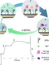
In vitro Assessment of Pathogen Effector Binding to Host Proteins by Surface Plasmon Resonance

利用表面等离子体共振技术评价病原菌与宿主蛋白的结合

MF Marina Franceschetti
MB Mark J. Banfield
CS Clare E. M. Stevenson
JD Juan Carlos De la Concepcion
4208 Views
Jul 5, 2020
The mechanisms of virulence and immunity are often governed by molecular interactions between pathogens and host proteins. The study of these interactions has major implications on understanding virulence activities, and how the host immune system recognizes the presence of pathogens to initiate an immune response. Frequently, the association between pathogen molecules and host proteins are assessed using qualitative techniques. As small differences in binding affinity can have a major biological effect, in vitro techniques that can quantitatively compare the binding between different proteins are required. However, these techniques can be manually intensive and often require large amounts of purified proteins. Here we present a simplified Surface Plasmon Resonance (SPR) protocol that allows a reproducible side-by-side quantitative comparison of the binding between different proteins, even in cases where the binding affinity cannot be confidently calculated. We used this method to assess the binding of virulence proteins (termed effectors) from the blast fungus Magnaporthe oryzae, to a domain of a host immune receptor. This approach represents a rapid and quantitative way to study how pathogen molecules bind to host proteins, requires only limited quantities of proteins, and is highly reproducible. Although this method requires the use of an SPR instrument, these can often be accessed through shared scientific services at many institutions. Thus, this technique can be implemented in any study that aims to understand host-pathogen interactions, irrespective of the expertise of the investigator.

生物物理学

NMR waterLOGSY应用于药物开发项目中蛋白质-配体相互作用的检测

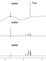
NMR waterLOGSY as An Assay in Drug Development Programmes for Detecting Protein-Ligand Interactions–NMR waterLOGSY

NMR waterLOGSY应用于药物开发项目中蛋白质-配体相互作用的检测

CB Carole J. R. Bataille
TR Terence H. Rabbitts
TC Timothy D. W. Claridge
5824 Views
Jul 5, 2020
In drug development programmes, multiple assays are needed for the determination of protein-compound interactions and evaluation of potential use in assays with protein-protein interactions. In this protocol we describe the waterLOGSY NMR method for confirming protein-ligand binding events.

癌症生物学

FAST蛋白的生成与检测

Generation and Testing of Fluorescent Adaptable Simple Theranostic (FAST) Proteins

FAST蛋白的生成与检测

Andrew S. Flies Andrew S. Flies
JD Jocelyn M. Darby
PM Peter R. Murphy
TP Terry L. Pinfold
AP Amanda L. Patchett
Patrick R. Lennard Patrick R. Lennard
7115 Views
Jul 5, 2020
This protocol provides a step-by-step method to create recombinant fluorescent fusion proteins that can be secreted from mammalian cell lines. This builds on many other recombinant protein and fluorescent protein techniques, but is among the first to harness fluorescent fusion proteins secreted directly into cell culture supernatant. This opens new possibilities that are not achievable with proteins produced in bacteria or yeast, such as direct use of the fluorescent protein-secreting cells in live co-culture assays. The Fluorescent Adaptable Simple Theranostic (FAST) protein system includes a histidine purification tag and a tobacco etch virus (TEV) cleavage site, allowing the purification tag and fluorescent protein to be removed for therapeutic use. This protocol is split into five parts: (A) In silico characterization of the gene-of-interest (GOI) and protein-of-interest (POI); (B) design of the expression vector; (C) assembly of the expression vector; (D) transfection of a eukaryotic cell line with the expression vector; (E) testing of the recombinant protein. This extensive protocol can be completed with only polymerase chain reaction (PCR) and cell culture training. Additionally, each part of the protocol can be used independently.
哺乳动物细胞脂筏的分离及其脂质组学分析

Isolation of Lipid Rafts from Cultured Mammalian Cells and Their Lipidomics Analysis

哺乳动物细胞脂筏的分离及其脂质组学分析

NM Nigora Mukhamedova
KH Kevin Huynh
HL Hann Low
PM Peter J. Meikle
DS Dmitri Sviridov
4737 Views
Jul 5, 2020
Lipid rafts are distinct liquid-ordered domains of plasma membranes of most eukaryotic cells providing platform for signaling pathways. Lipid composition of rafts is critical for their structural integrity and for regulation of signaling pathways originating from rafts. Here we provide a protocol to isolate lipid rafts from cultured human and animal cells and comprehensively analyse their lipid composition.
酪胺信号放大的人体组织标本和细胞系培养物中的MYCN和MYC免疫荧光

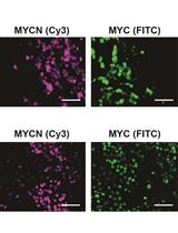
Tyramide Signal-Amplified Immunofluorescence of MYCN and MYC in Human Tissue Specimens and Cell Line Cultures

酪胺信号放大的人体组织标本和细胞系培养物中的MYCN和MYC免疫荧光

JS Johanna M. Schafer
JP Jennifer A. Pietenpol
3481 Views
Jul 5, 2020
MYC family members, MYC, MYCN, and MYCL, are oncogenic transcription factors that regulate the expression of genes involved in normal development, cell growth, proliferation, metabolism, and survival. While MYC is amplified and/or overexpressed across a variety of tissue types, MYCN is often overexpressed in tumors of the nervous system (neuroblastoma and medulloblastoma) or with neuroendocrine features (neuroendocrine prostate cancer). Given recent reports that MYCN expression is also deregulated in a variety of non-neuronal tissue types, we investigated whether MYCN was also deregulated in triple-negative breast cancer (TNBC). In contrast to previous individual immuno-fluorescence (IF) stains against higher expressing MYC family isoform protein, we developed an IF stain to simultaneously detect both MYCN- and MYC-expressing cells within the same tumor cell population. Our methodology allows for the detection of low level MYCN and MYC expression and can be multiplexed with additional protein probes. Herein, using tyramide signal amplification (TSA), we present two protocols for the IF detection of MYCN and MYC on formalin-fixed paraffin embedded (FFPE) tumor sections and in cell lines fixed in situ after growth as adherent cultures on chambered microscope slides.

免疫学

从人类和非人类灵长类动物多能干细胞中产生T细胞

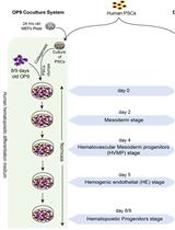
Generation of T cells from Human and Nonhuman Primate Pluripotent Stem Cells

从人类和非人类灵长类动物多能干细胞中产生T细胞

Akhilesh Kumar Akhilesh Kumar
SD Saritha S. D’Souza
GU Gene Uenishi
MP Mi Ae Park
JL Jeong Hee Lee
IS Igor I. Slukvin
5834 Views
Jul 5, 2020
Pluripotent stem cells (PSCs) have the potential to provide homogeneous cell populations of T cells that can be grown at a clinical scale and genetically engineered to meet specific clinical needs. OP9-DLL4, a stromal line ectopically expressing the Notch ligand Delta-like 4 (DLL4) is used to support differentiation of PSCs to T-lymphocytes. This article outlines several protocols related to generation of T cells from human and non-human primate (NHP) PSCs, including initial hematopoietic differentiation of PSC on OP9 feeders or defined conditions, followed by coculture of the OP9-DLL4 cells with the PSC-derived hematopoietic progenitors (HPs), leading to efficient differentiation to T lymphocytes. In addition, we describe a protocol for robust T cell generation from hPSCs conditionally expressing ETS1. The presented protocols provide a platform for T cell production for disease modeling and evaluating their use for immunotherapy in large animal models.
小鼠肾小球ICAM-1、ICAM-2和CD31的联合免疫染色

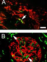
Co-immunostaining of ICAM-1, ICAM-2, and CD31 in Mouse Kidney Glomeruli

小鼠肾小球ICAM-1、ICAM-2和CD31的联合免疫染色

SS Sun-sang J. Sung
4224 Views
Jul 5, 2020
Glomerulonephritis (GN) is a common pathological condition in chronic kidney diseases that often leads to end stage renal failure. Mac-1 (CD11b/CD18)-mediated neutrophil, macrophage, and dendritic cell glomerular infiltration leading to cellular dysfunction and destruction is an important disease mechanism. The cellular distribution and dynamics of the expression of Mac-1 ligands ICAM-1 and ICAM-2 in GN have not been well studied because of the difficulties in tissue staining and colocalizing glomerular cells with surface antigens. To improve the visualization of cell surface marker and antigen expression in kidney compartments, we have devised an even but mild fixation procedure employing p-formaldehyde-lysine-periodate (PLP) perfusion. A large panel of antibodies (Ab) against cell surface markers was used to identify kidney cell types and adhesion molecules. When confocal microscopy was used in visualizing glomerular adhesion molecule staining, the endothelial cells were found to specifically express CD31, and these cells express ICAM-2 constitutively. Though ICAM-1 was not expressed by glomerular endothelial cells in homeostasis, it was highly upregulated in mice with chronic GN and severe proteinuria. VCAM-1, a ligand for VLA-4 important in leukocyte migration, was not expressed in the glomerulus. The results highlight the importance of ICAM-1 in the infiltration of macrophages and dendritic cells in cGN. This report will provide a widely applicable procedure for yielding high quality confocal images and for the identification and quantitation of receptors and other cellular antigens expressed in different kidney compartments and cell types.

微生物学

使用“酵母机”微流控设备观察营养梯度,基因表达和生长变异

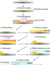
Observing Nutrient Gradients, Gene Expression and Growth Variation Using the "Yeast Machine" Microfluidic Device

使用“酵母机”微流控设备观察营养梯度,基因表达和生长变异

ZM Zoran S. Marinkovic
CV Clément Vulin
MA Mislav Acman
XS Xiaohu Song
JD Jean Marc Di Meglio
AL Ariel B. Lindner
PH Pascal Hersen
4223 Views
Jul 5, 2020
The natural environment of microbial cells like bacteria and yeast is often a complex community in which growth and internal organization reflect morphogenetic processes and interactions that are dependent on spatial position and time. While most of research is performed in simple homogeneous environments (e.g., bulk liquid cultures), which cannot capture full spatiotemporal community dynamics, studying biofilms or colonies is complex and usually does not give access to the spatiotemporal dynamics at single cell level. Here, we detail a protocol for generation of a microfluidic device, the “yeast machine”, with arrays of long monolayers of yeast colonies to advance the global understanding of how intercellular metabolic interactions affect the internal structure of colonies within defined and customizable spatial dimensions. With Saccharomyces cerevisiae as a model yeast system we used the “yeast machine” to demonstrate the emergence of glucose gradients by following expression of fluorescently labelled hexose transporters. We further quantified the expression spatial patterns with intra-colony growth rates and expression of other genes regulated by glucose availability. In addition to this, we showed that gradients of amino acids also form within a colony, potentially opening similar approaches to study spatiotemporal formation of gradients of many other nutrients and metabolic waste products. This approach could be used in the future to decipher the interplay between long-range metabolic interactions, cellular development, and morphogenesis in other same species or more complex multi-species systems at single-cell resolution and timescales relevant to ecology and evolution.
基于显微技术的疟疾玫瑰花环试验

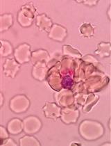
Microscopy-based Methods for Rosetting Assay in Malaria Research

基于显微技术的疟疾玫瑰花环试验

Wenn-Chyau Lee Wenn-Chyau Lee
Laurent Rénia Laurent Rénia
4097 Views
Jul 5, 2020
In malaria, rosetting phenomenon is a condition where a Plasmodium-infected erythrocyte stably adheres to at least an uninfected erythrocyte. This phenomenon that occurs in all species of human malaria parasite is likely to be an immune escape mechanism for the parasite. However, it has been associated with malaria pathogenesis, possibly by facilitating microvasculature occlusion along with direct endothelial cytoadherence by the infected erythrocytes. There are different microscopy-based techniques to visualize rosettes but neither of these techniques has yet to qualify as the official “gold standard” method. We have found that these techniques can be used interchangeably, provided that the conditions of the experiments are properly controlled. Here, we presented three methods as options for rosetting assay, i.e., the unstained wet mount technique, acridine orange based-fluorescence microscopy technique and Giemsa stained wet mount method, with preparation steps that enable consistent performance in rosetting experiments.

神经科学

胚胎小鼠大脑的器官型切片培养

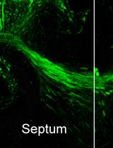
Organotypic Slice Culture of the Embryonic Mouse Brain

胚胎小鼠大脑的器官型切片培养

JC James M. Clegg
TP Thomas Pratt
4853 Views
Jul 5, 2020
Organotypic slice culture is a powerful technique for exploring the embryonic development of the mammalian brain. In this protocol we describe a basic slice culture technique we have used for two sets of experiments: axon guidance transplant assays and bead culture assays.
利用T迷宫评价小鼠工作记忆的非连续配对变换延迟任务

The Discrete Paired-trial Variable-delay T-maze Task to Assess Working Memory in Mice

利用T迷宫评价小鼠工作记忆的非连续配对变换延迟任务

GL Gian Marco Leggio
Sebastiano Alfio Torrisi Sebastiano Alfio Torrisi
FP Francesco Papaleo
3571 Views
Jul 5, 2020
Working memory abnormalities involving the prefrontal cortex (PFC) dramatically contribute to poor functional outcomes in patients with schizophrenia and still represent an unmet therapeutic need. Studies in rodents might provide essential tools to understand the mechanisms underlying PFC-dependent working memory dysfunctions, as well as precious tools for genetic and pharmacological testing. However, proper tests assessing working memory and sensitive to PFC-dependent functions must be used. In this regard, the discrete paired-trial variable-delay T-maze task, equivalent to delayed non-match to sample tasks used in humans, has proved to be an effective paradigm to test PFC-dependent working memory dysfunctions with high predictive validity in human studies.

植物科学

利用甲基化敏感扩增多态性分析野生马铃薯杂交种

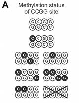
Methylation-sensitive Amplified Polymorphism as a Tool to Analyze Wild Potato Hybrids

利用甲基化敏感扩增多态性分析野生马铃薯杂交种

Nicolás Cara Nicolás Cara
CM Carlos F. Marfil
María V. Bertoldi María V. Bertoldi
RM Ricardo W. Masuelli
4199 Views
Jul 5, 2020
Methylation-Sensitive Amplification Polymorphism (MSAP) is a versatile marker for analyzing DNA methylation patterns in non-model species. The implementation of this technique does not require a reference genome and makes it possible to determine the methylation status of hundreds of anonymous loci distributed throughout the genome. In addition, the inheritance of specific methylation patterns can be studied. Here, we present a protocol for analyzing DNA methylation patterns through MSAP markers in potato interspecific hybrids and their parental genotypes.

干细胞

小鼠体内乳腺形成

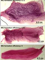
In vivo Mouse Mammary Gland Formation

小鼠体内乳腺形成

HS Heng Sun
XZ Xin Zhang
UC Un In Chan
SS Sek Man Su
SG Sen Guo
XX Xiaoling Xu
CD Chuxia Deng
3528 Views
Jul 5, 2020
For years, the mammary gland serves as a perfect example to study the self-renew and differentiation of adult stem cells, and the regulatory mechanisms of these processes as well. To assess the function of given genes and/or other factors on stemness of mammary cells, several In vitro assays were developed, such as mammospheres formation assay, detection of stem cell markers by mRNA expression or flow cytometry and so on. However, the capacity of reconstruction of whole mount in the cleared fat pad of recipient female mice is a golden standard to estimate the stemness of the cells. Here we described a step-by-step protocol for in vivo mammary gland formation assay, including preparation of “cleared” recipients and mammary cells for implantation, the surgery process and how to assess the experimental results. Combined with manipulation of mammary cells via gene editing and /or drug treatment, this protocol could be very useful in the researches of mammary stem cells and mammary development.