往期刊物2015

卷册: 5, 期号: 13

left

Dec

20

Dec

5

Nov

20

Nov

5

Oct

20

Oct

5

Sep

20

Sep

5

Aug

20

Aug

5

Jul

20

Jul

5

Jun

20

Jun

5

May

20

May

5

Apr

20

Apr

5

Mar

20

Mar

5

Feb

20

Feb

5

Jan

20

Jan

5

right

生物化学

免疫沉淀法识别调节蛋白质泛素化的因子:TRF2在人REST4泛素化中的案例研究

Identification of Factors in Regulating a Protein Ubiquitination by Immunoprecipitation: a Case Study of TRF2 on Human REST4 Ubiquitination

免疫沉淀法识别调节蛋白质泛素化的因子:TRF2在人REST4泛素化中的案例研究

WC Wei Cui
Patrick Ovando-Roche Patrick Ovando-Roche
11853 Views
Jul 5, 2015
Ubiquitination is the first step of the ubiquitin-proteasome pathway that regulates cells for their homeostatic functions and is an enzymatic, protein post-translational modification process in which ubiquitin is transferred to a target protein substrate by a set of three ubiquitin enzymes (Weissman et al., 2011; Bhattacharyya et al., 2014; Ristic et al., 2014). Given the importance of this process, it is plausible that ubiquitination is under strict control by many factors and that the regulatory machineries are protein-specific. An assay for the detection of a specific protein ubiquitination will enable us to examine whether a factor has a function to regulate the ubiquitination of this protein. Here we describe a protocol that detects the ubiquitination status of the human REST4 protein in cultured cells, a neural alternative splicing isoform of REST (RE-1 silencing transcription factor), that antagonizes the repressive function of REST on neural differentiation and neuron formation. Using this protocol, we show that the telomere binding protein TRF2 stabilizes the expression of the human REST4 by inhibiting its ubiquitination. This indicates that TRF2 plays a positive role in neural differentiation (Ovando-Roche et al., 2014). This protocol is also useful for the detection of ubiquitination of other proteins of interest.

免疫学

小鼠角膜基质中骨髓细胞的活体共聚焦成像

Confocal Imaging of Myeloid Cells in the Corneal Stroma of Live Mice

小鼠角膜基质中骨髓细胞的活体共聚焦成像

Matilda F. Chan Matilda F. Chan
Zena Werb Zena Werb
11844 Views
Jul 5, 2015
The accessibility and transparency of the cornea makes it a good tissue model for monitoring immunological responses using in vivo real time imaging analysis (Lee et al., 2010; Tan et al., 2013). These corneal qualities have also allowed for high-resolution in vivo imaging of non-ocular tissue transplanted into the anterior chamber of the mouse eye (Speier et al., 2008a; Speier et al., 2008b). This protocol was adapted from Speier (2008) to successfully assess real-time in vivo myeloid cell dynamics in wounded corneas of c-fms-EGFP mice (Chan et al., 2013). Macrophage colony-stimulating factor (CSF-1) regulates the differentiation and proliferation of cells of the mononuclear phagocyte system. The activity of CSF-1 is mediated by the CSF-1 receptor that is encoded by c-fms (Csf1r) protooncogene. The c-fms gene is expressed in macrophage, trophoblast cell lineages, and to some extent granulocytes. In the c-fms-EGFP mice EGFP, enhanced green fluorescent protein, is driven under the Csf1r, colony stimulating factor 1 receptor, promoter and highlights myeloid cells (Sasmono et al., 2003). This protocol can be further adapted to image other transgenic mice expressing fluorescent proteins.
角膜损伤动物模型

Animal Models of Corneal Injury

角膜损伤动物模型

Matilda F. Chan Matilda F. Chan
Zena Werb Zena Werb
10456 Views
Jul 5, 2015
The cornea is an excellent model system to use for the analysis of wound repair because of its accessibility, lack of vascularization, and simple anatomy. Corneal injuries may involve only the superficial epithelial layer or may penetrate deeper to involve both the epithelial and stromal layers. Here we describe two well-established in vivo corneal wound models: a mechanical wound model that allows for the study of re-epithelialization and a chemical wound model that may be used to study stromal activation in response to injury (Stepp et al., 2014; Carlson et al., 2003).

微生物学

无并发症和致命疟疾的小鼠模型

Mouse Models of Uncomplicated and Fatal Malaria

无并发症和致命疟疾的小鼠模型

BH Brian W. Huang
EP Emily Pearman
Charles C. Kim Charles C. Kim
15332 Views
Jul 5, 2015
Mouse models have demonstrated utility in delineating the mechanisms underlying many aspects of malaria immunology and physiology. The most common mouse models of malaria employ the rodent-specific parasite species Plasmodium berghei, P. yoelii, and P. chabaudi, which elicit distinct pathologies and immune responses and are used to model different manifestations of human disease. In vitro culture methods are not well developed for rodent Plasmodium parasites, which thus require in vivo maintenance. Moreover, physiologically relevant immunological processes are best studied in vivo. Here, we detail the processes of infecting mice with Plasmodium, maintaining the parasite in vivo, and monitoring parasite levels and health parameters throughout infection.
通过同源重组进行复合体的体内连接及在酿酒酵母中进行高效质粒拯救

Complex in vivo Ligation Using Homologous Recombination and High-efficiency Plasmid Rescue from Saccharomyces cerevisiae

通过同源重组进行复合体的体内连接及在酿酒酵母中进行高效质粒拯救

Gregory C. Finnigan Gregory C. Finnigan
Jeremy Thorner Jeremy Thorner
13599 Views
Jul 5, 2015
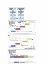
The protocols presented here allow for the facile generation of a wide variety of complex multipart DNA constructs (tagged gene products, gene fusions, chimeric proteins, and other variants) using homologous recombination and in vivo ligation in budding yeast (Saccharomyces cerevisiae). This method is straightforward, efficient and cost-effective, and can be used both for vector creation and for subsequent one-step, high frequency integration into a chromosomal locus in yeast. The procedure utilizes PCR with extended oligonucleotide “tails” of homology between multiple fragments to allow for reassembly in yeast in a single transformation followed by a method for highly efficient plasmid extraction from yeast (for transformation into bacteria). The latter is an improvement on existing methods of yeast plasmid extraction, which, historically, has been a limiting step in recovery of desired constructs. We describe the utility and convenience of our techniques, and provide several examples.[Introduction] Homologous recombination (HR) in S. cerevisiae has long been recognized as an extremely convenient method for assembling DNA fragments in vivo (Szostak et al., 1983; Ma et al., 1987; Oldenburg et al., 1997). Given the efficiency of HR in yeast, it has been exploited in ways that have both increased its utility, enhanced its versatility, and permitted its application to a broad range of experimental objectives. Improvements to this general approach include using in vivo ligation as a platform for directed mutagenesis (Muhlrad et al., 1992), introducing counterselection to aid in plasmid creation (Gunyuzlu et al., 2001; Anderson and Haj-Ahmad, 2003), and adapting in vivo assembly to vectors that cannot be propagated in yeast (Iizasa and Nagano, 2006; Joska et al., 2014). However, previous studies have not fully harnessed the power and utility of HR. We have found that HR in vivo allows for very efficient assembly and recovery of plasmids containing numerous (>5 separate pieces) fragments of DNA in a single transformation step. Thereby, we have been readily able to construct a wide variety of gene disruption cassettes and integration cassettes, to clone exceptionally large genes, to assemble multi-part chimeric genes, and to generate multiply-tagged gene products. The primary utility and power of our methods are the capability to correctly and efficiently assemble multiple DNA fragments transformed into yeast in a single step. Our approach (Figure 1A) has been used successfully: (i) to assemble in-frame chimeras between two or more different genes; (ii) to fuse gene products to fluorescent probes and/or epitope tags at either their N- and/or C-termini, or both; (iii) to create gene deletion cassettes with large amounts of untranslated flanking sequence; (iv) to introduce one or more short linker sequences or epitope tag(s) between assembled genes or gene fragments; (v) to utilize a variety of transcriptional promoters and terminators; and, importantly, (vi) in one step, to generate constructs marked with a drug resistance gene cassette or a selectable nutritional gene cassette that integrate into the genome at the desired locus. Because HR in the yeast cell carries out the in vivo construction process (and subsequent integration, if desired), no kit or proprietary system is required and the assembly of collections of plasmids can be done in a massively parallel manner. In this regard, our system is considerably less expensive than the in vitro enzyme-driven “Gibson cloning” (Gibson, 2011) procedure, yet still remarkably efficient. Also, our system (unlike those requiring restriction enzyme digests to insert gene fragments) does not result in the insertion (or loss) of any nucleotides, which can sometimes occur in classical restriction site cloning. In our method, precise control over both the coding sequence and the flanking untranslated regions (UTRs) can be achieved. Lastly, constructs generated using this system can be coupled with the haploid yeast genome deletion collection (Winzeler et al., 1999; Giaever et al., 2002) to allow for simple and efficient one-step integration at any designed locus. The only rate-limiting factor in our method is the need for the transformed yeast cells to grow for a few days before the construct (or genetically altered cell) can be recovered.Although similar overall methods may exist (Andersen, 2011), in our protocol, we developed several important improvements, which greatly enhance efficient recovery of the DNA constructs from yeast cells, including: (i) a specific yeast genotype that is much easier to lyse than standard laboratory strains, such as S288C (and its derivatives, e.g. BY4741); (ii) a spheroplasting step (to destroy the yeast cell wall); (iii) glass bead beating for better nucleic acid extraction; and, (iv) bacteria chemically treated for ultra-efficient DNA transformation.
地衣型真菌肺衣(地卷目、子囊菌门)菌丝体的无菌培养

Axenic Cultivation of Mycelium of the Lichenized Fungus, Lobaria pulmonaria (Peltigerales, Ascomycota)

地衣型真菌肺衣(地卷目、子囊菌门)菌丝体的无菌培养

Carolina Cornejo Carolina Cornejo
CS Christoph Scheidegger
Rosmarie Honegger Rosmarie Honegger
10207 Views
Jul 5, 2015
Lichens are symbiotic organisms consisting of a fungal partner (the mycobiont) and one or more algal or cyanobacterial partners (the photobiont); moreover lichen thalli comprise a plethora of epi- and endobiotic bacteria and non-lichenized fungi. Genetic markers are the most promising tools for the study of fungal diversity. However, applying genetic methods to intimately admixed symbiotic organisms typically requires the development of species-specific genetic markers, since DNA extraction from environmental specimens implicates the acquirement of total DNA of all symbionts and their cohabitants. While the cultivation of the alga is straight forward, the axenic cultivation of lichen-forming fungi is more difficult due to their very slow growth, as compared with the majority of non-lichenized taxa, and the presence of saprophytic, endophytic and parasitic fungi within the lichen thallus. Moreover, lichen-forming fungi (predominantly ascomycetes, few basidiomycetes) are oligotrophic organisms and thus adapted to nutrient poor conditions; in axenic culture on nutrient-rich media, as normally used for mass production of fast-growing saprophytic fungi, they often autointoxicate. Most lichen-forming fungi are not obligately biotrophic and thus can be cultured in the non-symbiotic state. Here, we present a protocol for the isolation of the lichen-forming ascomycete Lobaria pulmonaria into axenic culture and for mycelial mass culture as a source of pure fungal DNA. We describe the initiation of axenic cultures on agar plates from germinating ascospores and explain the optimization of the in vitro growth in liquid medium. By grinding the few dense, only centrifugally growing fungal colonies with a homogenizer we obtain lots of smaller, well growing colonies and thus higher amounts of mycelium for DNA or RNA isolation (Honegger and Bartnicki-Garcia, 1991).
木薯中地毯草黄单胞菌毒性的实验评估

Assays to Assess Virulence of Xanthomonas axonopodis pv. manihotis on Cassava

木薯中地毯草黄单胞菌毒性的实验评估

MC Megan Cohn
MS Mikel Shybut
DD Douglas Dahlbeck
BS Brian Staskawicz
9556 Views
Jul 5, 2015
Cassava (Manihot esculenta) is a root crop that provides calories for people living in more than 100 tropical and subtropical countries and serves as a raw material for processing into starch and biofuels as well as feed for livestock (Howeler et al., 2013). Xanthomonas axonopodis pv. manihotis (Xam), the causal agent of cassava bacterial blight (CBB), can cause extensive crop damage (reviewed in Lopez et al., 2012; Lozano, 1986). Bacterial movement, growth in planta and the ability to cause disease symptoms are all important measures of bacterial fitness and plant susceptibility to CBB. Here we present a protocol for visualizing the movement of Xam within the plant. We also provide a detailed method of assaying bacterial growth in the cassava leaf midvein, and bacterial growth and disease symptom development in the leaf apoplast. These methods will be important tools for determining Xam strain pathogenicity and for developing cassava varieties that are resistant to CBB.

神经科学

小鼠嗅觉行为的评估;气味检测以及习惯化与去习惯化测试

Assessment of Olfactory Behavior in Mice: Odorant Detection and Habituation-Dishabituation Tests

小鼠嗅觉行为的评估;气味检测以及习惯化与去习惯化测试

Ana Perez-Villalba Ana Perez-Villalba
MP Mª José Palop
Francisco Pérez-Sánchez Francisco Pérez-Sánchez
Isabel Fariñas Isabel Fariñas
12383 Views
Jul 5, 2015
Olfaction has adaptive value for rodents as it is essential for feeding and mating, the establishment of social and territorial relationships, or the detection of potential predators, among others (Apfelbach et al., 2005). Sensory input from the olfactory mucosa is first processed in the main olfactory bulb (MOB), a telencephalic structure that exhibits neurogenesis during the lifespan of the animal. Changes in MOB circuitry due to neuronal dysfunction or changes in interneuron turnover rate affect olfactory performance in different ways (Fleming et al., 2008; Breton-Provencher et al., 2009; Attems et al., 2014). Alterations in adult MOB neurogenesis, in particular, result in changes in odorant discrimination which can be assayed in habituation-dishabituation behavioral paradigms (Mouret et al., 2009; Delgado et al., 2014). Here, we present a simple protocol for the quantitative assessment of two olfactory tasks that can be used to detect neurogenic alterations in the MOB (Delgado et al., 2014). The procedure has been optimized to require little time and can, therefore, be used to analyze genetically modified mice that are housed in an isolated specific pathogen-free (SPF) mouse facility.
缺氧缺血后早产儿脑部损伤的小鼠模型

A Mouse Model of Preterm Brain Injury after Hypoxia-Ischemia

缺氧缺血后早产儿脑部损伤的小鼠模型

Anna-Maj Albertsson Anna-Maj Albertsson
XW Xiaoyang Wang
11402 Views
Jul 5, 2015
The rodent hypoxia-ischemia (HI) model, referred to as the Vannucci model, is the most commonly used model for studying perinatal hypoxic-ischemic brain injury (Zhu et al., 2009; Vannucci and Hagberg, 2004). In the Vannucci model, brain injury is acquired by combining a permanent unilateral common carotid artery ligation with subsequent exposure to hypoxia (Rice et al., 1981). The Vannucci model was originally developed in rat pups at postnatal day (PND)7 (Rice et al., 1981), an age at which the development of the rat brain corresponds to that of the human infant at gestational weeks 32-34 (Zhu et al., 2009; Vannucci et al., 2009). The Vannucci HI model has since been adapted to mouse models of perinatal brain injury, and this has allowed the technique to be used with a broader array of genetically modified animals. Mice at PND9 have been the most commonly used, and these correspond to the human near-term infant (Zhu et al., 2009). In the present protocol, the Vannucci model has been adapted to serve as a model of preterm brain injury in C57bl/6J mice at PND5, an age where the development of the mouse brain corresponds to the brain of a human preterm infant (Zhu et al., 2009; Albertsson et al., 2014). The injury acquired with this protocol is characterized by local white matter injury combined with small areas of focal cortical injury and hippocampal atrophy (Albertsson et al., 2014).

植物科学

采用本氏烟草瞬时表达系统进行蛋白质免疫共沉淀实验

Protein Immunoprecipitation Using Nicotiana benthamiana Transient Expression System

采用本氏烟草瞬时表达系统进行蛋白质免疫共沉淀实验

Fang Xu Fang Xu
Charles Copeland Charles Copeland
XL Xin Li
22274 Views
Jul 5, 2015
Nicotiana benthamiana (N. benthamiana) is a useful model system to transiently express protein at high level. This protocol describes in detail how to transiently express protein in N. benthamiana and how to carry out protein immunoprecipitation in this expression system. This protocol can be broadly used for investigation on protein-protein interaction, protein purification and other related protein assay.
菜豆中豆荚斑驳病毒(BPMV)的接种步骤

Bean Pod Mottle Virus (BPMV) Viral Inoculation Procedure in Common Bean (Phaseolus vulgaris L.)

菜豆中豆荚斑驳病毒(BPMV)的接种步骤

SP Stéphanie Pflieger
Sophie Blanchet Sophie Blanchet
Chouaib Meziadi Chouaib Meziadi
Manon M.S. Richard Manon M.S. Richard
Valérie Geffroy Valérie Geffroy
17604 Views
Jul 5, 2015
Viral vectors derived from the Bean pod mottle virus (BPMV) were shown to be highly efficient tools for functional studies in soybean (Glycine max) and common bean (Phaseolus vulgaris) (Zhang et al., 2010; Diaz-Camino et al., 2011; Pflieger et al., 2013; Pflieger et al., 2014). Indeed, BPMV-derived vectors enable successful foreign gene expression analysis as well as virus-induced gene silencing (VIGS) but the delivery procedure of the viral vector into plants (i.e. primary inoculation) is a critical step. It can be achieved by various techniques such as Agrobacterium-mediated infiltration (agro-inoculation), mechanical inoculation of in vitro transcribed RNA, or biolistic delivery of infectious plasmid DNA (i.e. a DNA plasmid carrying a cDNA copy of the modified viral genome under the control of a 35S promoter). These delivery methods may be incompatible with large-scale functional studies (Pflieger et al., 2013). Here, we present the protocol for rapid, cheap and simple mechanical inoculation of BPMV vectors by direct rubbing of infectious plasmid DNA (direct DNA rubbing). Once infected plants are obtained, we used a classical protocol of mechanical inoculation using infected leaf sap to inoculate new healthy common bean plants (i.e. secondary inoculation).
拟南芥幼苗的化学遗传筛选步骤

A Chemical Genetic Screening Procedure for Arabidopsis thaliana Seedlings

拟南芥幼苗的化学遗传筛选步骤

Marta Bjornson Marta Bjornson
XS Xingshun Song
AD Abhaya Dandekar
AF Annaliese Franz
GD Georgia Drakakaki
Katayoon  Dehesh Katayoon Dehesh
11781 Views
Jul 5, 2015
Unbiased screening approaches are powerful tools enabling identification of novel players in biological processes. Chemical genetic screening refers to the technique of using a reporter response, such as expression of luciferase driven by a promoter of interest, to discover small molecules that affect a given process when applied to plants. These chemicals then act as tools for identification of regulatory components that could not otherwise be detected by forward genetic screens due to gene family redundancy or mutant lethality. This protocol describes a chemical genetic screen using Arabidopsis thaliana seedlings, which has led to recognition of novel players in the plant general stress response.
哈茨木霉在拟南芥中的根部定植

Trichoderma harzianum Root Colonization in Arabidopsis

哈茨木霉在拟南芥中的根部定植

AA Ana Alonso-Ramírez
JP Jorge Poveda
IM Ignacio Martín
RH Rosa Hermosa
EM Enrique Monte
Carlos Nicolás Carlos Nicolás
9696 Views
Jul 5, 2015
Trichoderma is a soil-borne fungal genus that includes species with a significant impact on agriculture and industrial processes. In this article we show a detailed protocol of Trichoderma harzianum (T. harzianum) root invasion procedure described by Alonso-Ramírez et al. (2014). Some Trichoderma strains exert beneficial effects in plants through root colonization. They promote growth and development, modify root architecture, facilitate efficient nutrient use, or stimulate defenses against pathogens, although little is known about how this interaction takes place. For this purpose, Trichoderma-Arabidopsis hydroponic cultures were grown inside Phytatray II boxes, using mycelia obtained from spores of T. harzianum and Arabidopsis thaliana (A. thaliana) seedlings. In this way changes in root architecture, such as callose deposition, promoted by the fungus can be analyzed.
检测铝处理的拟南芥原生质体中交替氧化酶的表达

Detection of Alternative Oxidase Expression in Arabidopsis thaliana Protoplasts Treated with Aluminium

检测铝处理的拟南芥原生质体中交替氧化酶的表达

Jian Liu Jian Liu
Zhe Li Zhe Li
DX Da Xing
9274 Views
Jul 5, 2015
Aluminium (Al), a non-essential metal widespread in the environment that is known to be toxic to humans as well as to plants, can cause damage not only to the roots but also to the aerial parts of plants. Its toxicity has been recognized as one of the major factors that limit crop production on acid soil. Alternative oxidase, the respiratory terminal oxidase in plants, which contributes to maintain the electron flux and reduce mitochondrial ROS levels, is often dramatically induced to make plants to adapt better to stress conditions like Al stress. In this protocol, the expression of alternative oxidase induced by Al treatment was detected in Arabidopsis protoplasts using an adaptation of previous methods (Yamamoto et al., 2002; Li et al., 2011; Liu et al., 2014), which contribute to research on the mechanism of alternative oxidase in Al treatment.
植物样本中尿酸或黄嘌呤的定量测定

Quantification of Uric Acid or Xanthine in Plant Samples

植物样本中尿酸或黄嘌呤的定量测定

OH Oliver K. Hauck
CW Claus-Peter Witte
7828 Views
Jul 5, 2015
We developed this protocol to assay and quantify the content of uric acid or xanthine in various tissues of Arabidopsis thaliana mutant lines with defective urate oxidase or xanthine dehydrogenase1 and in their complementation and suppressor lines (Hauck et al., 2014). The protocol is based on a method developed by Invitrogen Life Technologies for measuring uric acid or xanthine in human serum (see References 2 and 3). That protocol though required two adaptions for its use in plant science. Firstly by heating the plant samples, the activity of urate oxidase and xanthine dehydrogenase in the wild type samples is eliminated. Wild type extracts always serve as the proper pigmentation background when calculating the standard curves of uric acid and xanthine. Secondly, all samples are measured with and without the addition of urate oxidase or xanthine dehydrogenase to correct for any H2O2 in the samples induced by previous stress.The assay is based on the following pair of coupled reactions:1) Uric acid + O2 → Hydroxyisourate + H2O2 (urate oxidase reaction)2) AR + H2O2 → Resorufin + O2 (horse radish peroxidase reaction)Accordingly for Xanthine:1) Xanthine + H2O + O2 → Uric acid + H2O2 (xanthine oxidase reaction)2) AR + H2O2 → Resorufin + O2 (horse radish peroxidase reaction)