往期刊物2020

卷册: 10, 期号: 9

left

Dec

20

Dec

5

Nov

20

Nov

5

Oct

20

Oct

5

Sep

20

Sep

5

Aug

20

Aug

5

Jul

20

Jul

5

Jun

20

Jun

5

May

20

May

5

Apr

20

Apr

5

Mar

20

Mar

5

Feb

20

Feb

5

Jan

20

Jan

5

right

生物化学

含氟硅-罗丹明适配体用于活性菌中RNA的激光共聚焦超分辨率成像

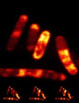
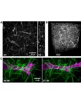
Confocal and Super-resolution Imaging of RNA in Live Bacteria Using a Fluorogenic Silicon Rhodamine-binding Aptamer

含氟硅-罗丹明适配体用于活性菌中RNA的激光共聚焦超分辨率成像

RW Regina Wirth
Peng Gao Peng Gao
G. Ulrich Nienhaus G. Ulrich Nienhaus
Murat Sunbul Murat Sunbul
AJ Andres Jäschke
7038 Views
May 5, 2020
Genetically encoded light-up RNA aptamers have been shown to be promising tools for the visualization of RNAs in living cells, helping us to advance our understanding of the broad and complex life of RNA. Although a handful of light-up aptamers spanning the visible wavelength region have been developed, none of them have yet been reported to be compatible with advanced super-resolution techniques, mainly due to poor photophysical properties of their small-molecule fluorogens. Here, we describe a detailed protocol for fluorescence microscopy of mRNA in live bacteria using the recently reported fluorogenic silicon rhodamine binding aptamer (SiRA) featuring excellent photophysical properties. Notably, with SiRA, we demonstrated the first aptamer-based RNA visualization using super-resolution (STED) microscopy. This imaging method can be especially valuable for visualization of RNA in prokaryotes since the size of a bacterium is only a few times greater than the optical resolution of a conventional microscope.
气相色谱-氢火焰离子化检测法测定哺乳动物组织中的脂肪酸

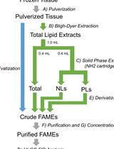
Quantification of Fatty Acids in Mammalian Tissues by Gas Chromatography–Hydrogen Flame Ionization Detection

气相色谱-氢火焰离子化检测法测定哺乳动物组织中的脂肪酸

FH Fumie Hamano
ST Suzumi M Tokuoka
TH Tomomi Hashidate-Yoshida
HS Hideo Shindou
TS Takao Shimizu
YK Yoshihiro Kita
5137 Views
May 5, 2020
In mammalian organisms, fatty acids (FAs) exist mostly in esterified forms, as building blocks of phospholipids, triglycerides, and cholesteryl esters, while some exist as non-esterified free FAs. The absolute quantification of FA species in total lipids or in a specific lipid class is critical in lipid-metabolism studies. To quantify FAs in biological samples, gas chromatography–hydrogen flame ionization detection (GC-FID)-based methods have been used as highly robust and reliable techniques. Prior to GC-FID analysis, FAs need to be derivatized to volatile FA methyl esters (FAMEs). The derivatization of unsaturated FAs using classical derivatization methods that rely on high reaction temperature requires skill; consequently, the quantification results are often unreliable. The recently available FA-methylation procedure rapidly and reliably derivatizes a variety of FA species, including poly-unsaturated FAs (PUFAs). To analyze FAs in mammalian tissue samples, lipid extraction and fractionation are also critical for robust analysis. In this report, we describe a whole protocol for the GC-FID-based FA quantification of mammalian tissue samples, including lipid extraction, fractionation, derivatization, and quantification. The protocol is useful when various FAs, especially unsaturated FAs, need to be reliably quantified.

癌症生物学

比色法测定RhoB-GTPase活性

Colorimetric RhoB GTPase Activity Assay

比色法测定RhoB-GTPase活性

KZ Kossay Zaoui
SD Stephanie Duhamel
4264 Views
May 5, 2020
The Ras homologous protein (Rho) GTPase subfamily, including RhoA, RhoB, and RhoC are small molecules (~21 kDa) that act as molecular switches in a wide range of signaling pathways to orchestrate biological processes associated with both physiological and tumorigenic cellular states. The Rho GTPases are crucial regulators of actin cytoskeleton rearrangements and FA dynamics and are required for effective cell migration and invasion, as well as cell cycle progression and apoptosis. The Rho GTPases activity is regulated by conformational switching between GTP-bound (active) and GDP-bound (inactive) states. This GTP/GDP cycling is tightly controlled by the guanine nucleotide exchange factors (GEFs), which function as activators by catalyzing the exchange of GDP for GTP and by the GTPase-activating proteins (GAPs), which enable hydrolysis of GTP leading to the Rho GTPase inactivation. Here, we describe a detailed protocol to perform a RhoB G-LISA activation assay to detect the level of GTP-loaded RhoB in vitro. This is the first colorimetric assay designed to specifically measure RhoB activation. This method was developed by adapting the RhoA G-LISA Activation Assay Kit (Cytoskeleton, Inc.) and allow the precise measurement of RhoB activity in less than 3 hours. This rapid methodology can be broadly used to assess the level of GTP-loaded RhoB in any kind of cellular models, to appreciate either the role RhoB activation in physiological processes, diseases, oncogenic transformation or for drug discovery in high throughput screens.

发育生物学

地标系统在拟南芥雄性减数分裂活体细胞成像中的应用

Live Cell Imaging of Male Meiosis in Arabidopsis by a Landmark-based System

地标系统在拟南芥雄性减数分裂活体细胞成像中的应用

MP Maria Ada Prusicki
EK Emma Mathilde Keizer
Rv Rik Peter van Rosmalen
CF Christian Fleck
AS Arp Schnittger
5772 Views
May 5, 2020
Live cell imaging has tremendously promoted our understanding of cellular and subcellular processes such as cell division. Here, we present a step-by-step protocol for a robust and easy-to-use live cell imaging approach to study male meiosis in the plant Arabidopsis thaliana as recently established. Our method relies on the concomitant analysis of two reporter genes that highlight chromosome configurations and microtubule dynamics. In combination, these reporter genes allowed the discrimination of five cellular parameters: cell shape, microtubule array, nucleus position, nucleolus position, and chromatin condensation. These parameters can adopt different states, e.g., the nucleus position can be central or lateral. Analyzing how tightly these states are associated gives rise to landmark stages that in turn allow a quantitative and qualitative dissection of meiotic progression. We envision that such an approach can also provide valuable criteria for the analysis of cell differentiation processes outside of meiosis.
机械力应用于果蝇胚胎的磁性粒子显微注射操作

Application of Mechanical Forces on Drosophila Embryos by Manipulation of Microinjected Magnetic Particles

机械力应用于果蝇胚胎的磁性粒子显微注射操作

AD Arturo D’Angelo
JS Jérôme Solon
3606 Views
May 5, 2020
Cells generate mechanical forces to shape tissues during morphogenesis. These forces can activate several biochemical pathways and trigger diverse cellular responses by mechano-sensation, such as differentiation, division, migration and apoptosis. Assessing the mechano-responses of cells in living organisms requires tools to apply controlled local forces within biological tissues. For this, we have set up a method to generate controlled forces on a magnetic particle embedded within a chosen tissue of Drosophila embryos. We designed a protocol to inject an individual particle in early embryos and to position it, using a permanent magnet, within the tissue of our choice. Controlled forces in the range of pico to nanonewtons can be applied on the particle with the use of an electromagnet that has been previously calibrated. The bead displacement and the epithelial deformation upon force application can be followed with live imaging and further analyzed using simple analysis tools. This method has been successfully used to identify changes in mechanics in the blastoderm before gastrulation. This protocol provides the details, (i) for injecting a magnetic particle in Drosophila embryos, (ii) for calibrating an electromagnet and (iii) to apply controlled forces in living tissues.
改良半固态克隆培养鉴定小鼠胚胎造血内皮细胞B-1和B-2祖细胞集落形成能力

A Modified Semisolid Clonal Culture for Identification of B-1 and B-2 Progenitor Colony Forming Ability of Mouse Embryonic Hemogenic Endothelial Cells

改良半固态克隆培养鉴定小鼠胚胎造血内皮细胞B-1和B-2祖细胞集落形成能力

MK Michihiro Kobayashi
Momoko Yoshimoto Momoko Yoshimoto
3317 Views
May 5, 2020
The search for the origin of the first hematopoietic stem cells (HSCs) in the mouse embryo has been a hot topic in the field of developmental hematopoiesis. Detecting lymphoid potential is one of the supportive evidence to show the definitive hematopoietic activity of HSCs. However, the first B-lymphoid potential in the mouse embryos are reported to be biased to innate-like B-1 cell lineage that can develop from hemogenic endothelial cells (HECs) independently of HSCs. On the other hand, conventional adaptive immune B cells (B-2) cells are considered to be exclusively derived from HSCs. Therefore, segregating B-1 and B-2 progenitor potential is important to understand the developmental process of HSCs that are also produced from HECs through intermediate precursors referred to as pre-HSCs. Both HECs and pre-HSCs show endothelial surface phenotype and require stromal support to detect their hematopoietic activity. The method utilizing stromal cell culture followed by modified semisolid clonal culture enables us to detect the number of colony forming units for B-1/B-2 progenitors originally derived from HECs/pre-HSCs, which will reflect the potential of B-1 biased or multi-lineage repopulating HSCs.

免疫学

HIV-CRISPR:一种CRISPR/Cas9筛选HIV复制相关基因的方法

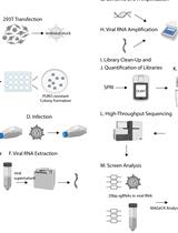
HIV-CRISPR: A CRISPR/Cas9 Screening Method to Identify Genes Affecting HIV Replication

HIV-CRISPR:一种CRISPR/Cas9筛选HIV复制相关基因的方法

FR Ferdinand Roesch
MO Molly OhAinle
8620 Views
May 5, 2020
Screening with CRISPR/Cas9 technology has already led to significant discoveries in the fields of cancer biology, cell biology and virology. Because of the relatively low false discovery rates and the ability to perform high-throughput, pooled approaches, it has rapidly become the assay of choice for screening studies, including whole-genome screens. Here, we describe a CRISPR screening protocol that allows for efficient screening of the entire life cycle of HIV-1 through packaging of the HIV-CRISPR lentiviral genomes by infecting HIV-1 virus in trans.
通过EdU掺入法评估体内B细胞增殖

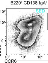
Evaluation of B Cell Proliferation in vivo by EdU Incorporation Assay

通过EdU掺入法评估体内B细胞增殖

AB Adi Biram
ZS Ziv Shulman
5840 Views
May 5, 2020
Generation of antibodies is crucial for establishing enduring protection from invading pathogens, as well as for maintaining homeostasis with commensal bacteria at mucosal surfaces. Chronic exposure to microbiota- and dietary- derived antigens results in continuous production of antibody producing cells within the Peyer’s patch germinal center structures. Recently, we have shown that B cells responding to gut-derived antigens colonize the subepithelial dome (SED) in Peyer’s patches and rapidly proliferate independently of their relative BCR affinity. To evaluate B cell proliferation within different niches in Peyer’s patches, we applied in vivo EdU incorporation assay as described in this protocol.
用小鼠CD4+T细胞评价体内外胞啃作用

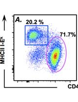
Assessing in vitro and in vivo Trogocytosis By Murine CD4+ T cells

用小鼠CD4+T细胞评价体内外胞啃作用

JR Jim Reed
Scott A. Wetzel Scott A. Wetzel
4845 Views
May 5, 2020
Recognition of antigens by lymphocytes (B, T, and NK) on the surface of an antigen-presenting cell (APC) leads to lymphocyte activation and the formation of an immunological synapse between the lymphocyte and the APC. At the immunological synapse APC membrane and associated membrane proteins can be transferred to the lymphocyte in a process called trogocytosis. The detection of trogocytosed molecules provides insights to the activation state, antigen specificity, and effector functions and differentiation of the lymphocytes. Here we outline our protocol for identifying trogocytosis-positive CD4+ T cells in vitro and in vivo. In vitro, antigen presenting cells are surface biotinylated and pre-loaded with magnetic polystyrene beads before incubating for a short time with in vitro activated CD4+ T cell blasts (90 min) or naïve T cells (3-24 h). After T cell recovery and APC depletion by magnetic separation trogocytosis positive (trog+) cells are identified by streptavidin staining of trogocytosed, biotinylated APC membrane proteins. Their activation phenotype, effector function, and effector differentiation are subsequently analyzed by flow cytometry immediately or after subsequent incubation. Similarly, trogocytosis-positive cells can be identified and similarly analyzed by flow cytometry. Previous studies have described methods for analyzing T cell trogocytosis to identify antigen-specific cells or the antigenic epitopes recognized by the cells. With the current protocol, the effects of trogocytosis on the individual T cell or the ability of trog+ T cells to modulate the activation and function of other immune cells can be assessed over an extended period of time.

微生物学

使用ADP特异性己糖激酶测量激酶和ATPase动力学的灵敏耦合酶活测定

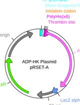
A Sensitive Coupled Enzyme Assay for Measuring Kinase and ATPase Kinetics Using ADP-Specific Hexokinase

使用ADP特异性己糖激酶测量激酶和ATPase动力学的灵敏耦合酶活测定

Ciaran R. McFarlane Ciaran R. McFarlane
JM James W. Murray
6866 Views
May 5, 2020
Kinases and ATPases perform essential biological functions in metabolism and regulation. Activity of these enzymes is commonly measured by coupling ATP consumption to the synthesis of a detectable product. For most assay systems the ATP concentration during the reaction is unknown, compromising the precision of the assay.Using the ADP-specific hexokinase (ADP-HK) from the thermophilic archaeon Thermococcus litoralis the protocol outlined here allows real time coupling of ATP consumption to downstream signal change enabling accurate kinetic measurements. ADP-HK phosphorylates glucose that is then used by glucose-6-phosphate dehydrogenase to reduce NAD+ to NADH which can be measured at 340 nm. We have shown this assay to be sensitive to the detection of micromole quantities of ADP with no detectable background from ATP.
秀丽隐杆线虫肠道细菌数量测定

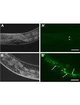
Quantification of Bacteria Residing in Caenorhabditis elegans Intestine

秀丽隐杆线虫肠道细菌数量测定

M. Fernanda Palominos M. Fernanda Palominos
AC Andrea Calixto
4756 Views
May 5, 2020
Quantification of intestinal colonization by pathogenic or commensal bacteria constitute a critical part of the analysis to understand host-microbe interactions during different time points of their interplay. Here we detail a method to isolate non-pathogenic and pathogenic bacteria from C. elegans intestines, and classify gut phenotypes induced by bacterial pathogens using fluorescently-tagged bacteria. Furthermore, these methods can be used to isolate and identify new culturable bacterial species from natural microbiomes of wild nematodes.
在利什曼原虫中利用DNase I和 Nuclease S1酶解进行病毒双链RNA鉴定

Viral Double-Stranded RNA Detection by DNase I and Nuclease S1 digestions in Leishmania parasites

在利什曼原虫中利用DNase I和 Nuclease S1酶解进行病毒双链RNA鉴定

NI Nathalie Isorce
NF Nicolas Fasel
4194 Views
May 5, 2020
Many RNA viruses are found in protozoan parasites. They can be responsible for more serious pathology or treatment failure. For the detection of viral double-stranded RNA (dsRNA), sequence-dependent and -independent methods are available, such as quantitative real-time PCR and immunofluorescence, dot blot, ELISA or sequencing. The technique presented here is sequence-independent and is well detailed in the following protocol, taking the example of Leishmania RNA virus (LRV) in Leishmania guyanensis (Lgy) species. To summarise, the protocol is divided into four major steps: RNA extraction from the parasites, RNA purification, enzymatic digestions with DNase I and Nuclease S1, and visualization by gel electrophoresis. This method can be used to detect other viral dsRNA in other parasites. It provides an additional tool, complementary to other techniques previously cited and it is easy and quite fast to achieve.

分子生物学

基于分割纳米荧光素酶的乙型脑炎病毒组装检测

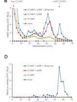
Split Nano Luciferase-based Assay to Measure Assembly of Japanese Encephalitis Virus

基于分割纳米荧光素酶的乙型脑炎病毒组装检测

SG Simon Goto
KI Kotaro Ishida
RS Ryosuke Suzuki
EM Eiji Morita
3546 Views
May 5, 2020
Cells infected with flavivirus release various forms of infectious and non-infectious particles as products and by-products. Comprehensive profiling of the released particles by density gradient centrifugation is informative for understanding viral particle assembly. However, it is difficult to detect low-abundance minor particles in such analyses. We developed a method for viral particle analysis that integrates a high-sensitivity split luciferase system and density gradient centrifugation. This protocol enables high-resolution profiling of particles produced by cells expressing Japanese encephalitis virus factors.

神经科学

快速建立能诱导表达功能研究所需蛋白的人神经细胞模型

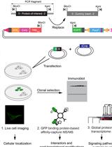
Rapid Generation of Human Neuronal Cell Models Enabling Inducible Expression of Proteins-of-interest for Functional Studies

快速建立能诱导表达功能研究所需蛋白的人神经细胞模型

XW Xinzhu Wang
EF Erik Friesen
IM Iris Müller
ML Mackenzie Lemieux
RD Ramona Dukart
IM Isabella BL Maia
SK Suneil Kalia
GS Gerold Schmitt-Ulms
4438 Views
May 5, 2020
CRISPR-Cas9 technology has transformed the ability to edit genomic sequences and control gene expression with unprecedented ease and scale. However, precise genomic insertions of coding sequences using this technology remain time-consuming and inefficient because they require introducing adjacent single-strand cuts through Cas9 nickase action and invoking the host-encoded homology-directed repair program through the concomitant introduction of large repair templates. Here, we present a system for the rapid study of any protein-of-interest in two neuronal cell models following its inducible expression from the human AAVS1 safe harbor locus. With lox-flanked foundation cassettes in the AAVS1 site and a tailor-made plasmid for accepting coding sequences-of-interest in place, the system allows investigators to produce their own neuronal cell models for the inducible expression of any coding sequence in less than a month. Due to the availability of preinserted enhanced green fluorescent protein (EGFP) coding sequences that can be fused to the protein-of-interest, the system facilitates functional investigations that track a protein-of-interest by live-cell microscopy as well as interactome analyses that capitalize on the availability of exquisitely efficient EGFP capture matrices.

系统生物学

基于模板的蛋白质结构精确预测的机器学习序列比对

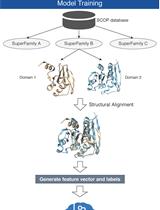
Sequence Alignment Using Machine Learning for Accurate Template-based Protein Structure Prediction

基于模板的蛋白质结构精确预测的机器学习序列比对

Shuichiro Makigaki Shuichiro Makigaki
Takashi Ishida Takashi Ishida
5052 Views
May 5, 2020
Template-based modeling, the process of predicting the tertiary structure of a protein by using homologous protein structures, is useful when good templates can be available. Indeed, modern homology detection methods can find remote homologs with high sensitivity. However, the accuracy of template-based models generated from the homology-detection-based alignments is often lower than that from ideal alignments. In this study, we propose a new method that generates pairwise sequence alignments for more accurate template-based modeling. Our method trains a machine learning model using the structural alignment of known homologs. When calculating sequence alignments, instead of a fixed substitution matrix, this method dynamically predicts a substitution score from the trained model.

更正

更正: 用于3D荧光成像的鼠尾胶的荧光标记

Correction Notice: Fluorescent Labeling of Rat-tail Collagen for 3D Fluorescence Imaging

更正: 用于3D荧光成像的鼠尾胶的荧光标记

Andrew D. Doyle Andrew D. Doyle
1538 Views
May 5, 2020