往期刊物2020

卷册: 10, 期号: 2

left

Dec

20

Dec

5

Nov

20

Nov

5

Oct

20

Oct

5

Sep

20

Sep

5

Aug

20

Aug

5

Jul

20

Jul

5

Jun

20

Jun

5

May

20

May

5

Apr

20

Apr

5

Mar

20

Mar

5

Feb

20

Feb

5

Jan

20

Jan

5

right

生物化学

核糖核蛋白免疫沉淀分析

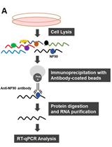
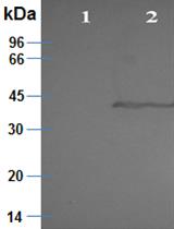
Ribonucleoprotein Immunoprecipitation (RIP) Analysis

核糖核蛋白免疫沉淀分析

JM Jennifer L. Martindale
MG Myriam Gorospe
MI Maria L. Idda
9106 Views
Jan 20, 2020
RNAs and RNA-binding proteins (RBPs) can interact dynamically in ribonucleoprotein (RNP) complexes that play important roles in controlling gene expression programs. One of the powerful ways to investigate changes in the association of RNAs with an RBP of interest is by immunoprecipitation (IP) analysis of native RNPs. RIP (RNP immunoprecipitation) analysis enables the rapid identification of endogenous RNAs bound to an RBP and to monitor time-dependent changes in this association, as well as changes in response to different metabolic and stress conditions. The protocol is based on the use of an antibody, typically an anti-RBP antibody, to immunoprecipitate the RNP complex. The RNA within the immunoprecipitated complex can then be isolated and further studied using different approaches such as PCR, microarray, Northern blot, and sequencing analyses. Among other advantages, RIP analysis (i) measures RNP associations in many samples relatively quickly, (ii) can be adapted easily to different endogenous RBPs, and (iii) provides extensive information at low cost. Among its limitations, RIP analysis does not inform on the specific sites of interaction of an RBP with a given target RNAs, although recent adaptations of RIP have been developed to overcome this problem. Here we provide an optimized protocol for RIP analysis that can be used to study RNA-protein interactions relevant to many areas of biology.
利用Bsoft处理和解释冷冻电镜数据方法:视网膜粘连蛋白,视网膜劈裂蛋白的实例研究

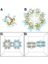
Protocols for Processing and Interpreting cryoEM Data Using Bsoft: A Case Study of the Retinal Adhesion Protein, Retinoschisin

利用Bsoft处理和解释冷冻电镜数据方法:视网膜粘连蛋白,视网膜劈裂蛋白的实例研究

JH J. Bernard Heymann
4008 Views
Jan 20, 2020
The goal of cryoEM is to determine the structures of biomolecules from electron micrographs. In many cases the processing is straightforward and can be handled with routine protocols. In other cases, the properties and behavior of the specimen require adaptions to properly interpret the data. Here I describe the protocols for examining the higher order assemblies of the retinal adhesion protein, retinoschisin (RS1), using the Bsoft package. The protocols for micrograph preprocessing, 2D classification and 3D alignment and reconstruction follow the usual patterns for the majority of cryoEM specimens. The interpretation of the results is specific to the branched network of RS1 filaments. The 2D class averages are used to determine the relative positions of the RS1 molecules, thus defining the interacting interfaces in the network. The major interface of the linear filament is then further examined by reconstructing the “unit cell” and fitting the molecular models.

生物物理学

利用荧光和原子力显微镜、荧光和浊度分析以及FRAP研究液-液相分离中的蛋白质聚集

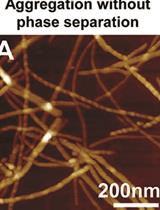
Studying Protein Aggregation in the Context of Liquid-liquid Phase Separation Using Fluorescence and Atomic Force Microscopy, Fluorescence and Turbidity Assays, and FRAP

利用荧光和原子力显微镜、荧光和浊度分析以及FRAP研究液-液相分离中的蛋白质聚集

WB W. Michael Babinchak
WS Witold K. Surewicz
8179 Views
Jan 20, 2020
Liquid-liquid phase separation (LLPS) underlies the physiological assembly of many membrane-less organelles throughout the cell. However, dysregulation of LLPS may mediate the formation of pathological aggregates associated with neurodegenerative diseases. Here, we present complementary experimental approaches to study protein aggregation within and outside the context of LLPS in order to ascertain the impact of LLPS on aggregation kinetics. Techniques described include imaging-based approaches [fluorescence microscopy, atomic force microscopy (AFM), fluorescence recovery after photobleaching (FRAP)] as well as plate reader assays [Thioflavin-T (ThT) fluorescence intensity and turbidity]. Data and conclusions utilizing these approaches were recently reported for the low complexity domain (LCD) of the transactive response DNA binding protein of 43 kDa (TDP-43).
680-1300 nm波段荧光分子双光子吸收特性的表征

Characterizing the Two-photon Absorption Properties of Fluorescent Molecules in the 680-1300 nm Spectral Range

680-1300 nm波段荧光分子双光子吸收特性的表征

MD Mikhail Drobizhev
RM Rosana S Molina
TH Thomas E Hughes
4069 Views
Jan 20, 2020
Two-photon laser scanning microscopy (2PLSM) is a state-of-the-art technique used for non-invasive imaging deep inside the tissue, with high 3D resolution, minimal out-of-focus photodamage, and minimal autofluorescence background. For optimal application of fluorescent probes in 2PLSM, their two-photon absorption (2PA) spectra, expressed in absolute cross sections must be characterized. Excitation at optimum wavelength will make it possible to reduce the laser power and therefore minimize photodamage. Obtaining 2PA spectra and cross sections requires correcting the two-photon excited fluorescence signals for a combination of laser properties, including the beam spatial profile, pulse duration, and absolute power, at each wavelength of the tuning range. To avoid such tedious day-to-day laser characterization required in the absolute measurement method, a relative method based on independently characterized 2PA reference standards is often used. By carefully analyzing the available literature data, we selected the most reliable standards for both the 2PA spectral shape and cross section measurements. Here we describe a protocol for measuring the 2PA spectral shapes and cross sections of fluorescent proteins and other fluorophores with the relative fluorescence method using these reference standards. Our protocol first describes how to build an optical system and then how to perform the measurements. In our protocol, we use Coumarin 540A in dimethyl sulfoxide and LDS 798 in chloroform for the spectral shape measurements to cover the range from 680 to 1300 nm, and Rhodamine 590 in methanol and Fluorescein in alkaline water (pH 11) for the absolute two-photon cross section measurements.

癌症生物学

人婴幼儿血管瘤中干细胞、内皮细胞和周细胞的分离

Isolation of Stem Cells, Endothelial Cells and Pericytes from Human Infantile Hemangioma

人婴幼儿血管瘤中干细胞、内皮细胞和周细胞的分离

LH Lan Huang
JB Joyce Bischoff
4079 Views
Jan 20, 2020
Infantile hemangioma (IH) is a vascular tumor noted for its excessive blood vessel formation during infancy, glucose-transporter-1 (GLUT1)-positive staining of the blood vessels, and its slow spontaneous involution over several years in early childhood. For most children, IH poses no serious threat because it will eventually involute, but a subset can destroy facial structures and impair vision, breathing and feeding. To unravel the molecular mechanism(s) driving IH-specific vascular overgrowth, which to date remains elusive, investigators have studied IH histopathology, the cellular constituents and mRNA expression. Hemangioma endothelial cells (HemEC) were first isolated from surgically removed IH specimens in 1982 by Mulliken and colleagues (Mulliken et al., 1982). Hemangioma stem cells (HemSC) were isolated in 2008, hemangioma pericytes in 2013 and GLUT1-positive HemEC in 2015. Indeed, as we describe here, it is possible to isolate HemSC, GLUT1-positive HemEC, GLUT1-negative HemEC and HemPericytes from a single proliferating IH tissue specimen. This is accomplished by sequential selection using antibodies against specific cell surface markers: anti-CD133 to select HemSC, anti-GLUT1 and anti-CD31 to select HemECs and anti-PDGFRβ to select HemPericytes. IH-derived cells proliferate well in culture and can be used for in vitro and in vivo vasculogenesis and angiogenesis assays.
体外骨碎片培养列阵模拟骨转移

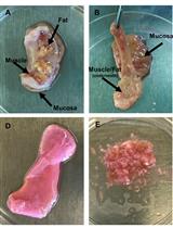
Bone-in-culture Array to Model Bone Metastasis in ex vivo Condition

体外骨碎片培养列阵模拟骨转移

HW Hai Wang
XZ Xiang H.-F. Zhang
4007 Views
Jan 20, 2020
Bone is the most frequently affected organ by metastases of breast cancer and prostate cancer. Our knowledge on bone metastasis is extremely limited due to the lack of potent and efficient experimental models. We developed the “Bone-In-Culture Array (BICA)” platform to model the bone colonization of cancer cells in ex vivo cultures. The use of the BICA platform will facilitate in-depth mechanistic studies and high-throughput screening of drugs for bone metastasis.

免疫学

细胞表面Ruffling的自动分析:Ruffle的宏观量化

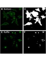
Automated Analysis of Cell Surface Ruffling: Ruffle Quantification Macro

细胞表面Ruffling的自动分析:Ruffle的宏观量化

Nicholas D. Condon Nicholas D. Condon
Jennifer L. Stow Jennifer L. Stow
Adam A. Wall Adam A. Wall
4483 Views
Jan 20, 2020
Cell surface protrusions include F-actin rich, wave-like ruffles that are erected transiently in response to stimuli and during cell migration. Macrophages are innate immune cells that ruffle constitutively and more dramatically in cells activated by pathogens. Dorsal ruffles and their resulting macropinosomes are key sites for environmental sampling, pathogen detection and immune signaling. Quantitative assessment of ruffling is important for assessing pathogen responses in macrophages and for analysis of growth factor responses in other cell types but automated and quantitative methods are lacking, and rely on manual and qualitative assessments. Here we present an automated ImageJ macro for quantifying dorsal cell surface protrusions from 3D microscope images. The assay presented here is suitable for high-throughput screening applications to detect drug, pathogen, or growth factor induced changes in cell ruffling by measuring ruffle area and intensity and providing normalized values in an easy to read combined spreadsheet.
固有层聚集培养模式中HIV-1介导的Gut CD4+ T细胞凋亡的量化分析

Quantifying HIV-1-Mediated Gut CD4+ T Cell Death in the Lamina Propria Aggregate Culture (LPAC) Model

固有层聚集培养模式中HIV-1介导的Gut CD4+ T细胞凋亡的量化分析

SD Stephanie M. Dillon
KG Kejun Guo
MC Moriah J. Castleman
MS Mario L. Santiago
CW Cara C. Wilson
4060 Views
Jan 20, 2020
Gut CD4 T cells are major targets of HIV-1 and are massively depleted early during infection. To better understand the mechanisms governing HIV-1-mediated CD4 T cell death, we developed the physiologically-relevant Lamina Propria Aggregate Culture (LPAC) model. The LPAC model is ideal for studying CD4 T cell death induced by clinically-relevant Transmitted/Founder (TF) HIV-1 strains and is also suitable for studying how enteric microbes and soluble factors (e.g., Type I Interferons) impact LP CD4 T cell death and function. Here, we detail the protocol to establish LP CD4 T cell infection using a process of spinoculation, the subsequent evaluation of infection levels using multicolor flow cytometry and the determination of overall LP CD4 T cell death using absolute LP CD4 T cell counts. We also describe the preparation of virus stocks of Transmitted/Founder (TF) HIV-1 infectious molecular clones that were successfully used in the LPAC model.

微生物学

硫酸酯酶法测定植物对土壤微生物活性的影响

Sulfatase Assay to Determine Influence of Plants on Microbial Activity in Soil

硫酸酯酶法测定植物对土壤微生物活性的影响

AK Anna Koprivova
AS Achim Schmalenberger
SK Stanislav Kopriva
4906 Views
Jan 20, 2020
Sulfatase activity is often used as a measure of the activity of soil microorganisms. It is thus a suitable tool to investigate the response of microbes to plants. Here we present a method to determine the influence of various Arabidopsis genotypes on the function of soil microbiota using the sulfatase as a quantitative measure. We grew the plants in soil/sand mix under control conditions and measured the sulfatase activity in soil using a spectrophotometric determination of the product. This protocol can be used to test the contribution of individual genes to control of microbiome assembly through analysis of mutants as well as the influence of environment on plant-microbe interactions.
微生物胞外囊泡的研究:密度梯度分离,保护性分析和活体示踪标记

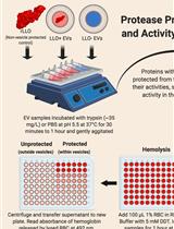
Study of Microbial Extracellular Vesicles: Separation by Density Gradients, Protection Assays and Labelling for Live Tracking

微生物胞外囊泡的研究:密度梯度分离,保护性分析和活体示踪标记

CC Carolina Coelho
RV Raghav Vij
DS Daniel Q. Smith
NB Nathan R. Brady
AH Anne Hamacher-Brady
AC Arturo Casadevall
4790 Views
Jan 20, 2020
Extracellular vesicles (EVs) are produced by all domains of life including Bacteria, Archaea and Eukarya. EVs are critical for cellular physiology and contain varied cargo: virulence factors, cell wall remodeling enzymes, extracellular matrix components and even nucleic acids and metabolites. While various protocols for isolating EVs have been established for mammalian cells, the field is actively developing tools to study EVs in other organisms. In this protocol we describe our methods to perform density gradient purification of EVs in bacterial cells, allowing for separation of EV subpopulations, followed by protection assays for EV cargo characterization. Furthermore, we devised a protocol which incorporates a fluorescent conjugate of fatty acids into EVs, the first to allow live-cell EV tracking to observe release of EVs, including during infection of mammalian cells by pathogenic bacteria. These protocols are powerful tools for EV researchers as they enable the observation of EV release and the study of the mechanisms of their formation and release.
利用荧光素酶报告系统检测大肠杆菌中启动子活性

Measurement of the Promoter Activity in Escherichia coli by Using a Luciferase Reporter

利用荧光素酶报告系统检测大肠杆菌中启动子活性

YY Yuki Yamanaka
HW Hiroki Watanabe
EY Erika Yamauchi
YM Yukari Miyake
KY Kaneyoshi Yamamoto
4688 Views
Jan 20, 2020
The reporter system is widely used technique for measuring promoter activity in bacterial cells. Until now, a number of reporter system have been developed, but the bioluminescent reporter constructed from the bacterial luciferase genes is one of the useful systems for measuring in vivo dynamics of gene expression. The introduced bioluciferase lux reporter enables easy, fast, and sensitive measurement of the promoter activity without cell lysis because the substrates of bioluminescent reaction are synthesized inside the bacterial cell, thereby allowing low-cost experiments. This protocol describes a high throughput technique to measure the promoter activity in Escherichia coli K-12 using the lux reporter system.
一种无放射性布氏锥虫糖原合成酶激酶-3抑制剂探究实验

A Radioactive-free Kinase Inhibitor Discovery Assay Against the Trypanosoma brucei Glycogen Synthase Kinase-3 short (TbGSK-3s)

一种无放射性布氏锥虫糖原合成酶激酶-3抑制剂探究实验

Antonia Efstathiou Antonia Efstathiou
DS Despina Smirlis
3781 Views
Jan 20, 2020
The identification of small molecules possessing inhibitory activity in vitro, against a given target kinase, is the first step in the drug discovery process. Herein, we describe a non radioactive protocol using luciferase-based ATP assay for the identification of inhibitors for the short isoform of the Trypanosoma brucei’s Glycogen Synthase Kinase-3 (TbGSK-3s). TbGSK-3s represents a potential drug target as it is essential for parasite survival. Small molecules used in our study are indirubin analogues possessing substitutions in different positions in the bis-indole backbone. Presently, the standard laboratory practice for the kinase assays is the incorporation of radiolabeled phosphate from [gamma-32P]ATP as the efforts for developing non-radioactive assays (ELISA-based assays, fluorescence quenching assays, etc.) exhibit limitations such as lack in sensitivity or limitations for broad applications. This protocol can be a useful starting point for lead discovery, as it surpasses the drawbacks of radioactive kinase assays and it allows for relatively sensitive measurements of kinase inhibition for TbGSK-3s.

神经科学

研究小鼠社交行为和重复自我修饰的单一试验

A Single Test to Study Social Behavior and Repetitive Self-grooming in Mice

研究小鼠社交行为和重复自我修饰的单一试验

NL Niraj V. Lawande
AU Ammar L. Ujjainwala
CC Catherine A. Christian
5041 Views
Jan 20, 2020
The ability to recognize and interact with members of the same species is essential for social communication. Investigating the neural substrates of social interest and recognition may offer insights into the behavioral differences present in disorders affecting social behavior. Assays used to study social interest in rodents include the 3-chamber test, a partition test, and a social interaction test. Here, we present a single protocol that can be used to quantify the level of social interest displayed by mice, the ability to distinguish between different individual mice (social recognition), and the level of repetitive self-grooming displayed. In the first part of the protocol, a social habituation/dishabituation test, the time spent by a test mouse sniffing a stimulus mouse is quantified over 9 trials. In the first 8 interactions, the same stimulus mouse is used repeatedly; on the ninth trial, a novel stimulus mouse is presented. Intact social recognition is indicated by a progressive decrease in the investigation time over trials 1-8, and an increase in trial 9. The interval between each social trial is used to quantify self-grooming, a stereotyped repetitive behavior in mice. We also present a method for randomized, blinded analysis of these behaviors to increase rigor and reproducibility of results. Therefore, this single behavioral test enables ready assessment of phenotypes of both social and repetitive behaviors in an integrated manner in the same animals. This feature can be advantageous in understanding interactions between these behaviors and phenotypes in mouse models with genetic variants associated with autism and other neurodevelopmental or neuropsychiatric disorders, which are often characterized by these behavioral differences.
一种简单有效的大鼠脊髓星形胶质细胞和小胶质细胞联合分离培养方法

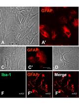
A Simple and Efficient Method for Concomitant Isolation and Culture of Enriched Astroglial and Microglial Cells from the Rat Spinal Cord

一种简单有效的大鼠脊髓星形胶质细胞和小胶质细胞联合分离培养方法

PM Pooja Shree Mishra
TR Trichur R Raju
4466 Views
Jan 20, 2020
Investigations into glial biology have contributed substantially in understanding the physiology and pathology of the nervous system. However, intricacies of the neuron-glial and glial-glial interactions in vivo present significant challenges while delineating the individual cell-type contributions, thus making the in vitro techniques exceedingly relevant to study glial biology. However, obtaining optimal yield along with high purity has been challenging for microglial cultures. Here we present a simple protocol to establish enriched astroglial as well as microglial cultures from the neonatal rat spinal cord. This method results in highly enriched astroglial and microglial cultures with maximal yield.
大鼠自由(低应力)探索测量方案

Protocol for Measuring Free (Low-stress) Exploration in Rats

大鼠自由(低应力)探索测量方案

Wojciech Pisula Wojciech Pisula
KM Klaudia Modlinska
3597 Views
Jan 20, 2020
Research on exploratory behavior plays a key part in behavioral science. Studying exploratory behavior of laboratory rodents may provide important data about many developmental and neurobiological processes occurring in animal ontogenesis. The proposed protocol for measuring the free (low-stress) exploration behavior in rats is straightforward, requires minimal resources and very little animal training. It can therefore be broadly applied to studying animal cognition, animal behavior in general, the aging processes, and several animal models of various phenomena.