往期刊物2019

卷册: 9, 期号: 24

left

Dec

20

Dec

5

Nov

20

Nov

5

Oct

20

Oct

5

Sep

20

Sep

5

Aug

20

Aug

5

Jul

20

Jul

5

Jun

20

Jun

5

May

20

May

5

Apr

20

Apr

5

Mar

20

Mar

5

Feb

20

Feb

5

Jan

20

Jan

5

right

生物化学

一种综合的实验方法:利用免疫组织化学染色和分析进行蛋白表达的半定量测定

Semi-quantitative Determination of Protein Expression Using Immunohistochemistry Staining and Analysis: An Integrated Protocol

一种综合的实验方法:利用免疫组织化学染色和分析进行蛋白表达的半定量测定

AC Alexandra R Crowe
Wei  Yue Wei Yue
15325 Views
Dec 20, 2019
Semi-quantitative immunohistochemistry (IHC) is a powerful method for investigating protein expression and localization within tissues that involves using software, such as the freely available Fiji (ImageJ), to conduct deconvolution and downstream analysis. Currently, there is lack of an integrated protocol that includes a detailed procedure on how to measure or compare protein expression. Publications that use semi-quantitative methods to ascertain protein expression often don’t provide enough details in their methods section, which makes it difficult for the reader to reproduce their data. The current protocol provides an example and detailed steps of conducting semi-quantitative analysis of IHC images using Fiji software.

细胞生物学

利用荧光团激活蛋白标记受体分析EGF受体的内吞作用

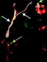
Analysis of EGF Receptor Endocytosis Using Fluorogen Activating Protein Tagged Receptor

利用荧光团激活蛋白标记受体分析EGF受体的内吞作用

MP Mireia Perez Verdaguer
ML Mads B. Larsen
MB Marcel P. Bruchez
SW Simon C. Watkins
SS Alexander Sorkin
4423 Views
Dec 20, 2019
Functional activities of many transmembrane proteins are controlled by their endocytosis. One of the most studied experimental models is the epidermal growth factor (EGF) receptor (EGFR). However, endocytic trafficking of EGFR has been predominantly analyzed using labeled EGF, whereas quantitative analyses of the endocytosis of the receptor itself have been sparse. The fluorescence microscopy methods described here are designed to directly quantify EGFR internalization in living cells without labeled EGFR ligands or antibodies. These methods utilize an engineered EGFR chimera in which the fluorogen activating protein (FAP) is fused to the receptor extracellular domain (FAP-EGFR). Binding of malachite green (MG) based dyes to FAP results in a strong far-red fluorescence of MG, thus efficiently labeling FAP-EGFR. In particular, binding of the cell impermeant MG-Bis-SA dye to FAP produces the pH-sensitive dual-excitation fluorescence, which allows differentiation of the cell-surface and internalized pools of FAP-EGFR. Two modifications of the methodology are described: 1) single-cell three-dimensional confocal imaging; and 2) high-throughput assay in multi-well plates. These methodologies can be adopted to study endocytosis of any other transmembrane protein extracellularly tagged with FAP.

免疫学

cDNA免疫PCR:一种新的基于PCR的抗原检测方法

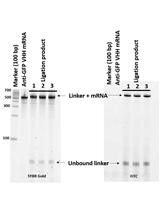
cDNA Display Mediated Immuno-PCR (cD-IPCR): A Novel PCR-based Antigen Detection Method

cDNA免疫PCR:一种新的基于PCR的抗原检测方法

Chathuni Jayathilake Chathuni Jayathilake
TT Takuya Terai
Naoto Nemoto Naoto Nemoto
5516 Views
Dec 20, 2019
Immuno-PCR (IPCR) is a powerful method in antigen detection where a PCR-amplifiable DNA reporter is conjugated to a specific antibody or an aptamer for the target molecule. In the development and application of IPCR, successful conjugation of a protein (an antibody) with a reporter DNA becomes challenging. To address this issue, we recently demonstrated the feasibility of IPCR based on cDNA display, a 1:1 covalent complex of a polypeptide and its encoding cDNA at the single molecule level. The cDNA display molecule for IPCR is generated first by transcribing the DNA that encodes the detection antibody into an mRNA by in vitro transcription. A puromycin DNA linker is then ligated to the mRNA and then in vitro translation and reverse-transcription are performed to generate the cDNA display molecule. The molecule is then directly used in antigen detection and subsequent qPCR. This method can be applied to detect various antigens in biological samples, if sequences of their single-domain antibodies (VHHs) or peptide aptamers are known.
提取和利用凋亡Jurkat细胞诱导腹腔巨噬细胞分泌白介素-10

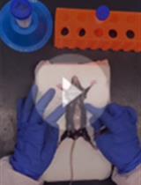
Isolation and Stimulation of Peritoneal Macrophages with Apoptotic Jurkat Cells to Produce IL-10

提取和利用凋亡Jurkat细胞诱导腹腔巨噬细胞分泌白介素-10

MS Mei Song
Xiaojing   Ma Xiaojing Ma
5391 Views
Dec 20, 2019
Clearance of apoptotic cells by macrophages is critical to ensuring cellular homeostasis and suppression of autoimmunity. Macrophage recognition of apoptotic cells triggers an anti-inflammatory response, which is mediated by the release of IL-10, TGF-β etc. with concurrent inhibition of pro-inflammatory cytokines (such as TNFα, IL-12, IL-1β). To characterize cytokine profile produced by macrophages during phagocytosis of apoptotic cells, we developed an effective, more physiologic system using isolated murine peritoneal macrophages and T-lymphocyte cell line Jurkat as a source of apoptotic cells. Apoptosis of Jurkat cells is induced with staurosporine, a protein kinase C (PKC) inhibitor and detected by Annexin V/propidium iodide staining. This in vitro assay demonstrates that murine peritoneal macrophages produce large amounts of IL-10 following exposure to apoptotic Jurkat cells.
溶酶体靶向红绿试验

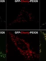
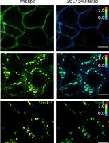
Lysosome Targeting RedGreen-assay: Selective Autophagy Sensing Assay for Mammalian Cells

溶酶体靶向红绿试验

VR Victoria Riccio
MV Miluska Vissa
AM Angus McQuibban
PK Peter Kijun Kim
5335 Views
Dec 20, 2019
The process of autophagy is an essential cellular mechanism, required to maintain general cell health through the removal of dysfunctional organelles, such as the ER, peroxisomes and mitochondria, as well as protein aggregates, and bacteria. Autophagy is an extremely dynamic process, and tools are constantly being developed to study the various steps of this process. This protocol details a method to study the end steps of autophagy-lysosomal fusion and the formation of the autolysosome. Many techniques have been used to study the various steps of the autophagy process. Here we describe the RedGreen-assay (RG-assay), an immunofluorescence-based technique used to visualize the targeting of substrates to the autolysosome in live cells. This technique takes advantage of the low lysosomal pH and over-expression of a tandem GFP-mCherry tagged protein targeted to an organelle of interest. While in the neutral cytosol or autophagosome, both GFP and RFP will fluoresce. However, within the autolysosome, the GFP signal is quenched due to the low pH environment and the RFP emission signal will predominate. This technique is readily quantifiable and amenable to high throughput experiments. Additionally, by tagging the GFP-RFP tandem fluorescent protein with organelle specific targeting sequences, it can be used to measure a wide range of substrates of autophagy.
沙眼衣原体血清型L2转化子和突变体的繁殖及纯化

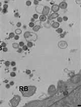
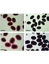
Propagation and Purification of Chlamydia trachomatis Serovar L2 Transformants and Mutants

沙眼衣原体血清型L2转化子和突变体的繁殖及纯化

RF Robert Faris
MW Mary M. Weber
3982 Views
Dec 20, 2019
Chlamydia trachomatis (C.t.) is an obligate intracellular pathogen that cannot be cultured axenically and must be propagated within eukaryotic host cells. There are at least 15 distinct chlamydial serovariants that belong to 2 major biovars commonly referred to as trachoma and lymphogranuloma venereum (LGV). The invasive chlamydia LGV serovar L2 is the most widely used experimental model for studying C.t. biology and infection and is the only strain with reliable genetic tools available. New techniques to genetically manipulate C.t. L2 have provided opportunities to make mutants using TargeTron and allelic exchange as well as strains overexpressing epitope-tagged proteins, in turn necessitating the regular purification of transformant and mutant clones. Purification of C.t. is a labor-intensive exercise and one of the most common reagents classically used in the purification process, Renografin, is no longer commercially available. A similar formulation of diatrizoate meglumine called Gastrografin is readily available and we as well as others have had great success using this in place of Renografin for chlamydial purifications. Here, we provide a detailed general protocol for infection, propagation, purification, and titering of Chlamydia trachomatis serovar L2 with additional notes specifically pertaining to mutants or recombinant DNA carrying clones.

微生物学

细菌核糖体印迹测序方法

Protocol for Ribosome Profiling in Bacteria

细菌核糖体印迹测序方法

FM Fuad Mohammad
AB Allen R. Buskirk
8645 Views
Dec 20, 2019
Ribosome profiling provides information on the position of ribosomes on mRNA on a genomic scale. Although this information is often used to detect changes in gene expression under different conditions, it also has great potential for yielding insight into the mechanism and regulation of protein synthesis itself. First developed in yeast, ribosome profiling involves the isolation and sequencing of ribosome-protected mRNA fragments generated by nuclease treatment. Since the application of ribosome profiling in bacteria has been problematic, we report here a systematically optimized protocol for E. coli that we have used with success for other bacteria as well. Cells are harvested by flash-freezing cultures directly in liquid nitrogen. After lysis, translation is arrested by high magnesium concentration without the use of antibiotics. These improvements eliminate artifacts induced by harvesting cells by centrifugation or filtration and by use of chloramphenicol to arrest translation. These improvements are especially appropriate for studies where the exact position of the ribosome is critical, and not merely the number of ribosomes per message, such as studies aimed at monitoring differences in local elongation rates.
用于宿主肠道微生物相互作用研究的红棕象甲(稻铁甲虫)(鞘翅目:椰象鼻虫科)无菌幼虫的制备

Preparation of Red Palm Weevil Rhynchophorus Ferrugineus (Olivier) (Coleoptera: Dryophthoridae) Germ-free Larvae for Host-gut Microbes Interaction Studies

用于宿主肠道微生物相互作用研究的红棕象甲(稻铁甲虫)(鞘翅目:椰象鼻虫科)无菌幼虫的制备

AM Abrar Muhammad
PH Prosper Habineza
YH Youming Hou
Zhanghong Shi Zhanghong Shi
4834 Views
Dec 20, 2019
Red palm weevil (RPW), Rhynchophorus ferrugineus Olivier, is a devastating pest of palm trees worldwide. RPW gut is colonized by diverse bacterial species which profoundly influence host development and nutritional metabolism. However, the molecular mechanisms behind the interactions between RPW and its gut microbiota remain mostly unknown. Antibiotics are usually employed to remove gut bacteria to investigate the impact of gut bacteria on insect fitness. However, administration of antibiotics cannot thoroughly remove gut bacteria for most insect species. Therefore, establishing germfree (GF) organisms is a powerful way to reveal the mutual interactions between gut bacteria and their insect hosts. Here, we describe a protocol to generate and maintain RPW GF larvae, being completely devoid of gut bacteria in laboratory. RPW GF larvae were established from the dechorionated fresh eggs which were reared on the sterilized artificial food under axenic conditions. The establishment of GF larvae set a solid foundation to deeply elucidate the molecular mechanisms behind the interactions between RPW and its gut microbiota.
密苏里游动放线菌孢子的制备及其固体表面黏附力检测

Preparation of Actinoplanes missouriensis Zoospores and Assay for Their Adherence to Solid Surfaces

密苏里游动放线菌孢子的制备及其固体表面黏附力检测

TT Takeaki Tezuka
DN Daisuke Nakane
TK Tomohiro Kimura
YO Yasuo Ohnishi
3557 Views
Dec 20, 2019
Spherical zoospores of a rare actinomycete, Actinoplanes missouriensis, adhere to various hydrophobic solid surfaces by means of type IV pili. The purpose of this protocol is to provide detailed descriptions of the preparation of A. missouriensis zoospores and an assay for the adhesion of the zoospores to solid surfaces. This adhesion assay, which measures numbers of zoospores that adhered to the dish surface and swimming zoospores in a tunnel chamber by using a phase-contrast microscope, can also be used for swimming cells of other microorganisms.

神经科学

髓鞘少突胶质细胞糖蛋白多肽( MOG35-55)诱发实验性自身免疫性脑脊髓炎:一种慢性多发性硬化模型

Myelin Oligodendrocyte Glycoprotein 35-55 (MOG 35-55)-induced Experimental Autoimmune Encephalomyelitis: A Model of Chronic Multiple Sclerosis

髓鞘少突胶质细胞糖蛋白多肽( MOG35-55)诱发实验性自身免疫性脑脊髓炎:一种慢性多发性硬化模型

SM Sakie Miyamura
NM Nagisa Matsuo
KN Kazuki Nagayasu
HS Hisashi Shirakawa
SK Shuji Kaneko
7344 Views
Dec 20, 2019
Multiple sclerosis (MS) is the common demyelinating disease of human central nervous system. Among mouse models available to study MS, including the cuprizone application and lysolecithin-injection models, experimental autoimmune encephalomyelitis (EAE) model is widely used so that chronic EAE model of C57BL/6J can reflect the autoimmune pathogenesis of MS well. Here we introduce the EAE model based on C57BL/6J mice, which is generated by injection of myelin oligodendrocyte glycoprotein 35-55 (MOG 35-55) as an antigen. After immunization with complete Freund's adjuvant, clinical signs and changes in body weight are observed one or two weeks later. The EAE model will continue to be useful for development of therapeutics for MS.
交感神经纤维精确计数的半自动模型

Semi-automated Model to Accurately Counting Sympathetic Nervous Fibers

交感神经纤维精确计数的半自动模型

DB Dennis Bleck
LE Lkham Erdene-Byambadoo
RB Ralph Brinks
MS Matthias Schneider
GP Georg Pongratz
4096 Views
Dec 20, 2019
In recent years, the role of sympathetic nervous fibers in chronic inflammation has become increasingly evident. At the onset of inflammation, sympathetic activity is increased in the affected tissue. However, sympathetic fibers are largely absent from chronically inflamed tissues. Apparently, there is a very dynamic relationship between sympathetic innervation and the immune system in areas of inflammation, and hence a rapid and easy method for quantification of nerve fiber density of target organs is of great value to answer potential research questions. Sympathetic nerve ends lie in close proximity to immune cells in lymphoid tissues and lymphoid cells are equipped with catecholamine receptors. Catecholamines such as dopamine and adrenaline are secreted by sympathetic nervous fibers and can influence immune cell activity directly. Thereby the sympathetic nervous system immediately participates in the regulation of inflammation. Changes in innervation density could therefore indicate dysregulation of inflammatory processes. Currently, nervous fiber densities are either determined by tedious manual counting, which is not suitable for high throughput approaches, or by expensive automated processes relying on specialized software and high-end microscopy equipment. Usually, tyrosine hydroxylase (TH) is used as the marker for sympathetic fibers. In order to overcome the current quantification bottleneck with a cost-efficient alternative, an automated process was established and compared to the classic manual approach of counting TH-positive sympathetic fibers. Since TH is not exclusively expressed on sympathetic fibers, but also in a number of catecholamine-producing cells, a prerequisite for automated determination of fiber densities is to reliably distinguish between cells and fibers. Therefore, an additional stain using peripherin which is exclusively expressed in nervous fibers as a secondary marker was established. This new and simple method can be used as a high-throughput approach to reliably and quickly estimate sympathetic nervous system (SNS) nerve fiber density in target tissues.

植物科学

利用HPAEC-PAD进行拟南芥种子黏液和整粒种子中单糖的测定

Analysis of Monosaccharides from Arabidopsis Seed Mucilage and Whole Seeds Using HPAEC-PAD

利用HPAEC-PAD进行拟南芥种子黏液和整粒种子中单糖的测定

GD Gillian H. Dean
KS Kresimir Sola
FU Faride Unda
SM Shawn D. Mansfield
GH George W. Haughn
4896 Views
Dec 20, 2019
Arabidopsis seed coat epidermal cells deposit a significant quantity of mucilage, composed of the cell wall components pectin, hemicellulose, and cellulose, into the apoplast during development. When mature seeds are hydrated, mucilage extrudes to form a gelatinous capsule around the seed. Determining the monosaccharide composition of both extruded mucilage and whole seeds is an essential technique for characterizing seed coat developmental processes and mutants with altered mucilage composition. This protocol covers growth of plants to produce seeds suitable for analysis, extraction of extruded mucilage using water and sodium carbonate (used for mutants with impaired mucilage release), and extraction of alcohol insoluble residue (AIR) from whole seeds. The prepared polysaccharides are then hydrolyzed using sulfuric acid, which hydrolyses all polysaccharides including cellulose. Sensitive and reproducible quantification of the resulting monosaccharides is achieved using high-performance anion exchange chromatography coupled with pulsed amperometric detection (HPAEC-PAD).

干细胞

人诱导多功能干细胞来源的视网膜色素上皮细胞的体外分化

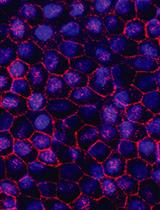
In vitro Differentiation of Human iPSC-derived Retinal Pigment Epithelium Cells (iPSC-RPE)

人诱导多功能干细胞来源的视网膜色素上皮细胞的体外分化

Agnieszka D’Antonio-Chronowska Agnieszka D’Antonio-Chronowska
MD Matteo D’Antonio
Kelly  A. Frazer Kelly A. Frazer
6894 Views
Dec 20, 2019
Induced Pluripotent Stem Cells (iPSCs) serve as an excellent model system for studying the molecular underpinnings of tissue development. Human iPSC-derived retinal pigment epithelium (iPSC-RPE) cells have fetal-like molecular profiles. Hence, biobanks like iPSCORE, which contain iPSCs generated from hundreds of individuals, are an invaluable resource for examining how common genetic variants exert their effects during RPE development resulting in individuals having different propensities to develop Age-related Macular Degeneration (AMD) as adults. Here, we present an optimized, cost-effective and highly reproducible protocol for derivation of human iPSC-RPE cells using small molecules under serum-free condition and for their quality control using flow cytometry and immunofluorescence. While most previous protocols have required laborious manual selection to enrich for iPSC-RPE cells, our protocol uses whole culture passaging and yields a large number of iPSC-RPE cells with high purity (88-98.1% ZO-1 and MiTF double positive cells). The simplicity and robustness of this protocol would enable its adaption for high-throughput applications involving the generation of iPSC-RPE samples from hundreds of individuals.
通过斑马鱼视网膜损伤和视网膜组织制备进行视力恢复研究

Retina Injury and Retina Tissue Preparation to Study Regeneration in Zebrafish

通过斑马鱼视网膜损伤和视网膜组织制备进行视力恢复研究

PS Poonam Sharma
RR Rajesh Ramachandran
4468 Views
Dec 20, 2019
Unlike mammals, primitive vertebrates have immense capability to regenerate almost all of their organs including the central nervous system. Among primitive organisms, zebrafish have been extensively used as a model system for regeneration studies. The retina is a part of the central nervous system and mammals lack the potential to repair any damage caused to it. Zebrafish have been used for retina regeneration studies because of ease in handling and maintenance. In zebrafish, Muller glia cells respond to damage and enter the regenerative cascade to maintain the retinal homeostasis. Zebrafish retinal damage can be induced by light, chemical or mechanical methods. Here we are describing the mechanical method of retinal injury, which ensures uniform damage to all retinal layers. Alongside this, we have also described in vivo manipulation strategies for the regeneration associated genes and preparation of retinal tissue for immunohistochemical analysis.

更正

更正:果蝇中CRISPR-Cas9介导的基因组编辑

Correction Notice: CRISPR-Cas9 Mediated Genome Editing in Drosophila

更正:果蝇中CRISPR-Cas9介导的基因组编辑

PP Ping Peng
XW Xia Wang
DS Da Shen
YJ Yu Jia
RX Rong-Gang Xu
LZ Li-Fei Zhu
Jian-Quan Ni Jian-Quan Ni
1686 Views
Dec 20, 2019