往期刊物2015

卷册: 5, 期号: 12

left

Dec

20

Dec

5

Nov

20

Nov

5

Oct

20

Oct

5

Sep

20

Sep

5

Aug

20

Aug

5

Jul

20

Jul

5

Jun

20

Jun

5

May

20

May

5

Apr

20

Apr

5

Mar

20

Mar

5

Feb

20

Feb

5

Jan

20

Jan

5

right

癌症生物学

代谢试验检测中性脂肪储存

Metabolic Assays for Detection of Neutral Fat Stores

代谢试验检测中性脂肪储存

JB Jan M. Baumann
LK Leila Kokabee
XW Xianhiu Wang
YS Yan Sun
JW Jason Wong
DC Douglas S. Conklin
14971 Views
Jun 20, 2015
Lipid droplets (LDs) are ubiquitous intracellular structures whose formation, growth, and maintenance are highly regulated (Wang et al., 2013; Ranall et al., 2011; Goodman, 2009). Lipid metabolism and droplet dynamics are of considerable interest to agriculture, biofuel production, viral pathology, nutrition, and cancer biology (Walther and Farese, 2009; Liu et al., 2010). Accumulation of fatty acids and neutral lipids in nonadipose tissues is cytotoxic (Kourtidis et al., 2009). BODIPY 493/503 (4,4-Difluoro-1,3,5,7,8-Pentamethyl-4-Bora-3a,4a-Diaza-s-Indacene) is the standard dye to study LDs within adipocytes. BODIPY 493/503 contains a nonpolar structure that, upon binding to neutral lipid, emits a green fluorescence signal with a narrow wavelength range, making it an ideal fluorophore for multi-labeling experiments. The hydrophobic nature of the dye molecules promotes rapid entry into the nonpolar environment of LDs (Listenberge and Brown, 2007). Gocze and Freeman showed that the lipid fluorescent variability is significantly lower when using BODIPY493/503 compared to Nile Red, suggesting that it may be more specific for the LD (Gocze and Freeman, 1994). Here, we describe a BODIPY 493/503 assay for the detection of neural fat stores in cultured cells (Figure 1) (Wang et al., 2013).

免疫学

小鼠无创气管滴注法

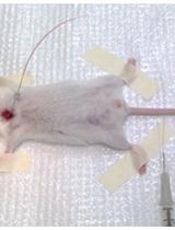
Non-invasive Intratracheal Instillation in Mice

小鼠无创气管滴注法

GO Guadalupe Ortiz-Muñoz
ML Mark R. Looney
19332 Views
Jun 20, 2015
The intratracheal instillation technique is used to deliver a variety of agents to the lungs ranging from pathogens (bacteria, viruses), toxins, to therapeutic agents. To model lung inflammation and injury, LPS can be administrated via intranasal, intratracheal, or aerosol approaches. Each technique has its limitations. The intratracheal technique can involve the non-invasive instillation method (via the oro-tracheal route) or a direct injection into the trachea. Here, we describe an optimized method for direct visual instillation of LPS via the non-invasive oro-tracheal route.
人自然杀伤细胞(NK)的细胞体外刺激和 IFNγ 及CD107a 细胞因子胞内染色

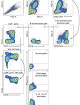
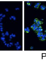
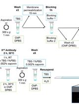
Ex vivo Human Natural Killer (NK) Cell Stimulation and Intracellular IFNγ and CD107a Cytokine Staining

人自然杀伤细胞(NK)的细胞体外刺激和 IFNγ 及CD107a 细胞因子胞内染色

VY Vanessa A. York
Jeffrey M. Milush Jeffrey M. Milush
18267 Views
Jun 20, 2015
Natural killer (NK) cells comprise 5–20% of peripheral blood mononuclear cells (PBMC) in humans. In addition to their fundamental roles in the defense against viral infections and tumor surveillance, NK cells help shape adaptive immune responses through their production of cytokines. NK cells are traditionally identified as CD3neg, CD14neg, CD19neg lymphocytes expressing CD56. Using a combination of markers that includes CD56 and CD7 greatly increases the ability to define the phenotype and function of NK cell subsets. Two key markers of NK cell function are the production of IFNγ and the release of cytotoxic granules measured by the expression of CD107a. Here we describe a method to assess IFNγ and CD107a expression in NK cells following stimulation with target cells or cytokines. This method can be used to assess the general functional capacity of NK cells in peripheral blood mononuclear cells from a wide range of study participants.
通过双重免疫刺激建立输血相关急性肺损伤的小鼠模型

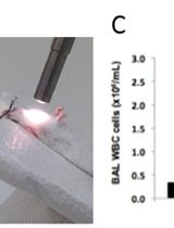
Two-event Transfusion-related Acute Lung Injury Mouse Model

通过双重免疫刺激建立输血相关急性肺损伤的小鼠模型

GO Guadalupe Ortiz-Muñoz
ML Mark R. Looney
9084 Views
Jun 20, 2015
Transfusion-related acute lung injury (TRALI) is defined as acute lung injury that occurs within 6 hours of a blood product transfusion. TRALI continues to be a leading cause of transfusion-related mortality and we have developed a mouse model of TRALI to better understand the mechanisms by which injury occurs and to test therapeutic approaches. Our model is a two-event model based on immune priming and the challenge of BALB/c wild-type mice with cognate MHC Class I monoclonal antibody (MHC I mAb). Immune priming with LPS mimics the primed state of recipients (first event) that is important for the development of TRALI. Donor HLA antibodies are frequently implicated in TRALI reactions, and cognate MHC Class I antibody (second event) produces acute lung injury in primed animals. Here, we describe a detailed protocol with high reproducibility within animals.

微生物学

引物延伸反应用于基于PCR的α-互补试验

Primer Extension Reactions for the PCR- based α- complementation Assay

引物延伸反应用于基于PCR的α-互补试验

Vasudevan Achuthan Vasudevan Achuthan
JD Jeffrey J. DeStefano
10499 Views
Jun 20, 2015
The PCR- based- α- complementation assay is an effective technique to measure the fidelity of polymerases, especially RNA-dependent RNA polymerases (RDRP) and Reverse Transcriptases (RT). It has been successfully employed to determine the fidelity of the poliovirus polymerase 3D-pol (DeStefano, 2010) as well as the human immunodeficiency virus Reverse Transcriptase (HIV RT) (Achuthan et al., 2014). A major advantage of the assay is that since the PCR step is involved, even the low yield of products obtained after two rounds of low yield of RNA synthesis (for RDRP) or reverse transcription (for RT) can be measured using the assay. The assay also mimics the reverse transcription process, since both RNA- and DNA- directed RT synthesis steps are performed. We recently used this assay to show that the HIV RT, at physiologically relevant magnesium concentration, has accuracy in the same range as other reverse transcriptases (Achuthan et al., 2014). Here, we describe in detail how to prepare the inserts using the primer extension reactions. The prepared inserts are then processed further in the PCR- based- α- complementation assay.
在高传染性和需要多种养分的革兰氏阴性菌-土拉弗朗西斯菌敲除突变株的获得方法

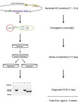
Generating Isogenic Deletions (Knockouts) in Francisella tularensis, a Highly-infectious and Fastidious Gram-negative Bacterium

在高传染性和需要多种养分的革兰氏阴性菌-土拉弗朗西斯菌敲除突变株的获得方法

Xiaojun Wu Xiaojun Wu
GR Guoping Ren
Jason F. Huntley Jason F. Huntley
9911 Views
Jun 20, 2015
Generating bacterial gene deletion mutants, also known as knockouts (KOs), is a powerful tool to investigate individual gene functions. However, fastidious bacteria such as Francisella tularensis (F. tularensis) often are difficult to genetically manipulate. Indeed, many different approaches have been tested to generate F. tularensis mutants. First, Tn5-based EZ::TN transposons have been successfully used to generate transposon libraries in F. tularensis (Qin and Mann, 2006; Weiss et al., 2007). However, creating a comprehensive transposon library with saturating mutations can be laborious, screening for gene disruption requires high-throughput assays where known phenotypes can be measured, and transposons may not completely inactivate the gene of interest or may alter downstream gene expression. Second, group II introns (also referred to as Targetron) have been used to inactivate F. tularensis genes of interest (Rodriguez et al., 2008; Rodriguez et al., 2009). Targetron functions by forming a complex between plasmid-encoded RNA and chromosomal DNA, followed by group II intron insertion into the gene of interest. The main advantage of Targetron is that it does not require an antibiotic resistance marker. However, as noted for transposons, targetron gene insertions may not eliminate all gene functions or may affect downstream gene expression. Third, homologous recombination can be used to completely replace the chromosomal target gene with a selectable marker, such as an antibiotic resistance marker. This classical genetic technique has been used in many F. tularensis studies (Ramakrishnan et al., 2008; Ren et al., 2014; Mohapatra et al., 2008; Robertson et al., 2013). To accomplish this, a suicide plasmid is engineered to include a selectable marker flanked by regions upstream and downstream of the gene of interest. This KO plasmid can be delivered into host bacteria by many methods, including electroporation, chemical transformation, or conjugation. Here, we describe an optimized procedure to generate KO plasmid constructs, use E. coli to conjugatively transfer the plasmid into F. tularensis, select for F. tularensis KOs using a series of kanamycin-, hygromycin-, and sucrose-resistance steps, and confirm that the gene of interest has been deleted (general overview of the knockout protocol diagramed in Figure 1). This optimized procedure is relatively simple, rapid, and, more importantly, includes a series of both positive and negative selection steps to increase the chances of deleting a target gene from F. tularensis.
大麦中镰刀菌属苗枯病(FSB)和镰刀菌属赤霉病(FHB)严重性的目测评估

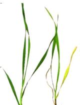
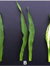
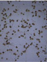
Visual Assessment of the Severity of Fusarium Seedling Blight (FSB) and Fusarium Head Blight (FHB) Disease in Barley

大麦中镰刀菌属苗枯病(FSB)和镰刀菌属赤霉病(FHB)严重性的目测评估

SA Shahin S. Ali
LG Lokanadha R. Gunupuru
Fiona M. Doohan Fiona M. Doohan
9794 Views
Jun 20, 2015
Fusarium pathogens are among the most damaging pathogens of cereals. These pathogens have the ability to attack the roots, seedlings, and flowering heads of barley and wheat plants (Simpson et al., 2004). Resulting in yield loss and head blight disease and also resulting in the contamination of grain with mycotoxins harmful to human and animal health (McMulen et al., 1997; Walter et al., 2010; Agostinelli et al., 2012). The study of Fusarium diseases, including host disease resistance and the effect of exogenous agents (chemicals, biocontrol agents, etc.), requires robust and effective methods for the assessment and quantification of visual disease symptoms. Here we describe the methods commonly used for the assessment and quantification of the severity of Fusarium seedling blight and Fusarium head blight disease.
错配引物延伸法

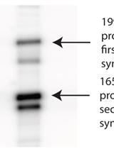
Mismatched Primer Extension Assays

错配引物延伸法

Vasudevan Achuthan Vasudevan Achuthan
JD Jeffrey J. DeStefano
7448 Views
Jun 20, 2015
Steady state kinetic assays have been a reliable way to estimate fidelity of several polymerases (Menendez-Arias, 2009; Rezende and Prasad, 2004; Svarovskaia et al., 2003). The ability to analyze the extension of primers with specific mismatches at the 3ʹ end is a major strength of the mismatched primer extension assays. Recently, we used the mismatched primer extension assays to show that the fidelity of HIV RT increases dramatically when concentration of Mg2+ is reduced to a physiologically relevant concentration (~0.25 mM) (Achuthan et al., 2014). Here, we describe in detail how to perform the mismatched primer extension assay to measure the standard extension efficiency using human immunodeficiency virus reverse transcriptase (HIV RT) at 2 mM Mg2+. The relative fidelity of the polymerase can then be estimated using the standard extension efficiency. The assay described here is based on the method published in Mendelman et al. (1990).

神经科学

鸡胚胎视网膜DNA电转移

Electroporation of Embryonic Chick Eyes

鸡胚胎视网膜DNA电转移

Agustín Luz-Madrigal Agustín Luz-Madrigal
Erika Grajales-Esquivel Erika Grajales-Esquivel
Katia Del Rio-Tsonis Katia Del Rio-Tsonis
13259 Views
Jun 20, 2015
The chick embryo has prevailed as one of the major models to study developmental biology, cell biology and regeneration. From all the anatomical features of the chick embryo, the eye is one of the most studied. In the chick embryo, the eye develops between 26 and 33 h after incubation (Stages 8-9, Hamburger and Hamilton, 1951). It originates from the posterior region of the forebrain, called the diencephalon. However, the vertebrate eye includes tissues from different origins including surface ectoderm (lens and cornea), anterior neural plate (retina, iris, ciliary body and retinal pigmented epithelium) and neural crest/head mesoderm (stroma of the iris and of the ciliary body as well as choroid, sclera and part of the cornea). After gastrulation, a single eye field originates from the anterior neural plate and is characterized by the expression of eye field transcriptional factors (EFTFs) that orchestrate the program for eye development. Later in development, the eye field separates in two and the optic vesicles form. After several inductive interactions with the lens placode, the optic cup forms. At Stages 14-15, the outer layer of the optic cup becomes the retinal pigmented epithelium (RPE) while the inner layer forms the neuroepithelium that eventually differentiates into the retina. One main advantage of the chick embryo, is the possibility to perform experiments to over-express or to down-regulate gene expression in a place and time specific manner to explore gene function and regulation. The aim of this protocol is to describe the electroporation techniques at Stages 8-12 (anterior neural fold and optic vesicle stages) and Stages 19-26 (eye cup, RPE and neuroepithelium). We provide a full description of the equipment, materials and electrode set up as well as a detailed description of the highly reproducible protocol including some representative results. This protocol has been adapted from our previous publications Luz-Madrigal et al. (2014) and Zhu et al. (2014).

植物科学

大麦幼苗叶的病毒诱导基因沉默(VIGS)

Virus-induced Gene Silencing (VIGS) in Barley Seedling Leaves

大麦幼苗叶的病毒诱导基因沉默(VIGS)

LG Lokanadha R. Gunupuru
SA Shahin S. Ali
Fiona M. Doohan Fiona M. Doohan
SS Steven R. Scofield
13302 Views
Jun 20, 2015
Virus induced gene silencing (VIGS) is one of the most potent reverse genetics technologies for gene functional characterisation. This method exploits a dsRNA-mediated antiviral defence mechanism in plants. Using this method allows researchers to generate rapid phenotypic data in a relatively rapid time frame as compared to the generation of stable transformants. Here we describe a simple method for silencing a target gene in barley seedling leaves using vectors based on the Barley Stripe Mosaic Virus (BSMV).
完整分离的葡萄果实体外培养系统的开发与实现

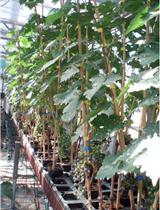
Development and Implementation of an in vitro Culture System for Intact Detached Grape Berries

完整分离的葡萄果实体外培养系统的开发与实现

Zhanwu Dai Zhanwu Dai
MM Messa Meddar
SD Serge Delrot
EG Eric Gomès
11201 Views
Jun 20, 2015
Grape composition depends on the metabolites accumulated and synthesized during grape development. It is of paramount importance for grape growers because of its major role in shaping wine quality. Therefore, understanding the regulation mechanisms that control the accumulation of quality-related metabolites in grape is of both scientific and agronomical interests. The composition of grape berry at harvest is under complex regulation and can be affected by many factors (Conde et al., 2007). The study of the effects of these factors on berries still attached to intact plants can be highly challenging because of the large size of the plants, interplant, intercluster and interberry variability; and because it is complicated to precisely control the nutrients and hormones imported by the berries, and the environment. Therefore, in vitro cultured grape berries are a good model system, which better represents berry anatomy structure (skin and flesh) than grape cell suspensions and nevertheless largely reduces the system complexity compared to whole plant (Bravdo et al., 1990; Pérez et al., 2000; Gambetta et al., 2010). To this end, an in vitro culture system of intact detached grape berries has been developed by coupling greenhouse fruiting-cuttings production and in vitro organ culture techniques (Dai et al., 2014). The cultured berries are able to actively absorb and utilize carbon and nitrogen from the culture medium, and exhibit fruit ripening features such as color changing and softening. This in vitro system may serve to investigate the response of berry composition to environmental and nutrient factors.
拟南芥花粉管膜和细胞壁聚合物的简单免疫标记法

A Simple Protocol for the Immunolabelling of Arabidopsis Pollen Tube Membranes and Cell Wall Polymers

拟南芥花粉管膜和细胞壁聚合物的简单免疫标记法

Marie Dumont Marie Dumont
CC Céline Cataye
AL Arnaud Lehner
EM Eric Maréchal
PL Patrice Lerouge
Denis Falconet Denis Falconet
Jean-Claude Mollet Jean-Claude Mollet
10900 Views
Jun 20, 2015
The pollen tube, a fast tip-growing cell, is an excellent model to study membrane and cell wall biosynthesis. Here, we describe a simple protocol using an easy to use device to perform immunofluorescence labelling of pollen tube membrane and cell wall. The use of the NucleoSpin column to perform all the steps of the immunolabelling procedure results in obtaining more intact pollen tubes.
采用 GC-EI-MS分析拟南芥花粉管中热水提取物的糖组分

Analysis of Sugar Component of a Hot Water Extract from Arabidopsis thaliana Pollen Tubes Using GC-EI-MS

采用 GC-EI-MS分析拟南芥花粉管中热水提取物的糖组分

Marie Dumont Marie Dumont
AL Arnaud Lehner
CL Corinne Loutelier-Bourhis
Jean-Claude Mollet Jean-Claude Mollet
PL Patrice Lerouge
8338 Views
Jun 20, 2015
Extraction with hot water is the oldest and simplest method used to recover pectin from an alcohol insoluble residue extract, although this method has not been widely used for the cell wall analysis of pollen tube, a model used to study cell wall. This protocol described this method applied for pectin extraction from 6 h-old Arabidopsis pollen tubes followed by a sugar composition analysis by gas chromatography mass spectrometry.
在龙胆越冬芽及越冬苗上体外施加龙胆二糖

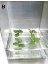
Gentiobiose Feeding in Gentian in vitro Overwintering Buds or Plantlets

在龙胆越冬芽及越冬苗上体外施加龙胆二糖

HT Hideyuki Takahashi
MN Masahiro Nishihara
7552 Views
Jun 20, 2015
To study the functions of sugars in plants, feeding experiment is one of the most common and easy methods. However, the traditional method, e.g., a floating of leaf discs on sugar-containing solution seems to have an insufficient efficiency of sugar uptake, despite of high osmotic and injury effects. This is a protocol to feed oligosaccharide gentiobiose into in vitro cultured tissues of gentian. This protocol enables to incorporate gentiobiose into intact tissues without exposure to osmotic stress and may be useful to other plant species that are able to propagate by shoot tip culture.