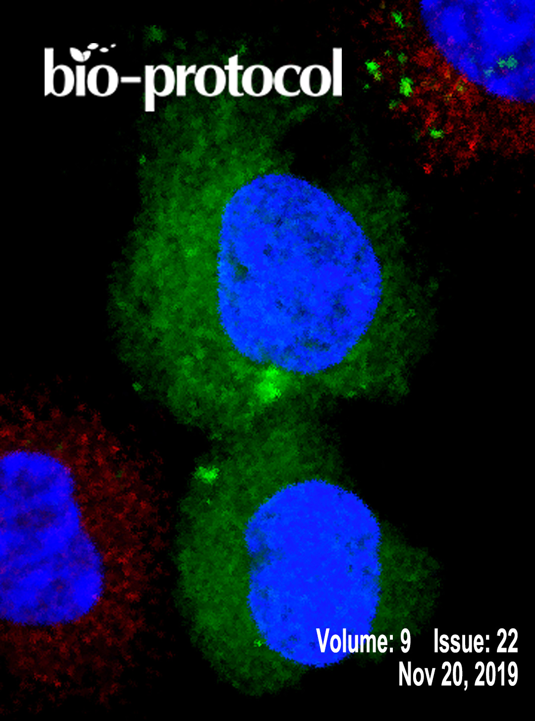
往期刊物2019

卷册: 9, 期号: 22

left

Dec

20

Dec

5

Nov

20

Nov

5

Oct

20

Oct

5

Sep

20

Sep

5

Aug

20

Aug

5

Jul

20

Jul

5

Jun

20

Jun

5

May

20

May

5

Apr

20

Apr

5

Mar

20

Mar

5

Feb

20

Feb

5

Jan

20

Jan

5

right

生物物理学

固体浸没透镜应用于低温条件下的超分辨率显微镜

Super-resolution Microscopy at Cryogenic Temperatures Using Solid Immersion Lenses

固体浸没透镜应用于低温条件下的超分辨率显微镜

BB Benji C. Bateman
LZ Laura C. Zanetti-Domingues
AM Amy N. Moores
SN Sarah R. Needham
DR Daniel J. Rolfe
LW Lin Wang
DC David T. Clarke
Marisa L. Martin-Fernandez Marisa L. Martin-Fernandez
5436 Views
Nov 20, 2019
Our mechanistic understanding of cell function depends on imaging biological processes in cells with molecular resolution. Super-resolution fluorescence microscopy plays a crucial role by reporting cellular ultrastructure with 20-30 nm resolution. However, this resolution is insufficient to image macro-molecular machinery at work. A path to improve resolution is to image under cryogenic conditions, which substantially increases the brightness of most fluorophores and preserves native ultrastructure much better than chemical fixatives. Cryogenic conditions are, however, underutilized because of the lack of compatible high numerical aperture (NA) objectives. Here we describe a protocol for the use of super-hemispherical solid immersion lenses (superSILs) to achieve super-resolution imaging at cryogenic temperatures with an effective NA of 2.17 and resolution of ~10 nm.
单细胞的光磁筛选及分离

Opto-magnetic Selection and Isolation of Single Cells

单细胞的光磁筛选及分离

LB Loïc Binan
JR Joannie Roy
SC Santiago Costantino
3834 Views
Nov 20, 2019
Capturing single cells from large heterogenous populations based solely on observable traits is necessary for many cell biology applications and remains a major technical challenge. The protocol we present allows the isolation of viable and metabolically active cells selected for their shape, migration speed, contact to other cells, or intracellular protein localization. We previously introduced a method termed Cell Labeling via Photobleaching (CLaP) for the efficient tagging of cells chosen for visual criteria. Here we describe a new protocol for capturing such cells using ferromagnetic beads termed single-cell magneto-optical capture (scMOCa). This technology is especially useful when the number of target cells represents an extremely low fraction of the total population (potentially one single cell), a situation in which conventional sorting techniques like fluorescent or magnetic activated cell sorting (F/MACS) cannot provide satisfactory results in terms of capture efficiency and specificity. scMOCa uses the lasers of a confocal microscope to photobleach and crosslink biotin-4-fluorecein molecules to cell membranes. Streptavidin coated magnetic beads then adhere to biotin moieties and a magnet allows the capture of illuminated cells. By precisely controlling liquid volumes and spacing between the different parts of a simple setup, high cell selectivity and capture efficacy can be achieved. scMOCA allows visual selection and isolation of any number of cells in a microscopy field and captured cells remain viable to generate new colonies of chosen phenotypes for downstream analyses.

癌症生物学

LC/MS法对小鼠肿瘤间质液中代谢产物的分离和定量

Isolation and Quantification of Metabolite Levels in Murine Tumor Interstitial Fluid by LC/MS

LC/MS法对小鼠肿瘤间质液中代谢产物的分离和定量

MS Mark R Sullivan
CL Caroline A Lewis
AM Alexander Muir
9528 Views
Nov 20, 2019
Cancer is a disease characterized by altered metabolism, and there has been renewed interest in understanding the metabolism of tumors. Even though nutrient availability is a critical determinant of tumor metabolism, there has been little systematic study of the nutrients directly available to cancer cells in the tumor microenvironment. Previous work characterizing the metabolites present in the tumor interstitial fluid has been restricted to the measurement of a small number of nutrients such as glucose and lactate in a limited number of samples. Here we adapt a centrifugation-based method of tumor interstitial fluid isolation readily applicable to a number of sample types and a mass spectrometry-based method for the absolute quantitation of many metabolites in interstitial fluid samples. In this method, tumor interstitial fluid (TIF) is analyzed by liquid chromatography-mass spectrometry (LC/MS) using both isotope dilution and external standard calibration to derive absolute concentrations of targeted metabolites present in interstitial fluid. The use of isotope dilution allows for accurate absolute quantitation of metabolites, as other methods of quantitation are inadequate for determining nutrient concentrations in biological fluids due to matrix effects that alter the apparent concentration of metabolites depending on the composition of the fluid in which they are contained. This method therefore can be applied to measure the absolute concentrations of many metabolites in interstitial fluid from diverse tumor types, as well as most other biological fluids, allowing for characterization of nutrient levels in the microenvironment of solid tumors.
利用胞嘧啶脱氨酶表达进行氟尿嘧啶标记RNA标记和分离

Labeling and Isolation of Fluorouracil Tagged RNA by Cytosine Deaminase Expression

利用胞嘧啶脱氨酶表达进行氟尿嘧啶标记RNA标记和分离

HB Harihar Basnet
JM Joan Massague
4339 Views
Nov 20, 2019
Tissues are comprised of different cell types whose interactions elicit distinct gene expression patterns that regulate tissue formation, regeneration, homeostasis and repair. Analysis of these gene expression patterns require methods that can capture as closely as possible the transcriptomes of cells of interest in their tissue microenvironment. Current technologies designed to study in situ transcriptomics are limited by their low sensitivity that require cell types to represent more than 1% of the total tissue, making it challenging to transcriptionally profile rare cell populations rapidly isolated from their native microenvironment. To address this problem, we developed fluorouracil-tagged RNA sequencing (Flura-seq) that utilizes cytosine deaminase (CD) to convert the non-natural pyrimidine fluorocytosine to fluorouracil. Expression of S. cerevisiae CD and exposure to fluorocytosine generates fluorouracil and metabolically labels newly synthesized RNAs specifically in cells of interest. Fluorouracil-tagged RNAs can then be immunopurified and used for downstream analysis. Here, we describe the detailed protocol to perform Flura-seq both in vitro and in vivo. The robustness, simplicity and lack of toxicity of Flura-seq make this tool broadly applicable to many studies in developmental, regenerative, and cancer biology.

发育生物学

果蝇幼虫固定脑组织中有丝分裂纺锤体角度和有丝分裂细胞距离测定

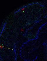
Measurement of Mitotic Spindle Angle and Mitotic Cell Distance in Fixed Tissue of Drosophila Larval Brains

果蝇幼虫固定脑组织中有丝分裂纺锤体角度和有丝分裂细胞距离测定

MF Maribel Franco
AC Ana Carmena
3319 Views
Nov 20, 2019
The positioning and the cleavage plane orientation of mitotic cells in pseudostratified epithelia (PSE) must be tightly regulated since failures in any of these processes might have fatal consequences during development. Here we present a simple method to determine the spindle orientation as well as the positioning of neuroepithelial mitotic cells within the Outer Proliferation Center (OPC) of Drosophila larval brains.

免疫学

骨髓衍生巨噬细胞中的细菌同步转移测定实验

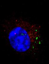
Bacterial Synchronized Transfer Assays in Bone Marrow Derived Macrophages

骨髓衍生巨噬细胞中的细菌同步转移测定实验

SD Sedelia R Dominguez
TK Tom Kawula
SS Shaun P Steele
4465 Views
Nov 20, 2019
Merocytophagy (“mero”, Greek for partial; “cytophagy” for cell eating) is a process by which cells acquire microbes and cytosolic material through phagocytosis of a small portion of neighboring cells upon cell-cell contact. Cell-cell contact dependent transfer events can be assessed through co-incubation of differently labeled cells. With these assays, it is difficult to analyze the recipient cells by microscopy or bacterial burden within only recipient cells. Therefore, we established a synchronized transfer assay that allows for recipient cells to be isolated from donor cells following transfer events at a high purity. Here, we present this assay in context of bacterial infections and cytosolic cellular staining. With this protocol, mechanisms of cell-cell contact dependent transfer events and the events following merocytophagy can easily be investigated.

微生物学

枯草芽孢杆菌在非生物表面传播途径的不同检测方法

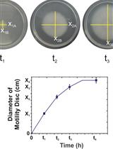
Assessing Different Ways of Bacillus subtilis Spreading over Abiotic Surfaces

枯草芽孢杆菌在非生物表面传播途径的不同检测方法

Marco  Bartolini Marco Bartolini
RG Roberto Grau
4745 Views
Nov 20, 2019
Surface-associate motility on biotic and abiotic environments is a key mechanism used by the model bacterium Bacillus subtilis and its closest relatives (i.e., B. amyloliquefaciens, B. thuringiensis, B. cereus, B. pumilus) for surface colonization and spreading across surfaces. The study of this mechanism in a research, industrial or clinic laboratory is essential; however, precautions should be taken for the reproducibility of the results, for example, the procedure to inoculate the bacteria on the testing plate, the humidity of the plate and the agar concentration. In this protocol, we describe, using Bacillus subtilis, how to perform these assays and, in addition, we show how by varying the agar concentration in the plate, you can make a first approximation of what type of motility has other bacterial species.
在小鼠模型中利用生物发光进行利什曼原虫的定量分析

Leishmania Parasite Quantification by Bioluminescence in Murine Models

在小鼠模型中利用生物发光进行利什曼原虫的定量分析

MR Marta Reverte
NF Nicolas Fasel
3886 Views
Nov 20, 2019
Leishmaniasis remains a major public health problem worldwide with a prevalence of 12 million, an incidence of 1 million persons, and 350 million people being at risk. Murine models have been largely used for studying the host-pathogen relationship and developing effective chemotherapies against Leishmania parasites. Thus, preclinical imaging is crucial for monitoring the disease outcome. The aim of this protocol is to quantify parasite burden using bioluminescence in vivo imaging. Here, we describe a high-throughput imaging workflow, together with data acquisition and analysis ideal to assess in vivo parasite load in mouse models.
利用 Percoll®在微分干涉显微镜下对隐球菌荚膜成像

Imaging Cryptococcus spp. Capsule by Differential Interference Contrast Microscopy Using Percoll®

利用 Percoll®在微分干涉显微镜下对隐球菌荚膜成像

HP Hugo Costa Paes
SF Stefânia de Oliveira Frazão
MF Maria Sueli Soares Felipe
AC Arturo Casadevall
AN André Moraes Nicola
3531 Views
Nov 20, 2019
The most important virulence factor in the Cryptococcus genus is the polysaccharide capsule. This genus includes several species that cause life-threatening invasive disease. An increase in capsule thickness is important during fungal infection. The capsule is usually imaged using India ink, and crucial insights on the dynamics of its growth have been obtained using capsule-binding proteins such as specific antibodies or complement. We have developed an alternative method that allows both static and time-lapse imaging of the capsule using Percoll®, a suspension of nanometric spheres that do not penetrate the capsule. Given that these particles have a higher refractive index than the capsule, the latter can be imaged by differential interference contrast (DIC) microscopy. Static observation of the capsule with DIC and Percoll® results in capsule thickness measurements that match those made with India ink. Using capsule-inducing media, a glass-bottom incubation chamber and a live-imaging system equipped for DIC microscopy, this method allows time-lapse imaging of capsule growth. In contrast with India ink staining, DIC imaging of Percoll® exclusion halos result in crisp images. The greatest advantage of this method, though, is that unlike India ink, the Percoll® particles are non-toxic and unlike opsonins they do not bind the capsule, resulting in observations of capsule growth that are free from interference of bound proteins on capsule physiology.

分子生物学

基于ELISA的蛋白质泛素化检测

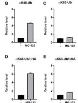
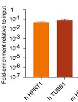
ELISA Based Protein Ubiquitylation Measurement

基于ELISA的蛋白质泛素化检测

YK Yuka Kamada
RF Ryosuke Fukuda
TO Tsukasa Okiyoneda
5903 Views
Nov 20, 2019
Ubiquitylation is a common post-translational modification of cellular proteins that results in proteasomal and lysosomal degradations. Ubiquitylation is generally measured by methods such as immunoblotting using anti-ubiquitin antibodies after isolating the protein-of-interest by denaturing immunoprecipitation. The following protocol can be used to easily quantify the ubiquitylation of the protein-of-interest tagged with biotin by ELISA.
利用原位5mC标记进行果蝇中肠细胞染色质开放性的鉴定

Determination of Chromatin Accessibility in Drosophila Midgut Enterocytes by in situ 5mC Labeling

利用原位5mC标记进行果蝇中肠细胞染色质开放性的鉴定

Eliya Bitman-Lotan Eliya Bitman-Lotan
HR Hector Rincon-Arano
Gal  Raz Gal Raz
AO Amir Orian
3614 Views
Nov 20, 2019
Regulation of gene expression involves dynamic changes in chromatin organization, where in many cases open chromatin structure correlates with gene activation. Several methods enable monitoring changes in chromatin accessibility, including ATAC-seq, FAIRE-seq, MNase-seq and DNAse-seq methods, which involve Next-generation-sequencing (NGS). Focusing on the adult Drosophila differentiated gut enterocytes (ECs) we used a sequencing-free method that enables visualizing and semi-quantifying large-scale changes in chromatin structure using in vitro methylation assay with the bacterial CpG Methyltransferase, M. Sssl, that determine chromatin accessibility. In brief, as CpG methylation is minimal in differentiated somatic Drosophila cells, we used the bacterial M. SssI enzyme to methylate CpG dinucleotides in situ depending on their chromatin accessibility. The methylated dinucleotides are detected using 5mCytosine monoclonal antibody and nuclei are visualized microscopically. Thus, the 5mC method enables to monitor large-scale chromatin changes in heterogenic cellular tissue focusing on the cell type of interest and without the need for cell purification or NGS.

神经科学

SarkoSpin:一种用于生化分离和鉴定病理性TDP-43聚集体的方法

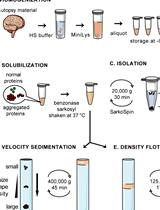
SarkoSpin: A Technique for Biochemical Isolation and Characterization of Pathological TDP-43 Aggregates

SarkoSpin:一种用于生化分离和鉴定病理性TDP-43聚集体的方法

MP Manuela Pérez-Berlanga
Florent Laferrière Florent Laferrière
MP Magdalini Polymenidou
6026 Views
Nov 20, 2019
TDP-43 is the main aggregating protein in neurodegenerative diseases such as amyotrophic lateral sclerosis (ALS) and frontotemporal lobar degeneration (FTLD). Aggregated TDP-43 is resistant to diverse detergent solubilization, yet physiological TDP-43 and other abundant proteins commonly co-purify with pathological TDP-43. This mixed isolation has precluded the elucidation of the biochemical and structural features of the pathological TDP-43 and its role in disease. Here we describe SarkoSpin, a method for the isolation of pure pathological TDP-43 from patient autopsy brain by sample solubilization with Sarkosyl after nuclease treatment. This purification, which is also applicable to cell culture material, permits the study of biochemical properties of exclusively pathological TDP-43, allowing for the first time the determination of their link to the clinical presentation of FTLD. This method opens up a path for the study of pathological TDP-43 at the molecular and structural level in the heterogeneous spectrum of ALS and FTLD cases.
有髓神经纤维的Gallyas银浸染法

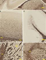
Gallyas Silver Impregnation of Myelinated Nerve Fibers

有髓神经纤维的Gallyas银浸染法

SJ Sabitha Joseph
HW Hauke B. Werner
JS Judith Stegmüller
4271 Views
Nov 20, 2019
In the nervous system of vertebrates, nerve impulse propagation is accelerated by the ensheathment of neuronal axons with myelin. Myelin sheaths are molecularly specialized, lipid-rich plasma membrane extensions of Schwann cells in the peripheral nervous system and oligodendrocytes in the central nervous system (CNS). To visualize myelinated nerve fibers and to allow for the morphological analyses of myelin in the brain and the spinal cord, an efficient method for silver impregnation of myelin has originally been developed by Ferenc Gallyas in 1979, referred to as Gallyas silver impregnation. Gallyas’ method is based on the agyrophilic characteristic of myelin to form and bind silver particles, while this process is suppressed in tissues other than myelin. The silver particles are finally enhanced in a developing step (“physical developer”). The main advantage of this method is that it efficiently visualizes both large myelinated fiber tracts and individual myelinated axons. Here we provide our laboratory protocol that is suitable for paraffin embedded sections and the use of light microscopy based on Gallyas’ original protocol and subsequent modifications by Pistorio and colleagues.

植物科学

应用损伤面积的自动化检测和测量评价炭疽菌对拟南芥叶片致病性的方法

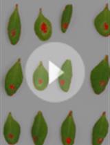
Method for Assessing Virulence of Colletotrichum higginsianum on Arabidopsis thaliana Leaves Using Automated Lesion Area Detection and Measurement

应用损伤面积的自动化检测和测量评价炭疽菌对拟南芥叶片致病性的方法

Ayako Tsushima Ayako Tsushima
PG Pamela Gan
KS Ken Shirasu
6074 Views
Nov 20, 2019
The plant pathogenic fungus, Colletotrichum higginsianum is widely used to understand infection mechanisms, as it infects the model plant Arabidopsis thaliana. To determine the virulence of C. higginsianum, several methods have been developed, such as disease reaction scoring, lesion measurement, entry rate assays, and relative fungal biomass assays using real-time quantitative PCR. Although many studies have taken advantage of these methods, they have shortcomings in terms of objectivity, time, or cost. Here, we show a lesion area detection method applying ImageJ color thresholds to images of A. thaliana leaves infected by C. higginsianum. This method can automatically detect multiple lesions in a short time without the requirement for special equipment and measures lesion areas in a standardized way. This high throughput technique will aid better understanding of plant immunity and pathogenicity and contribute to reproducibility of assays.