往期刊物2019

卷册: 9, 期号: 20

left

Dec

20

Dec

5

Nov

20

Nov

5

Oct

20

Oct

5

Sep

20

Sep

5

Aug

20

Aug

5

Jul

20

Jul

5

Jun

20

Jun

5

May

20

May

5

Apr

20

Apr

5

Mar

20

Mar

5

Feb

20

Feb

5

Jan

20

Jan

5

right

生物化学

利用FLIM-FRET进行小分子抑制蛋白相互作用的活细胞检测

Measuring Small-molecule Inhibition of Protein Interactions in Live Cells Using FLIM-FRET

利用FLIM-FRET进行小分子抑制蛋白相互作用的活细胞检测

JP James M. Pemberton
QL Qian Liu
DA David W. Andrews
6083 Views
Oct 20, 2019
This protocol was designed to quantitatively measure small-molecule displacement of proteins in live mammalian cells using fluorescence lifetime imaging microscopy–Förster resonance energy transfer (FLIM-FRET). Tumour cell survival is often dependent on anti-apoptotic proteins, which bind to and inhibit pro-apoptotic proteins, thus preventing apoptosis. Small-molecule inhibitors that selectively target these proteins (termed BH3-mimetics) are therefore a promising avenue for the treatment of several cancers. Previous techniques used to study the efficacy of these drugs often use truncated versions of both pro- and anti-apoptotic proteins, as they are membrane bound and hydrophobic in nature. As a result, the true efficacy of these drugs to displace full-length pro-apoptotic proteins in their native environment within a cell is poorly understood. This protocol describes FLIM-FRET methods to directly measure the displacement (or lack of displacement) of full-length Bcl-2 family proteins in live mammalian cells.

癌症生物学

基于免疫荧光法的组织样本中心体数目确定

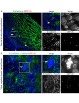
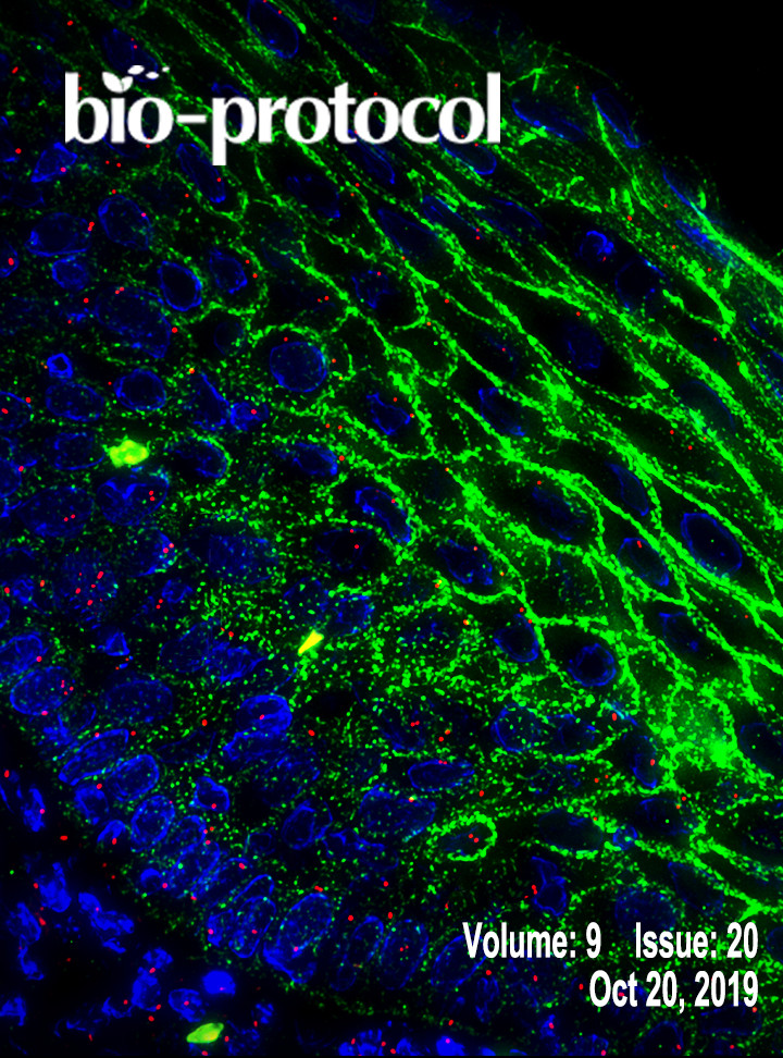
Immunofluorescence-based Determination of Centrosome Number in Tissue Samples

基于免疫荧光法的组织样本中心体数目确定

MW Mengdie Wang
GR Gregory C. Rogers
Anne E. Cress Anne E. Cress
4169 Views
Oct 20, 2019
Centrosome numerical abnormalities have been reported in a variety of tumors. Centrosome numbers in cancer cells display both inter-tumor and intra-tumor heterogeneity. The over production of centrosomes (centrosome amplification) is unique in cancer cells and is a promising target for therapy. Thus, a method to quantify centrosome numbers on a single cell level is needed. Here, we describe a protocol to quantify centrosome numbers in formalin fixed paraffin embedded (FFPE) tissue samples by multiplexing antibodies to define bona fide centrosomes and cell borders. Centrosomes in single cells are identified using high resolution immunofluorescent microscopy with Z-sectioning. This protocol is easy to perform and has been used to quantify centrosome numbers on a single cell level in a variety of human tissue samples.

细胞生物学

小鼠离体肺组织切片中肺泡生成的延时成像

Time-lapse Imaging of Alveologenesis in Mouse Precision-cut Lung Slices

小鼠离体肺组织切片中肺泡生成的延时成像

KA Khondoker M. Akram
LY Laura L.Yates
RM Róisín Mongey
SR Stephen Rothery
DG David C. A. Gaboriau
JS Jeremy Sanderson
MH Matthew Hind
MG Mark Griffiths
CD Charlotte H. Dean
6825 Views
Oct 20, 2019
Alveoli are the gas-exchange units of lung. The process of alveolar development, alveologenesis, is regulated by a complex network of signaling pathways that act on various cell types including alveolar type I and II epithelial cells, fibroblasts and the vascular endothelium. Dysregulated alveologenesis results in bronchopulmonary dysplasia in neonates and in adults, disrupted alveolar regeneration is associated with chronic lung diseases including COPD and pulmonary fibrosis. Therefore, visualizing alveologenesis is critical to understand lung homeostasis and for the development of effective therapies for incurable lung diseases. We have developed a technique to visualize alveologenesis in real-time using a combination of widefield microscopy and image deconvolution of precision-cut lung slices. Here, we describe this live imaging technique in step-by-step detail. This time-lapse imaging technique can be used to capture the dynamics of individual cells within tissue slices over a long time period (up to 16 h), with minimal loss of fluorescence or cell toxicity.
利用流式细胞术进行贴壁细胞的磷酸化蛋白分析

Phospho-protein Analysis in Adherent Cells Using Flow Cytometry

利用流式细胞术进行贴壁细胞的磷酸化蛋白分析

RS Renu Sharma
AS Amit Sharma
AK Atul Kumar
BJ Bithiah Grace Jaganathan
5240 Views
Oct 20, 2019
Protein phosphorylation is one of the most important post-translational modifications, which acts as a reversible on or off switch for the activity of a large number of proteins. Analyzing the phosphorylation status of different proteins can reveal the alterations in the state of the cells in response to cellular damage, cancer and pharmaceutical drugs. Techniques such as mass spectrometry, radiolabeling, 2D-gel electrophoresis and western blotting are used to quantify protein phosphorylation. These assays can quantify phosphorylation in the bulk population of cells, however, flow cytometry can couple cell surface marker expression data with phosphorylation data to understand differential signaling in a sub-population within a heterogeneous population of cells. Our protocol describes the use of flow-cytometry for rapid and single cell-based quantification of intracellular phospho-protein with the help of anti-phospho protein specific antibody.

发育生物学

小鼠胫骨前肌切片中肌纤维类型的免疫组化鉴定

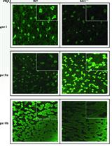
Immunohistochemical Identification of Muscle Fiber Types in Mice Tibialis Anterior Sections

小鼠胫骨前肌切片中肌纤维类型的免疫组化鉴定

VR Vanitha V. Rao
AM Abhishek Mohanty
6187 Views
Oct 20, 2019
Mammalian skeletal muscle is a metabolically active tissue that is made up of different types of muscle fibers. These myofibers are made up of important contractile proteins that provide force during contraction of the muscle like actin and myosin. Murine myofibers have been classified into 4 types: Type I, Type IIa, Type IIb and Type IIX. Each muscle fiber has been identified with specific type of MyHC expressed, which in turn gives differential contractility to the muscle.There have been well-known methodologies to identify different myofibers: histochemical myosin ATPase staining which uses the differential ATPase activity between slow and fast fibers, quantification of metabolic enzymes like malate dehydrogenase and lactate dehydrogenase on specific fragments of muscle fibers. The drawback of these techniques is that they cannot differentiate the subtypes of myofibers, for example, Type IIa and Type IIb. They should be used in conjunction with other known histochemical staining techniques. Here, we devise a direct and robust immunohistochemical staining methodology that utilizes the differential expression of MyHC isoforms in different myofibers types, thus efficiently distinguishing the heterogeneity of the muscle fibers. We use antibodies that specifically recognize Type I, Type IIa and Type IIb fibers on serially cut frozen mouse tibialis anterior sections that can be quantified by ImageJ software.

微生物学

全血HIV-2 DNA的定量分析

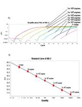
Quantification of HIV-2 DNA in Whole Blood

全血HIV-2 DNA的定量分析

Zsófia  Szojka Zsófia Szojka
Sara  Karlson Sara Karlson
MJ Marianne Jansson
Patrik Medstrand Patrik Medstrand
5040 Views
Oct 20, 2019
Time to AIDS infection is longer with HIV-2, compared to HIV-1, but without antiretroviral therapy both infections will cause AIDS-related mortality. In HIV-2 infection, monitoring of antiretroviral treatment (ART) efficacy is challenging since a large proportion of HIV-2-infected individuals displays low or undetectable plasma RNA levels. Hence, quantification of cellular DNA load may constitute an alternative method for monitoring ART efficacy. Moreover, sensitive HIV-2 DNA quantification protocols are also important for the characterization of the HIV-2 reservoirs, and ultimately for the development of HIV-2 cure strategies. We have developed a sensitive and robust HIV-2 DNA quantification protocol based on whole blood as DNA source, including normalization of leukocyte cell numbers using parallel quantification of the single copy porphobilinogen deaminase gene. The specificity and sensitivity of the assay was 100%. The limit of detection was 1 copy and limit of quantification was 5 copies. When applying this protocol to HIV-2 infected, it was found that HIV-2 viral DNA was detectable in individuals in whom viral RNA was undetectable or under quantification level. Thus, this method provides a sensitive approach to HIV-2 DNA viral quantification from whole blood of HIV-2 infected patients.
用于预测新的适应性表型突变路径的概率模型

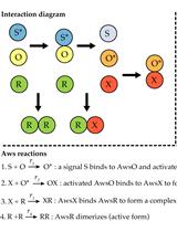
Probabilistic Models for Predicting Mutational Routes to New Adaptive Phenotypes

用于预测新的适应性表型突变路径的概率模型

EL Eric Libby
PL Peter A. Lind
3763 Views
Oct 20, 2019
Understanding the translation of genetic variation to phenotypic variation is a fundamental problem in genetics and evolutionary biology. The introduction of new genetic variation through mutation can lead to new adaptive phenotypes, but the complexity of the genotype-to-phenotype map makes it challenging to predict the phenotypic effects of mutation. Metabolic models, in conjunction with flux balance analysis, have been used to predict evolutionary optimality. These methods however rely on large scale models of metabolism, describe a limited set of phenotypes, and assume that selection for growth rate is the prime evolutionary driver. Here we describe a method for computing the relative likelihood that mutational change will translate into a phenotypic change between two molecular pathways. The interactions of molecular components in the pathways are modeled with ordinary differential equations. Unknown parameters are offset by probability distributions that describe the concentrations of molecular components, the reaction rates for different molecular processes, and the effects of mutations. Finally, the likelihood that mutations in a pathway will yield phenotypic change is estimated with stochastic simulations.One advantage of this method is that only basic knowledge of the interaction network underlying a phenotype is required. However, it can also incorporate available information about concentrations and reaction rates as well as mutational biases and mutational robustness of molecular components. The method estimates the relative probabilities that different pathways produce phenotypic change, which can be combined with fitness models to predict evolutionary outcomes.

分子生物学

血小板的分离和活化实验

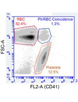
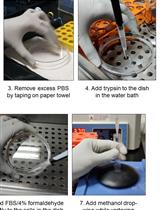
Platelet Isolation and Activation Assays

血小板的分离和活化实验

Laura C. Burzynski Laura C. Burzynski
NP Nicholas Pugh
MC Murray C.H. Clarke
10862 Views
Oct 20, 2019
Platelets regulate hemostasis and are the key determinants of pathogenic thrombosis following atherosclerotic plaque rupture. Platelets circulate in an inactive state, but become activated in response to damage to the endothelium, which exposes thrombogenic material such as collagen to the blood flow. Activation results in a number of responses, including secretion of soluble bioactive molecules via the release of alpha and dense granules, activation of membrane adhesion receptors, release of microparticles, and externalization of phosphatidylserine. These processes facilitate firm adhesion to sites of injury and the recruitment and activation of other platelets and leukocytes, resulting in aggregation and thrombus formation. Platelet activation drives the hemostatic response, and also contributes to pathogenic thrombus formation. Thus, quantification of platelet-associated responses is key to many pathophysiologically relevant processes. Here we describe protocols for isolating, counting, and activating platelets, and for the rapid quantification of cell surface proteins using flow cytometry.
用于核酸纯化和操作的功能化顺磁性磁珠的简单合成方法

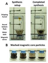
Simple Synthesis of Functionalized Paramagnetic Beads for Nucleic Acid Purification and Manipulation

用于核酸纯化和操作的功能化顺磁性磁珠的简单合成方法

PO Phil Oberacker
PS Peter Stepper
DB Donna Bond
KH Katharina Hipp
TH Timothy A. Hore
TJ Tomasz P. Jurkowski
10373 Views
Oct 20, 2019
The purification of nucleic acids is one of the most common procedures employed in modern molecular biology laboratories. Typically, commercial column-based protocols are utilized to isolate DNA or RNA from various sources. However, these methods not only require specialized equipment, but are also extremely expensive for high-throughput applications. Although an elegant answer to this issue can be provided by paramagnetic beads, bead-based open-source protocols have been limited in the past. Here, we provide an easy to follow step-by-step manual for the synthesis of paramagnetic beads, as well as their functionalization with either a silica- or a carboxyl-surface that can be used to replace the commercial columns with self-made magnetic beads. Together with a variety of detailed protocols for their use in high-throughput nucleic acids extractions, this bead synthesis method forms the recently published open platform Bio-On-Magnetic-Beads (BOMB), which is available on PLOS Biology (Oberacker et al., 2019). Updated protocols can be found on the associated webpage (https://bomb.bio).

神经科学

一种用于链脲佐菌素诱导的大鼠阿尔兹海默症建模的立体定向手术

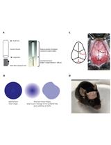
Intracerebral Injection of Streptozotocin to Model Alzheimer Disease in Rats

一种用于链脲佐菌素诱导的大鼠阿尔兹海默症建模的立体定向手术

Daniel Moreira-Silva Daniel Moreira-Silva
Robson C. L. Vizin Robson C. L. Vizin
TM Talita M. S. Martins
TF Tatiana L. Ferreira
MA Maria C. Almeida
DC Daniel C. Carrettiero
7247 Views
Oct 20, 2019
Animal models have promoted meaningful contribution to science including Alzheimer’s disease (AD) research. Several animal models for AD have been used, most of them related to genetic mutations observed in familial AD. However, sporadic form of AD, also named late-onset is the most frequent form of the disease, which is multifactorial, being influenced by genetic, environmental and lifestyle factors. Here, we describe a protocol of an AD-like pathology of the sporadic form using Wistar rats by a single bilateral intracerebroventricular (icv) injection of streptozotocin (STZ, 2 mg/kg). Icv injection of STZ induces brain resistance to insulin and other pathological alterations related to those observed in AD, such as cognitive impairment and accumulation of phosphorylated tau protein and β-amyloid in the brain. Thus, icv injection of STZ is a useful tool to investigate the pathological mechanisms and the metabolic alterations involved in AD and to propose new therapeutic approaches and neuroprotective drugs.
光遗传学在食品避臭实验中的应用

Optogenetic Food Odor Avoidance Assay

光遗传学在食品避臭实验中的应用

JP Jay M. Patel
Jessica Swanson Jessica Swanson
Benjamin R. Arenkiel Benjamin R. Arenkiel
4217 Views
Oct 20, 2019
Appetite is tightly linked to the sensory experience of feeding, including the smell, taste, and sight of food. Sensory perception can affect the palatability of food, modulating appetite beyond homeostatic requirements. Hypothalamic neurons that govern feeding are responsive to sensory cues associated with food, including food odors. However, the circuit mechanisms by which sensory information is processed and relayed to feeding nodes to affect feeding behavior is not well understood. Recent work has identified a population of excitatory basal forebrain neurons that modulate potent appetite suppression, as well as respond to food-associated and innately aversive odorants. To investigate this circuitry, we stereotaxically targeted virus expressing Cre-dependent channelrhodopsin to the basal forebrain and implanted fiber optic cannulas over the injection site. Mice were allowed to recover and underwent training to form a passive association of chow with a unique monomolecular odorant. After training, mice were fasted overnight, and were then presented with both the food-associated odor as well as a similar, novel odor in zones of an arena with and without photostimulation. To evaluate whether stimulation of this circuitry influenced sensory modulation of feeding behavior, video recording and behavioral tracking analysis were used to compare time spent investigating either odor. Thus, this protocol provides a useful paradigm to assay the contribution of different circuits in appetitive and aversive behaviors.
利用Vsoc-maze研究啮齿动物的社交能力和社交新奇偏好

Use of the Vsoc-maze to Study Sociability and Preference for Social Novelty in Rodents

利用Vsoc-maze研究啮齿动物的社交能力和社交新奇偏好

SM Sara Martínez-Torres
MG Maria Gomis-González
AN Alba Navarro-Romero
RM Rafael Maldonado
AO Andrés Ozaita
3616 Views
Oct 20, 2019
Studying social behavior in mouse models empowers the understanding of the neurobiological mechanisms involved, which are affected in neuropsychiatric disorders, allowing the evaluation of therapeutic strategies. Behavioral methods available are time-consuming and reducing the length of behavioral sessions may render more manageable experiments and reduce animal stress. We validated a new reliable and sensitive method to study two features of social behavior (sociability and preference for social novelty) in two strains of male mice, the C57BL/6J inbreed strain and the CD1 (ICR) outbreed strain, using a modified version of the V-shaped maze (Vsoc-maze). The Vsoc-maze for sociability and preference for social novelty improves time performance by shortening the length of the sessions, and reduces variability compared to the classical approach performed in the three-chamber apparatus. Altogether, the Vsoc-maze allows evaluating the specific alterations of social behavior in mice in a time-efficient and reproducible manner.

植物科学

小麦幼苗中一氧化氮可视化以及亚硝基硫醇含量和NOS、NR活性测定

Visualization of Nitric Oxide, Measurement of Nitrosothiols Content, Activity of NOS and NR in Wheat Seedlings

小麦幼苗中一氧化氮可视化以及亚硝基硫醇含量和NOS、NR活性测定

SA Sandeep B. Adavi
Lekshmy Sathee Lekshmy Sathee
BP Birendra K. Padhan
OS Ompal Singh
HM Hari S. Meena
KD Kumar Durgesh
SJ Shailendra K. Jha
5292 Views
Oct 20, 2019
Nitric oxide (NO), is a redox-active, endogenous signalling molecule involved in the regulation of numerous processes. It plays a crucial role in adaptation and tolerance to various abiotic and biotic stresses. In higher plants, NO is produced either by enzymatic or non-enzymatic reduction of nitrite and an oxidative pathway requiring a putative nitric oxide synthase (NOS)-like enzyme. There are several methods to measure NO production: mass spectrometry, tissue localization by DAF-FM dye. Electron paramagnetic resonance (EPR) also known as electron spin resonance (ESR) and spectrophotometric assays. The activity of NOS can be measured by L-citrulline based assay and spectroscopic method (NADPH utilization method). A major route for the transfer of NO bioactivity is S-nitrosylation, the addition of a NO moiety to a protein cysteine thiol forming an S-nitrosothiol (SNO). This experimental method describes visualization of NO using DAF-FM dye by fluorescence microscopy (Zeiss AXIOSKOP 2). The whole procedure is simplified, so it is easy to perform but has a high sensitivity for NO detection. In addition, spectrophotometry based protocols for assay of NOS, Nitrate Reductase (NR) and the content of S-nitrosothiols are also described. These spectrophotometric protocols are easy to perform, less expensive and sufficiently sensitive assays which provide adequate information on NO based regulation of physiological processes depending on the treatments of interest.
非水体系分离(NAF)用于拟南芥叶组织亚细胞组分的代谢研究

Non-aqueous Fractionation (NAF) for Metabolite Analysis in Subcellular Compartments of Arabidopsis Leaf Tissues

非水体系分离(NAF)用于拟南芥叶组织亚细胞组分的代谢研究

DM David B. Medeiros
SA Stéphanie Arrivault
JA Jessica Alpers
AF Alisdair R. Fernie
FA Fayezeh Arabi
4777 Views
Oct 20, 2019
The accurate determination of metabolite distribution in subcellular compartments is still challenging in plant science. Various methodologies, such as fluorescence resonance energy transfer-based technology, nuclear magnetic resonance spectroscopy and protoplast fractionation allow the study of metabolite compartmentation. However, large changes in metabolite levels occur during such procedures. Therefore, the non-aqueous fractionation (NAF) technique is currently the best method for the study of in-vivo metabolite distribution. Our protocol presents a detailed workflow including the NAF procedure and quantification of compartment-specific markers for three subcellular compartments: ADP glucose pyrophosphorylase (AGPase) as plastidic marker, phosphoenolpyruvate carboxylase (PEPC) as cytosolic marker, and nitrate and acid invertase as vacuolar markers.
水稻茎杆的细胞壁组成分析

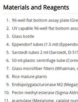
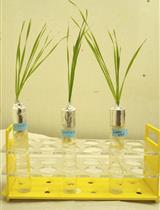
Cell Wall Compositional Analysis of Rice Culms

水稻茎杆的细胞壁组成分析

LZ Lanjun Zhang
BZ Baocai Zhang
YZ Yihua Zhou
4586 Views
Oct 20, 2019
The plant cell wall is a complicated network that is mainly constituted of polysaccharides, such as cellulose, hemicellulose and pectin. Many noncellulosic polysaccharides are further acetylated, which confers these polymers flexible physicochemical properties. Due to the significance of cell wall in plant growth and development, the analytic platform has been the focus for a long time. Here, we use internodes/culms, an important organ to provide mechanical support for rice plants, as an experimental sample to explore the method for cell wall composition analysis. The method includes preparation of cell wall residues, sequential extraction of polysaccharides, and measurement of cellulose. The procedure for acetate examination is also described. This method is applicable to determine the composition of individual cell wall polymers and the modifier acetates, and is suitable to identify cell wall relevant mutants based on the advantages in high throughput, precision and repeatability.