往期刊物2019

卷册: 9, 期号: 16

left

Dec

20

Dec

5

Nov

20

Nov

5

Oct

20

Oct

5

Sep

20

Sep

5

Aug

20

Aug

5

Jul

20

Jul

5

Jun

20

Jun

5

May

20

May

5

Apr

20

Apr

5

Mar

20

Mar

5

Feb

20

Feb

5

Jan

20

Jan

5

right

生物化学

体外ERK3激酶的放射性分析

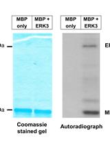
A Radioactive in vitro ERK3 Kinase Assay

体外ERK3激酶的放射性分析

LE Lobna Elkhadragy
WL Weiwen Long
5778 Views
Aug 20, 2019
Mitogen-activated protein kinases (MAPKs) are serine/threonine kinases that have an important role in signal transduction. Extracellular signal-regulated kinase 3 (ERK3), also known as MAPK6, is an atypical MAPK. Here, we describe in detail an in vitro assay for the kinase activity of ERK3 using myelin basic protein (MBP) or steroid receptor coactivator-3 (SRC-3) as substrates. The assay is carried out in the presence of [γ-32P]-ATP which results in radiolabeling of phosphorylated substrates. Separation of the reaction components by gel electrophoresis followed by autoradiography enables detection of the radiolabeled products, and hence determination of the kinase activity of ERK3. This assay can be used for several applications including identification of substrates, determination of the effect of molecules or mutations on kinase activity, and testing specific kinase inhibitors. Furthermore, the protocol outlined here can be adapted to measure the activity of other kinases by using their specific substrates.
以细胞为基础的DDR1向胶原包被微珠的招募分析

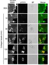
Cell-based Assay for Recruitment of DDR1 to Collagen-coated Beads

以细胞为基础的DDR1向胶原包被微珠的招募分析

VJ Victoria Juskaite
BL Birgit Leitinger
4352 Views
Aug 20, 2019
The discoidin domain receptors, DDR1 and DDR2, are key signaling receptors for the extracellular matrix protein collagen. The interactions of cells with collagen are difficult to study because of the difficulty to obtain native collagen fibers for in vitro studies. Thus, in vitro studies often use acid-soluble collagens in the form of single triple helices, which are not representative of the densely packed insoluble collagen fibers found in tissues. In this protocol, we describe a method that allows stimulating DDR1 locally with collagen-coated beads. Latex beads are first coated with acid-soluble collagen, then added to cells expressing DDR1. Recruitment of DDR1 to the beads and collagen-induced DDR1 phosphorylation is visualized by immunofluorescence microscopy on a widefield microscope. In this method, densely packed collagen is presented to cells in an insoluble form. Bead coating is easy to perform, and this method thus presents a straightforward protocol with which to study local recruitment of collagen receptors to insoluble collagen.

癌症生物学

培养细胞中蛋白和mRNA在单细胞水平上的三维重构和量化分析

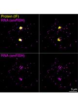
Three-dimensional Reconstruction and Quantification of Proteins and mRNAs at the Single-cell Level in Cultured Cells

培养细胞中蛋白和mRNA在单细胞水平上的三维重构和量化分析

XJ Xueer Jiang
IB Ingrid Brust-Mascher
LJ Li-En Jao
9477 Views
Aug 20, 2019
Gene expression is often regulated by the abundance, localization, and translation of mRNAs in both space and time. Being able to visualize mRNAs and protein products in single cells is critical to understand this regulatory process. The development of single-molecule RNA fluorescence in situ hybridization (smFISH) allows the detection of individual RNA molecules at the single-molecule and single-cell levels. When combined with immunofluorescence (IF), both mRNAs and proteins in individual cells can be analyzed simultaneously. However, a precise and streamlined quantification method for the smFISH and IF combined dataset is scarce, as existing workflows mostly focus on quantifying the smFISH data alone. Here we detail a method for performing sequential IF and smFISH in cultured cells (as described in Sepulveda et al., 2018) and the subsequent statistical analysis of the smFISH and IF data via three-dimensional (3D) reconstruction in a semi-automatic image processing workflow. Although our method is based on analyzing centrosomally enriched mRNAs and proteins, the workflow can be readily adapted for performing and analyzing smFISH and IF data in other biological contexts.

细胞生物学

利用改进的Sca/eS和机器学习用于螺旋器中毛细胞的成像和分析

A Novel Technique for Imaging and Analysis of Hair Cells in the Organ of Corti Using Modified Sca/eS and Machine Learning

利用改进的Sca/eS和机器学习用于螺旋器中毛细胞的成像和分析

SU Shinji Urata
TI Tadatsune Iida
YS Yuri Suzuki
SL Shiou-Yuh Lin
YM Yu Mizushima
CF Chisato Fujimoto
YM Yu Matsumoto
TY Tatsuya Yamasoba
5697 Views
Aug 20, 2019
Here, we describe a sorbitol-based optical clearing method, called modified Sca/eS that can be used to image all hair cells (HCs) in the mouse cochlea. This modification of Sca/eS is defined by three steps: decalcification, de-lipidation, and refractive index matching, which can all be completed within 72 h. Furthermore, we established automated analysis programs that perform machine learning-based pattern recognition. These programs generate 1) a linearized image of HCs, 2) the coordinates of HCs, 3) a holocochleogram, and 4) clusters of HC loss. In summary, a novel approach that integrates modified Sca/eS and programs based on machine learning facilitates quantitative and comprehensive analysis of the physiological and pathological properties of all HCs.
基于成纤维细胞间隙封闭显微成像的鼠成纤维细胞的体外迁移分析

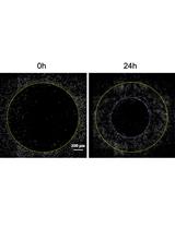
Fibroblast Gap-closure Assay-Microscopy-based in vitro Assay Measuring the Migration of Murine Fibroblasts

基于成纤维细胞间隙封闭显微成像的鼠成纤维细胞的体外迁移分析

AL Agnieszka P. Looney
MB Mallar Bhattacharya
5247 Views
Aug 20, 2019
Pulmonary fibrosis is characterized by pathological scaring of the lung. Similar to other fibrotic diseases, scar formation is driven by excessive extracellular matrix deposition by activated, proliferative, and migratory fibroblasts.Currently, the two most widely used chemotaxis and cell migration assays are the scratch assay and the transmembrane invasion assay. Here we present a gap closure assay that employs commercially available cell lines, equipment and reagents and is time efficient as well as straightforward. The protocol uses an Oris pro cell migration assay 96-well plate with a dissolvable plug in the center of each well to create a cell free area at the time of seeding. Cell repopulation of the empty zone is captured via light microscopy at different time points and quantified with free image analysis software. The clear advantages of this assay in comparison to similar protocols are the use of uncomplicated cell culture methods and the ability to image the experiment throughout.

发育生物学

果蝇翅膀成虫盘体内培养和弗林蛋白酶抑制剂检测

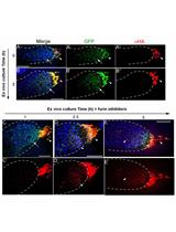
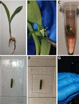
Ex vivo Drosophila Wing Imaginal Disc Culture and Furin Inhibitor Assay

果蝇翅膀成虫盘体内培养和弗林蛋白酶抑制剂检测

AS Alex Sohr
LD Lijuan Du
SR Sougata Roy
4760 Views
Aug 20, 2019
Furin is an evolutionarily conserved proprotein convertase (PC) family enzyme with a broad range of substrates that are essential for developmental, homeostatic, and disease pathways. Classical genetic approaches and in vitro biochemical or cell biological assays identified that precursor forms of most growth factor family proteins are processed by Furin. To quantitatively assess the potential role of Furin in cleaving and modulating intercellular dispersion of a Drosophila signaling protein, we developed a simple assay by combining genetics, ex vivo organ culture, pharmacological treatment, and imaging analyses. The protocol herein describes how to ex vivo culture Drosophila wing imaginal discs expressing a fluorescently tagged Drosophila Fibroblast Growth Factor (FGF, Branchless/Bnl) over a long period of time in the presence of Furin inhibitors and monitor the cleavage and intercellular dispersion of the truncated Bnl parts using microscopy. Although the assay described here is for assessing the effect of Furin inhibition on Bnl cleavage in the Drosophila larval wing imaginal disc, the principle and methodology can easily be adopted for any other signals, tissue systems, or organisms. This strategy and protocol provide an assay for examining Furin activity on a specific substrate by directly visualizing the spatiotemporal distribution of its truncated parts in an ex vivo-cultured organ.

免疫学

体外胸腺调节性T细胞祖细胞向成熟胸腺调节性T细胞的分化

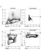
In vitro Differentiation of Thymic Treg Cell Progenitors to Mature Thymic Treg Cells

体外胸腺调节性T细胞祖细胞向成熟胸腺调节性T细胞的分化

DO David L. Owen
MF Michael A. Farrar
3976 Views
Aug 20, 2019
Thymic Treg cell differentiation occurs via a two-step process. Step one generates Treg cell progenitors (TregP) and is driven by strong TCR interactions with antigens presented in the thymus. Step two is initiated by activation of STAT5 via IL-2, or IL-15, leading to mature Treg cells capable of emigrating from the thymus and mediating immune tolerance and homeostasis in peripheral tissues. Herein we describe an in vitro TregP cell differentiation assay that models the second, cytokine dependent, step of thymic Treg cell development. It can be utilized with relative ease to determine if a population of thymocytes represents a potential progenitor population for Treg cells as well as test how different cytokines or chemical inhibitors modulate the differentiation of known TregP cell populations into mature Treg cells.

微生物学

细胞壁孔隙的荧光猝灭显微镜无创定量分析

Non-invasive Quantification of Cell Wall Porosity by Fluorescence Quenching Microscopy

细胞壁孔隙的荧光猝灭显微镜无创定量分析

XL Xiaohui Liu
TP Thomas Günther Pomorski
JL Johannes Liesche
5207 Views
Aug 20, 2019
All bacteria, fungi and plant cells are surrounded by a cell wall. This complex network of polysaccharides and glycoproteins provides mechanical support, defines cell shape, controls cell growth and influences the exchange of substances between the cell and its surroundings. Despite its name, the cell wall is a flexible, dynamic structure. However, due to the lack of non-invasive methods to probe the structure, relatively little is known about the synthesis and dynamic remodeling of cell walls. Here, we describe a non-invasive method that quantifies a key physiological parameter of cell walls, the porosity, i.e., the size of spaces between cell wall components. This method measures the porosity-dependent decrease of the plasma membrane-localized fluorescent dye FM4-64 in the presence of the extracellular quencher Trypan blue. This method is applied to bacteria, fungi and plant cell walls to detect dynamic changes of porosity in response to environmental cues.
浮游植物中半胱天冬酶活性检测

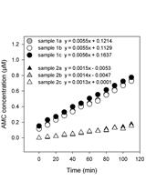
Assessment of Metacaspase Activity in Phytoplankton

浮游植物中半胱天冬酶活性检测

DS Dina Spungin
IB Ilana Berman-Frank
4124 Views
Aug 20, 2019
Programmed cell death (PCD) is an irreversible, genetically-controlled form of cell suicide in which an endogenous biochemical pathway leads to morphological changes and ultimately, cellular demise. PCD is accompanied by de-novo protein synthesis of a family of proteases-"caspases" that are often used as a diagnostic marker of PCD. Although phytoplankton do not contain true caspases, caspase-like activity (hypothetical proteins with analogous activity) has been traditionally used as a diagnostic marker of PCD in marine phytoplankton. Increased caspase-like proteolytic activity was demonstrated when synthetic fluorogenic activity substrates specific for caspases (with an Asp at the P1 position) were applied upon PCD induction. Metacaspases, cysteine proteases, share structural properties with those of caspases, yet they are highly specific for Arg and Lys cleavage site at the P1 position implying that caspase specific substrates are not indicative of metacaspase catalytic activity. This method specifically tests direct metacaspase activity in phytoplankton by the cleavage of the fluorogenic metacaspase substrate Ac-VRPR-AMC. Metacaspase activity was tested by the addition of a metacaspase specific peptide that is conjugated to the fluorescent reporter molecule. The cleavage of the peptide by the metacaspase releases the fluorochrome that, when excited by light, emits fluorescence. The level of metacaspase enzymatic activity in the cell lysate is directly proportional to the fluorescence signal detected. The use of specific standards in this test enables the quantification of the fluorescence results. This assay directly allows monitoring the metacaspase cleavage products and thereby tracing evidence for programmed cell death.
枯草芽孢杆菌自然遗传转化过程中完整供体DNA长度的测定

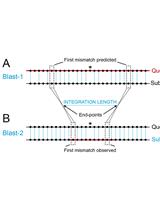
Measurement of the Length of the Integrated Donor DNA during Bacillus subtilis Natural Chromosomal Transformation

枯草芽孢杆菌自然遗传转化过程中完整供体DNA长度的测定

ES Ester Serrano
BC Begoña Carrasco
3485 Views
Aug 20, 2019
For natural transformation to occur, bacterial cells must first develop a programmed physiological state called competence. Competence in Bacillus subtilis, which occurs only in a fraction of cells, is a transient stress response that allows cells to take up DNA from the environment. During natural chromosomal transformation, the internalized linear single-stranded (ss) DNA recombines with the identical (homologous) or partially identical (homeologous) sequence of the resident duplex. The length of the integrated DNA, which can be measured, depends on the percentage of sequence divergence between the donor (internalized) and the recipient (chromosomal) DNAs.The following protocol describes how to induce the development of competence in B. subtilis cells, how to transform them with donor DNAs representing different percentages of sequence divergence compared with the recipient chromosomal DNA, how to calculate the chromosomal transformation efficiency for each of them, and how to amplify the chromosomal DNA from the transformants in order to measure the length in base pairs (bp) of the integrated donor DNA.

分子生物学

一种改良的Smart-seq2样品制备方案在小鼠卵母细胞中应用稀有细胞全长单细胞mRNA测序

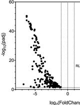
Application of a Modified Smart-seq2 Sample Preparation Protocol for Rare Cell Full-length Single-cell mRNA Sequencing to Mouse Oocytes

一种改良的Smart-seq2样品制备方案在小鼠卵母细胞中应用稀有细胞全长单细胞mRNA测序

RT Rebecca S. Treger
SP Scott D. Pope
XX Xiaojun Xing
AI Akiko Iwasaki
6001 Views
Aug 20, 2019
Endogenous retroviruses (ERV) are transposable retroelements that form ~10% of the murine genome and whose family members are differentially expressed throughout embryogenesis. However, precise regulation of ERV in germ cells remains unclear. To investigate ERV expression in oocytes, we adapted a single-cell mRNA-sequencing library preparation method to generate bulk sequencing libraries from growing oocytes in a time- and cost-efficient manner. Here, we present a modified Smart-seq2 protocol that yields full-length cDNA libraries from purified RNA obtained from low numbers of pooled immature or mature oocytes. Using this method, RNA-sequencing libraries can be generated from any rare or difficult-to-isolate populations for subsequent sequencing and retroelement expression analysis.
对FFPE组织样品中OATP1B1 c. 521 T>C多态性进行基因分型的优化实验方法

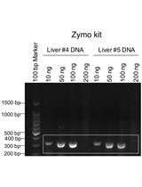
Genotyping of the OATP1B1 c. 521 T>C Polymorphism from the Formalin-Fixed Paraffin-Embedded (FFPE) Tissue Specimens: An Optimized Protocol

对FFPE组织样品中OATP1B1 c. 521 T>C多态性进行基因分型的优化实验方法

Alexandra R Crowe Alexandra R Crowe
JM Jonathan Miller
Wei  Yue Wei Yue
3629 Views
Aug 20, 2019
Organic anion transporting polypeptide (OATP) 1B1 is a liver-specific transport protein that plays an important role in hepatic drug disposition. It transports many drugs from the blood into the liver, including lipid-lowering statins. The c. 521 T>C polymorphism of OATP1B1 has reduced transport activity and is associated with statin-induced myopathy. Formalin-fixed paraffin-embedded (FFPE) liver tissues can be an enriched source for genotyping of this clinically significant OATP1B1 polymorphism in retrospective studies. The successfulness of genotyping using Sanger-sequencing of a PCR product from FFPE tissue relies on a successful PCR amplification using genomic DNA extracted from the FFPE tissues. Such PCR amplification is often limited by the quality of DNA extracted from the FFPE tissue. An optimized method for high-quality DNA extraction and efficient PCR amplification is highly needed in order to genotype polymorphisms such as the c. 521 T>C polymorphism using FFPE tissues. The current study established an optimized and reproducible method for a Sanger-sequencing-based genotyping method using FFPE human liver tissues that is applicable to even small FFPE tissues such as needle-core biopsy specimens.

神经科学

基于圆筒试验的小鼠帕金森综合症模型中感觉运动系统功能测试

Cylinder Test to Assess Sensory-motor Function in a Mouse Model of Parkinson’s Disease

基于圆筒试验的小鼠帕金森综合症模型中感觉运动系统功能测试

LM Luiz Alexandre Viana Magno
MC Mélcar Collodetti
HT Helia Tenza-Ferrer
MR Marco Aurélio Romano-Silva
12248 Views
Aug 20, 2019
Parkinson’s disease is a progressive neurodegenerative movement disorder that happens due to the loss of dopaminergic neurons in the substantia nigra. The deficiency of dopamine in the basal nuclei drives cardinal motor symptoms such as bradykinesia and hypokinesia. The current protocol describes the cylinder test, which is a relatively simple behavioral assessment that evaluates the motor deficits upon unilateral degeneration of the nigrostriatal pathway in experimental models of Parkinson’s disease. Since dopamine-depleted mice exhibit the preferential use of the forelimb ipsilateral to the lesion, here researchers perform the cylinder test to investigate the therapeutic effects of antiparkinsonian treatments on the performance of the contralateral (injured) limb.
用于成年小鼠脑组织的细胞特异性CRISPR基因编辑的病毒载体的构建

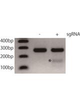
Construction of Viral Vectors for Cell Type-specific CRISPR Gene Editing in the Adult Mouse Brain

用于成年小鼠脑组织的细胞特异性CRISPR基因编辑的病毒载体的构建

HY Hiroshi Yamaguchi
Ld Luis de Lecea
5808 Views
Aug 20, 2019
Recently developed gene editing technologies based on engineered CRISPR/Cas9 systems enables researchers to disrupt genes in a cell type-specific manner in the adult mouse brain. Using these technologies, we recently showed that the dopamine beta-hydroxylase gene in Locus Coeruleus (LC) norepinephrine neurons plays a vital role in the maintenance of wakefulness. Our method consists of four steps, (1) crossing Cre-dependent spCas9 knockin mice with a Cre-driver mouse line to express spCas9 in the target neural populations, (2) cloning of sgRNA, (3) construction of an AAV (adeno associated virus) vector expressing dual sgRNA, and (4) virus packaging and stereotaxic injection of the virus into the target brain area. Here, we describe a detailed protocol of AAV vector construction for cell type-specific CRISPR gene editing in the adult mouse brain. The method adopts a dual-sgRNA strategy for efficient disruption of the target gene. At first, a few different sgRNAs targeting the same gene are cloned into a plasmid expressing spCas9. After evaluation of the sgRNAs by a T7 endonuclease assay, the two most efficient sgRNAs are cloned in tandem into an AAV vector using the Gibson Assembly method.
胡须损伤测试:一种检测小鼠触觉过敏症的有效方法

Whisker Nuisance Test: A Valuable Tool to Assess Tactile Hypersensitivity in Mice

胡须损伤测试:一种检测小鼠触觉过敏症的有效方法

LB Luigi Balasco
GC Gabriele Chelini
YB Yuri Bozzi
GP Giovanni Provenzano
5085 Views
Aug 20, 2019
Abnormal response to tactile stimulation, described as both hyper- and hypo-reactivity, is a common sensory impairment in multiple neuropsychiatric disorders. The neural bases of tactile sensitivity remain so far unknown. In the last years, animal studies have proven to be useful for shedding light on the cellular and molecular mechanism underlying sensory impairments. However, few behavioral tests have been developed in mice for assessing tactile perception abnormalities (e.g., the whisker nuisance [WN] test and the tactile prepulse inhibition assay). Here we provide a modified version of the WN test, which is based on the previously developed method by McNamara et al. (2010). The WN test permits to specifically detect tactile hypo/hyper-sensitivity relative to whisker stimulation in mice. The test starts with a habituation phase in which the mouse familiarizes itself with the experimental cage and the researcher/experimenter. After a sham session, the experimental session begins, consisting of bilateral whisker stimulation with a wooden stick. The advantages of using this protocol are many: it is relatively simple to set with no particular or expensive equipment needed, it is easily reproducible, it allows researchers to assess a variety of behavioral responses to a whisker-specific tactile perception in mice (i.e., fearful behavior, stance, hyperventilation, aggressive behavior and evasiveness) and provides important translational opportunities.