往期刊物2019

卷册: 9, 期号: 14

left

Dec

20

Dec

5

Nov

20

Nov

5

Oct

20

Oct

5

Sep

20

Sep

5

Aug

20

Aug

5

Jul

20

Jul

5

Jun

20

Jun

5

May

20

May

5

Apr

20

Apr

5

Mar

20

Mar

5

Feb

20

Feb

5

Jan

20

Jan

5

right

生物化学

通过体外RNA剪切检测IRE1依赖的RNA降解

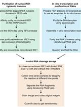
In vitro RNA Cleavage Assays to Characterize IRE1-dependent RNA Decay

通过体外RNA剪切检测IRE1依赖的RNA降解

GK G. Elif Karagöz
JP Jirka Peschek
PW Peter Walter
DA Diego Acosta-Alvear
7138 Views
Jul 20, 2019
The kinase/RNase IRE1 is a key effector of the cellular response to endoplasmic reticulum stress. The RNase activity of IRE1 can be measured in cells or in the test tube. Here we describe a protocol for the in vitro cleavage and analysis of RNA substrates of IRE1. The method consists of the in vitro transcription, purification and re-folding of IRE1 substrate RNAs followed by their cleavage using recombinant cytosolic kinase/RNase domains of IRE1 and the separation of the resulting fragments by denaturing polyacrylamide gel electrophoresis. This protocol allows the study of the cleavage kinetics of IRE1’s RNA substrates in vitro.
等电聚焦用于小鼠视网膜中磷酸化视紫红质的定量分析

Isoelectric Focusing to Quantify Rhodopsin Phosphorylation in Mouse Retina

等电聚焦用于小鼠视网膜中磷酸化视紫红质的定量分析

SL Sowmya Bekshe Lokappa
M. Carter Cornwall M. Carter Cornwall
JC Jeannie Chen
6036 Views
Jul 20, 2019
Rhodopsin is a G-protein coupled receptor (GPCR) that mediates vision under dim light. Upon light exposure, rhodopsin is phosphorylated at multiple serine and threonine sites at its carboxyl-terminus by rhodopsin kinase (GRK1). This, in turn, reduces its ability to activate the visual G-protein transducin. Binding of light-activated, phosphorylated rhodopsin by arrestin (ARR1) fully terminates the catalytic activity of rhodopsin. Quantification of the levels of the differentially phosphorylated rhodopsin species provides definitive information about the role of phosphorylated rhodopsin in visual functions. Isoelectric Focusing (IEF) is a technique which is used to separate ampholytic components, such as proteins, based on their isoelectric point (pI). It is a useful technique used to distinguish protein isoforms and post-translational modifications such as phosphorylation, glycosylation, deamination, and acetylation, due to their effects on the protein’s pI. Isoelectric Focusing can provide high resolution of differentially phosphorylated forms of a protein. Though other techniques such as kinase activity assays, phospho-specific antibodies, western blot, enzyme-linked immunosorbent assays (ELISA), radiolabeling and mass spectrometry are used to detect and quantify protein phosphorylation, IEF is a simple and cost-effective method to quantify rhodopsin phosphorylation, as it can readily detect individual phosphorylated forms. Here we provide a detailed protocol for determining phosphorylated rhodopsin species using the Isoelectric Focusing technique.

癌症生物学

利用微核体外分析系统和洋葱体内实验进行遗传毒性测试

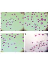
Evaluation of Genotoxicity by Micronucleus Assay in vitro and by Allium cepa Test in vivo

利用微核体外分析系统和洋葱体内实验进行遗传毒性测试

CB Christina N. Banti
SH Sotiris K. Hadjikakou
8363 Views
Jul 20, 2019
The in vitro and in vivo genotoxicity of new metallodrugs either as Small Bioactive Molecules (SBAMs) or Conjugates of Metals with Drugs (CoMeDs) is evaluated by the micronucleus test and the Allium cepa assay, respectively. Fetal lung fibroblast cells (MRC-5), normal human corneal epithelial cells (HCEC) and immortalized human keratinocytes cells (HaCaT) were incubated with solutions of SBAMs or CoMeDs at their IC50 values for 48 h (the concentration of a compound which is required to inhibit the cells growth by 50% in relation to the non-treated cells). The micronucleus abundance percentage towards the corresponding one, of the non-treated cells indicates the in vitro genotoxicity of the formulations. The in vivo Allium cepa test comprises the exposing of the plant Allium cepa roots to an SBAMs or a CoMeDs solution for 48 h. The percentages of the mitotic index, the chromosome aberrations, the nuclear abnormalities and the presence of the micronucleus are calculated indicating the in vivo genotoxicity of the agent.
单层水凝胶培养人胶质母细胞瘤细胞系的全RNA分离

Total RNA Isolation from Separately Established Monolayer and Hydrogel Cultures of Human Glioblastoma Cell Line

单层水凝胶培养人胶质母细胞瘤细胞系的全RNA分离

MJ Manasi P Jogalekar
ES Elba E Serrano
5415 Views
Jul 20, 2019
Astrocytoma is an invasive carcinoma occurring in the nervous system and currently lacks effective treatment options. A deeper understanding of the mechanisms of tumorigenesis and tumor progression is needed in order to develop novel therapeutic strategies. Recent advances in in vitro culture systems have demonstrated that the use of three-dimensional (3D) culture models could be more relevant for this purpose as compared to monolayer or two-dimensional (2D) models due to their resemblance to in vivo cancer pathology. High-throughput techniques such as RNA sequencing, microarray analyses and cloning could provide useful insights into the relevance of these systems to the native tissue. Previous studies have reported RNA extraction protocols needed for such applications. We have modified these protocols to suit the isolation of total RNA from monolayer and hydrogel cultures of astrocytoma established using basement membrane matrix, GeltrexTM. We have used this method to demonstrate the differences in the expression of genes involved in autophagy, a process deregulated in many cancer types, in monolayer and hydrogel cultures using quantitative polymerase chain reaction (qPCR). This protocol can be adopted by the researchers who wish to understand the molecular basis of gene expression in hydrogel cultures of normal as well as cancer cell lines.

细胞生物学

通过电镜样品制备实现的人体组织和器官中细胞连接体及其定位的纳米与介观图谱分析

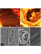
Electron Microscopy Sample Preparation Protocol Enabling Nano-to-mesoscopic Mapping of Cellular Connectomes and Their Habitats in Human Tissues and Organs

通过电镜样品制备实现的人体组织和器官中细胞连接体及其定位的纳米与介观图谱分析

LN Lucy Ngo
Anton D. Nathanson Anton D. Nathanson
TG Tomasz Garbowski
UK Ulf Knothe
DZ Dirk Zeidler
MK Melissa L. Knothe Tate
6898 Views
Jul 20, 2019
Multibeam scanning electron microscopy (multiSEM) provides a technical platform for seamless nano-to-mesoscale mapping of cells in human tissues and organs, which is a major new initiative of the U.S. National Institutes of Health. Such cross-length-scale imaging is expected to provide unprecedented understanding of relationships between cellular health and tissue-organ as well as organismal-scale health outcomes. For example, understanding relationships between loss in cell viability and cell network connectivity enables identification of emergent behaviors and prediction of degenerative disease onset, in organs as diverse as bone and brain, at early timepoints, providing a basis for future treatments and prevention. Developed for rapid throughput imaging of minute defects on semiconductor wafers, multiSEM has recently been adapted for imaging of human organs, their constituent tissues, and their respective cellular inhabitants. Through integration of geospatial approaches, statistical and network modelling, advances in computing and the management of immense datasets, as well as recent developments in machine learning that enable the automation of big data analyses, multiSEM and other cross- cutting imaging technologies have the potential to exert a profound impact on elucidation of disease mechanisms, translating to improvements in human health. Here we provide a protocol for acquisition and preparation of sample specimen sizes of diagnostic relevance for human anatomy and physiology. We discuss challenges and opportunities to integrate this approach with multibeam scanning electron microscopy workflows as well as multiple imaging modalities for mapping of organ and tissue structure and function.
活体细胞中脂滴融合的脂类交换速率检测

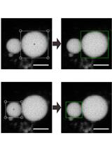
Lipid-exchange Rate Assay for Lipid Droplet Fusion in Live Cells

活体细胞中脂滴融合的脂类交换速率检测

Jia Wang Jia Wang
BC Boon Tin Chua
PL Peng Li
FC Feng-Jung Chen
6050 Views
Jul 20, 2019
Lipid droplets (LDs) are central organelles in maintaining lipid homeostasis. Defective LD growth often results in the development of metabolic disorders. LD fusion and growth mediated by cell death–inducing DNA fragmentation factor alpha (DFFA)-like effector (CIDE) family proteins are crucial for various biological processes including unilocular LD formation in the adipocytes, lipid storage in the liver, milk lipid secretion in the mammary epithelia cells, and lipid secretion in the skin sebocytes. Previous methodology by Gong et al. (2011) first reported a lipid-exchange rate assay to evaluate the fusion ability of each LD pair in the cells mediated by CIDE family proteins and their regulators, but photobleaching issue remains a problem and a detailed procedure was not provided. Here, we provide an improved and detailed protocol for the lipid-exchange rate measurement. The three key steps for this assay are cell preparation, image acquisition, and data analysis. The images of the fluorescence recovery are acquired after photobleaching followed by the measurement of the intensity changes in the LD pair. The difference in fluorescent intensity is used to obtain the lipid exchange rate between the LDs. The accuracy and repetitiveness of the calculated exchange rates are assured with three-cycle of photobleaching process and the linear criteria in data fitting. With this quantitative assay, we are able to identify the functional roles of the key proteins and the effects of their mutants on LD fusion.

免疫学

小鼠肺泡巨噬细胞的分离与长期培养

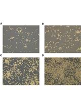
Isolation and Long-term Cultivation of Mouse Alveolar Macrophages

小鼠肺泡巨噬细胞的分离与长期培养

CB Clara Jana-Lui Busch
JF Jérémy Favret
LG Laufey Geirsdóttir
KM Kaaweh Molawi
MS Michael H. Sieweke
15194 Views
Jul 20, 2019
Alveolar macrophages (AM) are tissue-resident macrophages that colonize the lung around birth and can self-maintain long-term in an adult organism without contribution of monocytes. AM are located in the pulmonary alveoli and can be harvested by washing the lungs using the method of bronchoalveolar lavage (BAL). Here, we compared different conditions of BAL to obtain high yields of murine AM for in vitro culture and expansion of AM. In addition, we describe specific culture conditions, under which AM proliferate long-term in liquid culture in the presence of granulocyte-macrophage colony-stimulating factor. This method can be used to obtain large numbers of AM for in vivo transplantation or for in vitro experiments with primary mouse macrophages.

微生物学

利用HyperLOPIT绘制酿酒酵母空间蛋白质组图谱的方法

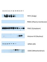
A Protocol to Map the Spatial Proteome Using HyperLOPIT in Saccharomyces cerevisiae

利用HyperLOPIT绘制酿酒酵母空间蛋白质组图谱的方法

DN Daniel J. H. Nightingale
KL Kathryn S. Lilley
SO Stephen G. Oliver
5920 Views
Jul 20, 2019
The correct subcellular localization of proteins is vital for cellular function and the study of this process at the systems level will therefore enrich our understanding of the roles of proteins within the cell. Multiple methods are available for the study of protein subcellular localization, including fluorescence microscopy, organelle cataloging, proximity labeling methods, and whole-cell protein correlation profiling methods. We provide here a protocol for the systems-level study of the subcellular localization of the yeast proteome, using a version of hyperplexed Localization of Organelle Proteins by Isotope Tagging (hyperLOPIT) that has been optimized for use with Saccharomyces cerevisiae. The entire protocol encompasses cell culture, cell lysis by nitrogen cavitation, subcellular fractionation, monitoring of the fractionation using Western blotting, labeling of samples with TMT isobaric tags and mass spectrometric analysis. Also included is a brief explanation of downstream processing of the mass spectrometry data to produce a map of the spatial proteome. If required, the nitrogen cavitation lysis and Western blotting portions of the protocol may be performed independently of the mass spectrometry analysis. The protocol in its entirety, however, enables the unbiased, systems-level and high-resolution analysis of the localizations of thousands of proteins in parallel within a single experiment.

神经科学

神经元突起RNA含量的Axon-seq 深入分析

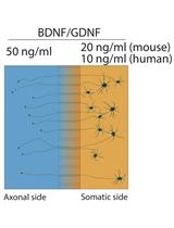
Axon-seq for in Depth Analysis of the RNA Content of Neuronal Processes

神经元突起RNA含量的Axon-seq 深入分析

Jik Nijssen Jik Nijssen
JA Julio Aguila
Eva Hedlund Eva Hedlund
11536 Views
Jul 20, 2019
Neuronal processes have an RNA composition that is distinct from the cell body. Therefore, to fully understand neuronal biology in health and disease we need to study both somas, dendrites and axons. Here we describe a detailed protocol of a newly refined method, Axon-seq, for RNA sequencing of axons (and dendrites) grown in isolation using single microfluidic devices. We also detail how to generate motor neurons from mouse and human pluripotent stem cells for sequencing, but Axon-seq is applicable to any neuronal cell. In Axon-seq, the axons are recruited through a growth factor gradient, lysed and directly processed to cDNA without RNA isolation. A careful bioinformatic step ensures that any soma-contaminated samples are easily identified and removed.
小鼠强迫性进食行为的检测步骤

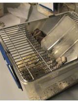
Protocol for Measuring Compulsive-like Feeding Behavior in Mice

小鼠强迫性进食行为的检测步骤

MF Marta Fructuoso
JE Jose Espinosa-Carrasco
IE Ionas Erb
CN Cedric Notredame
MD Mara Dierssen
5108 Views
Jul 20, 2019
Obesity is an important health problem with a strong environmental component that is acquiring pandemic proportion. The high availability of caloric dense foods promotes overeating potentially causing obesity. Animal models are key to validate novel therapeutic strategies, but researchers must carefully select the appropriate model to draw the right conclusions. Obesity is defined by an increased body mass index greater than 30 and characterized by an excess of adipose tissue. However, the regulation of food intake involves a close interrelationship between homeostatic and non-homeostatic factors. Studies in animal models have shown that intermittent access to sweetened or calorie-dense foods induces changes in feeding behavior. However, these studies are focused mainly on the final outcome (obesity) rather than on the primary dysfunction underlying the overeating of palatable foods. We describe a protocol to study overeating in mice using diet-induced obesity (DIO). This method can be applied to free choice between palatable food and a standard rodent chow or to forced intake of calorie-dense and/or palatable diets. Exposure to such diets is sufficient to promote changes in meal pattern that we register and analyze during the period of weight gain allowing the longitudinal characterization of feeding behavior in mice. Abnormal eating behaviors such as binge eating or snacking, behavioral alterations commonly observed in obese humans, can be detected using our protocol. In the free-choice procedure, mice develop a preference for the rewarding palatable food showing the reinforcing effect of this diet. Compulsive components of feeding are reflected by maintenance of feeding despite an adverse bitter taste caused by adulteration with quinine and by the negligence of standard chow when access to palatable food is ceased or temporally limited. Our strategy also enables to identify compulsive overeating in mice under a high-caloric regime by using limited food access and finally, we propose complementary behavioral tests to confirm the non-homeostatic food-taking triggered by these foods. Finally, we describe how to computationally explore large longitudinal behavioral datasets.
一种用于探究斑马鱼明暗节律的培养箱标准设计

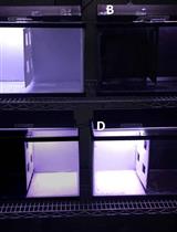
A Standardized Tank Design for the Light Dark Task in Zebrafish

一种用于探究斑马鱼明暗节律的培养箱标准设计

AF Amanda Facciol
Steven Tran Steven Tran
Robert Gerlai Robert Gerlai
4472 Views
Jul 20, 2019
The light dark paradigm is a common behavioral test used to screen a variety of pharmacological agents, including anxiogenics and anxiolytics. Although most often used in rodents, the light dark task has recently been adapted for use in zebrafish. However, a number of inconsistent findings have been reported for this species. Some have found zebrafish to prefer black, while others report a preference for light. Careful analysis of light dark preference experiments using zebrafish reveals significant variation in testing tank design and test conditions, including lighting and substrate color. Additionally, in some experiments the designated dark side of the testing tank is completely covered, producing a “cave-like” environment which further confounds results. Lastly, authors commonly use the terms “light vs. dark” interchangeably with “white vs. black”, when these are two separate factors that may influence preference: illumination level vs. background shade. To address these limitations, we designed testing tanks that differentiate illumination vs. background shade preference in zebrafish. This design allows for simple standardization of light dark testing apparatus in zebrafish, and facilitates more reliable comparison across studies.

植物科学

感染大麦中白粉病吸器的分离提取

Isolation of Powdery Mildew Haustoria from Infected Barley

感染大麦中白粉病吸器的分离提取

LL Linhan Li
BC Benjamin Collier
PS Pietro D. Spanu
6726 Views
Jul 20, 2019
Blumeria graminis is a fungus that causes powdery mildews on grasses, such as barley. Investigations of this pathogen present many challenges due to its obligate biotrophic nature. This means that the fungus can only grow in the presence of a living host plant. B. graminis forms epiphytic mycelia on the plant surface and feeding organs (haustoria) inside the epidermal cells of the host plant. Therefore, it is difficult to separate the fungus from plant tissues. This protocol shows how to obtain different fungal structures from powdery mildew infected barley leaves. The epiphytic mycelia including conidia and conidiophores can be separated after immersing the infected leaves into 5% cellulose acetate dissolved in acetone, and peeling off the cellulose acetate membrane. Then, the haustoria are isolated from dissected epidermis after cellulase degradation of plant cell walls. The isolated haustoria remain intact with few plant impurities. The haustoria may be visualized by epifluorescence microscopy after staining with the chitin-specific dye WGA-Alexa Fluor 488. Finally, dissected material can be either processed immediately or kept at -80 °C for long-term storage for studies on gene expression and protein identification, for example by mass spectrometry.
蓝甲醇衍生物作为丛枝菌根真菌与植物共生标志物的定量研究

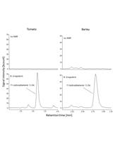
Quantification of Blumenol Derivatives as Leaf Biomarkers for Plant-AMF Association

蓝甲醇衍生物作为丛枝菌根真菌与植物共生标志物的定量研究

EM Elisabeth Mindt
MW Ming Wang
Martin Schäfer Martin Schäfer
RH Rayko Halitschke
IB Ian T. Baldwin
6015 Views
Jul 20, 2019
Symbiotic interactions between arbuscular mycorrhizal fungi (AMF) and plants are widespread among land plants and can be beneficial for both partners. The plant is provided with mineral nutrients such as nitrogen and phosphorous, whereas it provides carbon resources for the fungus in return. Due to the large economic and environmental impact, efficient characterization methods are required to monitor and quantify plant-AMF colonization. Existing methods, based on destructive sampling and elaborate root tissue analysis, are of limited value for high-throughput (HTP) screening. Here we describe a detailed protocol for the HTP quantification of blumenol derivatives in leaves by a simple extraction procedure and sensitive liquid chromatography mass spectrometry (LC/MS) analysis as accurate proxies of root AMF-associations in both model plants and economically relevant crops.

干细胞

成年C57BL/6J小鼠单个肌纤维的培养分离和卫星细胞的免疫染色

Isolation and Culture of Single Myofiber and Immunostaining of Satellite Cells from Adult C57BL/6J Mice

成年C57BL/6J小鼠单个肌纤维的培养分离和卫星细胞的免疫染色

SC Shujie Chen
HD Hongrong Ding
XY Xiangping Yao
Liwei Xie Liwei Xie
8449 Views
Jul 20, 2019
Myofiber isolation followed with ex vivo culture could recapitulate and visualize satellite cells (SCs) activation, proliferation, and differentiation. This approach could be taken to understand the physiology of satellite cells and the molecular mechanism of regulatory factors, in terms of the involvement of intrinsic factors over SCs quiescence, activation, proliferation and differentiation. Single myofiber culture has several advantages that the traditional approach such as FASC and cryosection could not compete with. For example, myofiber isolation and culture could be used to observe SCs activation, proliferation and differentiation at a continuous manner within their physiological “niche” environment while FACS or cryosection could only capture single time-point upon external stimulation to activate satellite cells by BaCl2, Cardiotoxin or ischemia. Furthermore, in vitro transfection with siRNA or overexpression vector could be performed under ex vivo culture to understand the detailed molecular function of a specific gene on SCs physiology. With these advantages, the physiological state of SCs could be analyzed at multiple designated time-points by immunofluorescence staining. In this protocol, we provide an efficient and practical protocol to isolate single myofiber from EDL muscle, followed with ex vivo culture and immunostaining.