往期刊物2019
卷册: 9, 期号: 13
生物化学
Oxygen Consumption Measurements in Caenorhabditis elegans Using the Seahorse XF24
利用海马细胞能量代谢实时测定仪XF24 进行秀丽隐杆线虫中耗氧量测定
Adhesive and Cohesive Peel Force Measurement of Human Airway Mucus
人气道粘液的黏附力和粘结剥离力测定
生物物理学
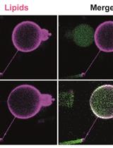
Dynamic and Sequential Protein Reconstitution on Negatively Curved Membranes by Giant Vesicles Fusion
巨型囊泡融合在负曲面膜上的顺序蛋白重建及动态

细胞生物学
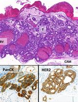
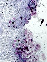
The Chick Chorioallantoic Membrane (CAM) Assay as a Three-dimensional Model to Study Autophagy in Cancer Cells
以鸡胚绒毛尿囊膜(CAM)为三维模型研究癌细胞自噬作用
免疫学
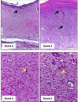
Morphological Evaluation of Wound Healing Events in the Excisional Wound Healing Model in Rats
在大鼠切除创面愈合模型中进行伤口愈合的形态学评估


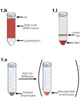
In vitro Infection of Primary Human Monocytes with HIV-1
原代人单核细胞的体外HIV-1侵染

微生物学
Gentamicin Protection Assay to Determine the Number of Intracellular Bacteria during Infection of Human TC7 Intestinal Epithelial Cells by Shigella flexneri
庆大霉素保护试验确定福氏志贺菌侵染人TC7肠上皮细胞的胞内寄生数目


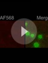
Fluorescence Microscopy Assay to Measure HIV-1 Capsid Uncoating Kinetics in vitro
HIV-1衣壳脱壳动力学的荧光显微体外检测
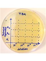
Plaque Assay to Determine Invasion and Intercellular Dissemination of Shigella flexneri in TC7 Human Intestinal Epithelial Cells
蚀斑测定弗氏志贺在TC7人肠上皮细胞中的侵袭和细胞间传播
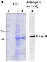
Preparation and Purification of Active Recombinant Human Pancreatic Lipase in Escherichia coli
重组人胰脂肪酶在大肠杆菌中的表达和纯化
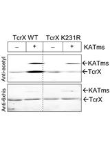
In vitro and in vivo Assessment of Protein Acetylation Status in Mycobacteria
分枝杆菌中蛋白乙酰化状态的体外和体内检测
分子生物学
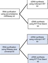
An Efficient Approach for RNA Extraction from Boar Sperm and Seminal Plasma
一种从公猪精子和精液中提取RNA的高效方法
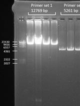
Detection of Heteroplasmic Variants in the Mitochondrial Genome through Massive Parallel Sequencing
大规模平行测序检测线粒体基因组异质粒

Optogenetic Inactivation of Transcription Factors in the Early Embryo of Drosophila
果蝇早期胚胎中的光遗传失活转录因子
干细胞
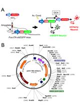
CRISPR/Cas9 + AAV-mediated Intra-embryonic Gene Knocking in Mice
小鼠体内由CRISPR / Cas9 + AAV介导的胚胎内基因敲除