往期刊物2019

卷册: 9, 期号: 10

left

Dec

20

Dec

5

Nov

20

Nov

5

Oct

20

Oct

5

Sep

20

Sep

5

Aug

20

Aug

5

Jul

20

Jul

5

Jun

20

Jun

5

May

20

May

5

Apr

20

Apr

5

Mar

20

Mar

5

Feb

20

Feb

5

Jan

20

Jan

5

right

癌症生物学

前列腺成纤维细胞中巨胞饮的可视检测

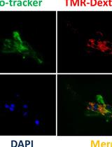
Visualization of Macropinocytosis in Prostate Fibroblasts

前列腺成纤维细胞中巨胞饮的可视检测

Rajeev   Mishra Rajeev Mishra
NB Neil A. Bhowmick
6028 Views
May 20, 2019
Macropinocytosis has emerged as an important mechanism for non-selective route to internalize extracellular fluids and dissolved molecules in eukaryotic cell. As fundamental cellular behavior, macropinocytosis plays specific and distinct roles in many physiological and pathological processes, such as nutrients uptake, antigen presentation, pathogen capture, and tumorigenesis. It supports tumorigenesis by providing metabolic needs to dividing cells in Ras driven cancer. In recent years, macropinocytosis has gained considerable interest in physiology and various diseases, including cancer, neurodegenerative diseases and atherosclerosis, which in turn has led to the discovery of new endocytic recycling systems. Approaches to assess macropinocytosis will provide insight into its underlying regulatory molecular mechanisms and enable the physiological control of macropinocytosis for controlled drug delivery and targeted cancer therapy. Macropinocytosis is an important phenomenon in Ras-expressing cancer cells and, recently, we have revealed a functional role for macropinocytosis in cancer associated fibroblasts (CAFs) fueling cancer cell growth. Here, we describe a protocol for detection of macropinocytosis in prostatic fibroblasts in vitro by utilizing fluorescently-labeled, lysine-fixable, 70 kDa high molecular weight dextran. Macropinosomes are visualized as fluorescent intracellular puncta either by confocal or fluorescent microscopy. To follow, subsequent intracellular events and their underlying mechanisms after macropinosomes formation, we perform co-localization of quenched BSA (DQTM-BSA) along with dextran labeling in cancer associated fibroblasts. Our protocol provides a consistent way to understand macropinocytosis in wild type or genetic manipulated prostatic fibroblast.

细胞生物学

利用流式细胞术测定转染效率

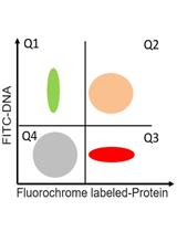
A Flow Cytometric Method to Determine Transfection Efficiency

利用流式细胞术测定转染效率

WM Wenli Mu
SH Stefanie Homann
CH Christian Hofmann
AG Alexandr Gorin
DH Diana Huynh
OY Otto Orlean Yang
TK Theodoros Kelesidis
11031 Views
May 20, 2019
Mammalian cell transfection is a powerful technique commonly used in molecular biology to express exogenous DNA or RNA in cells and study gene and protein function. Although several transfection strategies have been developed, there is a wide variation with regards to transfection efficiency, cell toxicity and reproducibility. Thus, a sensitive and robust method that can optimize transfection efficiency based not only on expression of the target protein of interest but also on the uptake of the nucleic acids, can be an important tool in molecular biology. Herein, we present a simple, rapid and robust flow cytometric method that can be used as a tool to optimize transfection efficiency while overcoming limitations of prior established methods that quantify transfection efficiency.
秀丽隐杆线虫活体呼吸速率的测定

Measurement of Respiration Rate in Live Caenorhabditis elegans

秀丽隐杆线虫活体呼吸速率的测定

LN Li Fang Ng
JG Jan Gruber
8026 Views
May 20, 2019
Mitochondrial function and dysfunction are at the core of aging and involved in many age-dependent diseases. Rate of oxygen consumption is a measure of mitochondrial function and energy production rate. The nematode Caenorhabditis elegans (C. elegans) offers an opportunity to study “living” mitochondria without the need for mitochondrial extraction, purification and associated artifacts. Oxygen consumption rate (OCR) is traditionally measured using single-chamber Clark electrodes with or without the addition of metabolic modulators. More recently, multi-well oxygen electrodes with automated injection system have been developed to enable rapid measurement of OCR under different conditions. Here, we describe a detailed protocol that we have adapted from existing protocols to measure coupled and uncoupled mitochondrial respiration (with and without metabolic modulators) in live respiring nematodes using a Seahorse XFe96 extracellular flux analyzer. We present details on our protocol, including preparation of nematode culture, use of metabolic modulators, execution of Seahorse XF assay as well as post-experimental data analysis. As a reference, we provide results of a series of experiments in which the metabolic activity of N2 wild-type nematodes was compared to N2 nematode treated with paraquat, a compound that generates reactive oxygen species (ROS), thus causing oxidative damage and mitochondrial dysfunction. These data illustrate the kind of insights that can be obtained even using a low number of nematodes (10 animals only per well).
哺乳动物细胞胞外ATP和胞内钙离子的协同荧光记录

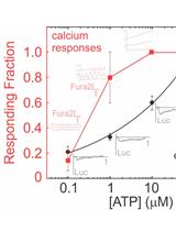
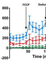
Simultaneous Fluorescent Recordings of Extracellular ATP and Intracellular Calcium in Mammalian Cells

哺乳动物细胞胞外ATP和胞内钙离子的协同荧光记录

NM Nicholas Mikolajewicz
SK Svetlana V. Komarova
5382 Views
May 20, 2019
Extracellular ATP is a potent signaling molecule that stimulates intracellular calcium responses through purinergic (P2) receptors in mammalian cells. While extracellular ATP and intracellular calcium can be measured separately, simultaneous monitoring can offer additional insights into P2 receptor physiology. This protocol takes advantage of the overlapping fluorescence spectra between the ATP-detection substrate luciferin and calcium indicator dye Fura2. Mammalian cells are loaded with Fura2-AM and live-cell recordings are acquired in the presence of a luciferin-luciferase imaging solution. This protocol allows to study stimulus-induced ATP release and directly relate changes in extracellular ATP concentration to observed calcium responses.

免疫学

TZA, 一种基于敏感报告因子细胞学检测方法, 可用于准确快速量化静息CD4+ T细胞中可诱导的且复制能力强的潜伏HV-1

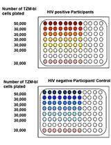
TZA, a Sensitive Reporter Cell-based Assay to Accurately and Rapidly Quantify Inducible, Replication-competent Latent HIV-1 from Resting CD4+ T Cells

TZA, 一种基于敏感报告因子细胞学检测方法, 可用于准确快速量化静息CD4+ T细胞中可诱导的且复制能力强的潜伏HV-1

AS Anwesha Sanyal
VR Vatsala S. Rangachar
PG Phalguni Gupta
7016 Views
May 20, 2019
The latent HIV-1 viral reservoir in resting CD4+ (rCD4+) T cells represents a major barrier to an HIV-1 cure. There is an ongoing effort to identify therapeutic approaches that will eliminate or reduce the size of this reservoir. However, clinical investigators lack an assay to determine whether or not a decrease in the latent reservoir has been achieved. Therefore, it is critical to develop assays that can reproducibly quantify the reservoir size and changes therein, in participant’s blood during a therapeutic trial. Quantification of the latent HIV viral reservoir requires a highly sensitive, cost-effective assay capable of measuring the low frequency of rCD4+ T cells carrying functional provirus. Preferably, such an assay should be such that it can be adopted for high throughput and could be adopted under conditions for use in large-scale clinical trials. While PCR-based assays are commonly used to quantify pro-viral DNA or intracellular RNA transcript, they cannot distinguish between replication-competent and defective proviruses. We have recently published a study where a reporter cell-based assay (termed TZA or TZM-bl based quantitative assay) was used to quantify inducible replication-competent latent HIV-1 in blood. This assay is more sensitive, cost-efficient, and faster than available technology, including the quantitative viral outgrowth assay or the Q-VOA. Using this assay, we show that the size of the inducible latent HIV-1 reservoir in virally suppressed participants on ART is approximately 70-fold larger than previous estimates. We describe here in detail an optimized method to quantitate latently infected cells using the TZA.
果蝇肠道摄入外来细菌后次氯酸R19-S染色

Hypochlorous Acid Staining with R19-S in the Drosophila Intestine upon Ingestion of Opportunistic Bacteria

果蝇肠道摄入外来细菌后次氯酸R19-S染色

SH Salma Hachfi
OB Olivia Benguettat
AG Armel Gallet
5747 Views
May 20, 2019
The intestine is endowed with an innate immune system that is required to fight any exogenous bacteria that are swallowed along with the food. The first line of defense that is mounted by the gut epithelium is the release of immune Reactive Oxygen Species (ROS), such as hypochlorous acid (HOCl), into the lumen. HOCl is produced within 1.5 h of bacterial ingestion and is very labile once released. Therefore, to monitor HOCl production upon ingestion of allochthonous bacteria, one needs a detection system that can quickly and efficiently detect HOCl production in the intestine. While most of the ROS-sensitive probes available in the market detect all kinds of ROS without any distinction, the R19-S fluorescent probe has been developed to specifically detect HOCl. Here, we describe a protocol to monitor HOCl production using this probe in the gut lumen of adult Drosophila upon ingestion of the opportunistic bacteria Bacillus thuringiensis.

微生物学

丁香假单胞菌的生物膜形成实验

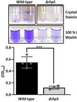
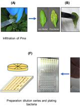
Biofilm Formation Assay in Pseudomonas syringae

丁香假单胞菌的生物膜形成实验

XS Xiaolong Shao
YX Yingpeng Xie
YZ Yingchao Zhang
Xin   Deng Xin Deng
12491 Views
May 20, 2019
Pseudomonas syringae is a model plant pathogen that infects more than 50 plant species worldwide, thus leading to significant yield loss. Pseudomonas biofilm always adheres to the surfaces of medical devices or host cells, thereby contributing to infection. Biofilm formation can be visualized on numerous matrixes, including coverslips, silicone tubes, polypropylene and polystyrene. Confocal laser scanning microscopy can be used to visualize and analyze biofilm structure. In this study, we modified and applied the current method of P. aeruginosa biofilm measurement to P. syringae, and developed a convenient protocol to visualize P. syringae biofilm formation using a borosilicate glass tube as the matrix coupled with crystal violet staining.
哺乳动物和昆虫细胞中的辛德比斯病毒的α病毒颗粒的纯化和蛋白质组分析

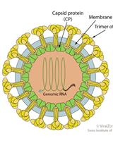
Purification and Proteomic Analysis of Alphavirus Particles from Sindbis Virus Grown in Mammalian and Insect Cells

哺乳动物和昆虫细胞中的辛德比斯病毒的α病毒颗粒的纯化和蛋白质组分析

Raquel  Hernandez Raquel Hernandez
TG Trevor Glaros
GR Gabrielle Rizzo
DF Davis F. Ferreira
6331 Views
May 20, 2019
Current mass spectrometry (MS) methods and new instrumentation now allow for more accurate identification of proteins in low abundance than previous protein fractionation and identification methods. It was of interest if this method could serve to define the virus proteome of a membrane-containing virus. To evaluate the efficacy of mass spec to determine the proteome of medically important viruses, Sindbis virus (SINV), the prototypical alphavirus was chosen for evaluation. This model system was chosen specifically because the alphaviruses contain members which are human pathogens, this virus is well defined biochemically and structurally, and grows to high titers in both vertebrate and non-vertebrate host cells. The SINV proteome was investigated using this method to determine if host proteins are specifically packaged into infectious virions. It was also of interest if the SINV proteome, when grown in multiple host cells representing vertebrate and mosquito hosts, incorporated specific host proteins from all hosts. Observation of recurrent or distinctive proteins in the virus proteome aided in the determination of proteins incorporated into the virion as opposed to those bound to the particle exterior. Mass spectrometry analysis identified the total protein content of purified virions within limits of detection. The most significant finding was that in addition to the host proteins, SINV non-structural protein 2 (nsP2) was detected within virions grown in all host cells examined. This analysis identified host factors not previously associated with alphavirus entry, replication, or egress, identifying at least one host factor integrally involved in alphavirus replication. Key to the success of this analysis is the method of virus purification which must deliver measurably infectious virus free of high levels of contaminants. For SINV and other members of the alphavirus family, this is accomplished by isopycnic centrifugation through potassium tartrate, followed by a high salt wash.
用于检测复制适应性的肠道病毒竞争实验

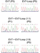
Enterovirus Competition Assay to Assess Replication Fitness

用于检测复制适应性的肠道病毒竞争实验

Valeria  Lulla Valeria Lulla
AF Andrew E. Firth
5446 Views
May 20, 2019
In virology the difference between the fitness of two viruses can be determined by using various methods, such as virus titer, growth curve analysis, measurement of virus infectivity, analysis of produced RNA copies and viral protein production. However, for closely performing viruses, it is often very hard to distinguish the differences. In vitro competition assays are a sensitive tool for determining viral replication fitness for many viruses replicating in cell culture. Relative viral replication fitness is usually measured from multiple cycle growth competition assays. Competition assays provide a sensitive measurement of viral fitness since the viruses are competing for cellular targets under identical growth conditions. This protocol describes a competition assay for enteroviruses and contains two alternative formats for initial infections, which can be varied depending on specific goals for each particular experiment. The protocol involves infection of cells with competing viruses, passaging, RNA extraction from infected cells, RT-PCR and Sanger sequencing followed by comparative analysis of resulting chromatograms obtained under various initial infection conditions. The techniques are applicable to members of many virus families, such as alphaviruses, flaviviruses, pestiviruses, and other RNA viruses with an established reverse genetics system.
用于恶臭假单胞菌生物膜扩散模式研究的定制设备的组装

Assembly of a Custom-made Device to Study Spreading Patterns of Pseudomonas putida Biofilms

用于恶臭假单胞菌生物膜扩散模式研究的定制设备的组装

DE David R. Espeso
EM Esteban Martínez-García
Víctor de Lorenzo Víctor de Lorenzo
4238 Views
May 20, 2019
Biofilms are bacterial communities in the shape of exopolysaccharide matrix-encased aggregates attached onto interphases able to resist environmental aggressions. The development of bacteria in the shape of biofilms deeply affects the performance of many industrial processes which work with fluidic systems, where bacteria may settle and prosper. As a consequence industrial equipment experiments low performance issues and substantial maintenance costs. The study of how bacteria of industrial interest such as Pseudomonas putida spread in these fluidic systems is highly dependent on the chosen experimental system to retrieve such data, thus using scaled prototypes becomes an essential step towards the design of a more efficient system to handle biofilms, either to control them or to prevent them. This protocol describes how to assemble, operate and maintain a device to grow and monitor the biofilm spreading pattern of this bacterium (as a function of the fluid hydrodynamics) in a custom-made chamber larger than those typically used in laboratory environments, and how to analyze the information gathered from it in a straightforward fashion. Description of the protocol was thought to be used as a working template not only for the presented case study but for any other potential experiment in different contexts and diverse scales following similar design principles.

神经科学

体外定量分析神经元周围基质网的结构完整性方法

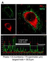
Protocol to Quantitatively Assess the Structural Integrity of Perineuronal Nets ex vivo

体外定量分析神经元周围基质网的结构完整性方法

BT Bhanu P. Tewari
HS Harald Sontheimer
5922 Views
May 20, 2019
Perineuronal nets (PNNs) are extracellular matrix assemblies of highly negatively charged proteoglycans that wrap around fast-spiking parvalbumin (PV) expressing interneurons in the cerebral cortex. PNNs play important roles in neuronal plasticity and modulate biophysical properties of the enclosed interneurons. Various central nervous system diseases including schizophrenia, Alzheimer disease and epilepsy present with qualitative alteration in PNNs, however prior studies failed to quantitatively assess such changes at single PNN level and correlate them with functional changes in disease. We describe a method to quantify the structural integrity of PNNs using high magnification image analysis of Wisteria Floribunda Agglutinin (WFA)-labeled PNNs in combination with cell-type-specific marker such as PV and NeuN. A polyline intensity profile of WFA along the entire perimeter of cell shows alternate segments with and without WFA labeling, indicating the intact chondroitin sulfate proteoglycan (CSPG) and holes of PNN respectively. This line intensity profile defines CSPG peaks, where intact PNN is present, and CSPG valleys (holes) where the PNN is missing. The average number of peaks reflect the integrity of the lattice assembly of PNN. The average size of PNN holes can be readily computed using image analysis software. Furthermore, degradation of PNNs using a bacterial-derived enzyme, Chondroitinase ABC (ChABC), allows to experimentally manipulate PNNs in situ brain slices during which biophysical properties can be assessed by patch-clamp recordings. We describe optimized experimental parameters to degrade PNNs in brain slices before as well as during recordings to study the possible change in function in real time. Our protocols provide effective and appropriate methods to modulate and quantify the PNN’s experimental manipulations.

植物科学

一种的可用于检测拟南芥中细菌性病原体和害虫诱导的系统抗性的改进生物学方法

An Improved Bioassay to Study Arabidopsis Induced Systemic Resistance (ISR) Against Bacterial Pathogens and Insect Pests

一种的可用于检测拟南芥中细菌性病原体和害虫诱导的系统抗性的改进生物学方法

NC Nicolás M. Cecchini
Yi   Song Yi Song
SR Suruchi Roychoudhry
JG Jean T. Greenberg
CH Cara H. Haney
7024 Views
May 20, 2019
The plant immune system is essential for plants to perceive and defend against bacterial, fungal and insect pests and pathogens. Induced systemic resistance (ISR) is a systemic immune response that occurs upon root colonization by beneficial microbes. A well-studied model for ISR is the association of specific beneficial strains of Pseudomonas spp. with the reference plant Arabidopsis thaliana. Here, we describe a robust, increased throughput, bioassay to study ISR against the bacterial pathogen Pseudomonas cannabina pv. alisalensis (formerly called Pseudomonas syringae pv. maculicola) strain ES4326 and the herbivore Trichoplusia ni by inoculating Pseudomonas simiae strain WCS417 (formerly called Pseudomonas fluorescens WCS417) on Arabidopsis plants grown in Jiffy-7® peat pellets. While most commonly used for Pseudomonas-triggered ISR on Arabidopsis, this assay is effective for diverse rhizosphere bacterial strains, plant species, pathogens and herbivores.
一种可调整的用于分析研究非无菌根际土壤化学特性的方法

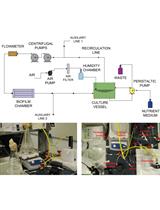
An Adjustable Protocol to Analyze Chemical Profiles of Non-sterile Rhizosphere Soil

一种可调整的用于分析研究非无菌根际土壤化学特性的方法

AW Alex Williams
Jurriaan  Ton Jurriaan Ton
Pierre   Pétriacq Pierre Pétriacq
5795 Views
May 20, 2019
The analysis of chemical diversity in non-sterile rhizosphere soil has been a pressing methodological challenge for years. Rhizosphere-enriched chemicals (i.e., rhizochemicals) include root exudation chemicals, (microbial) breakdown products thereof, and de novo produced metabolites by rhizosphere-inhabiting microbes, all of which can play an important role in plant-soil interactions. The power and resolution of analytical methods and statistical analysis pipelines allow for better acquisition, separation and identification of rhizochemicals, thus providing unprecedented insight into the biochemistry underpinning plant-soil interactions. The current protocol describes a recently developed method to characterize rhizochemical profiles from plants, including crops, and is modular and customizable, allowing for application across a range of different plant-soil combinations. The protocol provides in-depth details about the experimental system for sample collection, data acquisition by liquid chromatography coupled to mass spectrometry, and analytical pipeline, which statistically selects for rhizochemicals by statistical comparison between metabolite profiles from plant-containing soil and plant-free soil. Moreover, the optional addition of chemical standards permits a semi-targeted approach, which improves the annotation of chemical signatures and identification of single rhizochemicals.