往期刊物2019

卷册: 9, 期号: 8

left

Dec

20

Dec

5

Nov

20

Nov

5

Oct

20

Oct

5

Sep

20

Sep

5

Aug

20

Aug

5

Jul

20

Jul

5

Jun

20

Jun

5

May

20

May

5

Apr

20

Apr

5

Mar

20

Mar

5

Feb

20

Feb

5

Jan

20

Jan

5

right

生物化学

非放射性无细胞分析法测定分子伴侣对于AMP激活激酶活性的影响

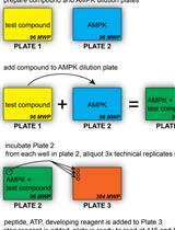
Non-radiometric Cell-free Assay to Measure the Effect of Molecular Chaperones on AMP-activated Kinase Activity

非放射性无细胞分析法测定分子伴侣对于AMP激活激酶活性的影响

EW Elizabeth B. Wilson
CR Carrie E. Rubel
Jonathan C. Schisler Jonathan C. Schisler
4896 Views
Apr 20, 2019
AMP-activated kinase (AMPK) is a trimeric protein holoenzyme with kinase activity. AMPK plays an important role in cellular metabolism and is thought to function as a fuel sensor within the cell, exerting kinase activity to activate energy-conserving pathways and simultaneously inhibit energy-consuming pathways. Traditional in vitro methods to measure AMPK activity to test potential agonists or antagonists utilize radiolabeled ATP with a peptide substrate. Although radiolabeling provides a high level of sensitivity, this approach is not ideal for medium to high-throughput screening, dose-response curves, or kinetic analyses. Our protocol utilizes Invitrogen’s Z’-LYTETM Kinase Assay Kit (Ser/Thr 23 Peptide) to measure changes in the enzymatic activity of AMPKɑ2β1γ1 in the presence of a molecular chaperone. The Z’-LYTETM platform is based on Fluorescence Resonance Energy Transfer (FRET). The AMPK peptide substrate (S/T 23 peptide: MRPRKRQGSVRRRV) is a self-contained FRET system, using coumarin as the donor and fluorescein as the acceptor. When the peptide is phosphorylated, it is sensitive to cleavage by a site-specific protease. The cleavage of the phospho-peptide eliminates the FRET pair, and the ratiometric analysis of FRET is used as an indirect measure of AMPK kinase activity. This method does not require the use of radiolabeling or antibodies and is used in a multi-well format, with high reproducibility and throughput capabilities.

癌症生物学

利用叠氮丙氨酸标记和流式细胞分析测定细胞周期中蛋白的合成

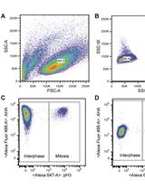
Measuring Protein Synthesis during Cell Cycle by Azidohomoalanine (AHA) Labeling and Flow Cytometric Analysis

利用叠氮丙氨酸标记和流式细胞分析测定细胞周期中蛋白的合成

KI Koshi Imami
TY Tomoharu Yasuda
8795 Views
Apr 20, 2019
Protein synthesis is one of the most fundamental biological processes to maintain cellular proteostasis. Azidohomoalaine (AHA) is a non-radioactive and “clickable” amino acid analog of methionine which can be incorporated into newly synthesized proteins. Thus, AHA-labeled nascent proteins can be detected and quantified through fluorescent labeling by "click" chemistry. Here we describe a protocol to measure protein synthesis by AHA labeling and flow cytometry. Taking advantage of gating different cell populations, we provide a typical example of the flow cytometric-based analysis of protein synthesis during the cell cycle. While we used mouse B cells in this protocol this method can be readily applied to any cell types and organisms.
利用小鼠Phospho-RTK Array试剂盒无偏筛选肿瘤提取物中激活受体酪氨酸激酶

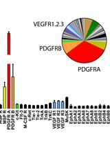
Unbiased Screening of Activated Receptor Tyrosine Kinases (RTKs) in Tumor Extracts Using a Mouse Phospho-RTK Array Kit

利用小鼠Phospho-RTK Array试剂盒无偏筛选肿瘤提取物中激活受体酪氨酸激酶

JN Julian Naipauer
LC Lucas E. Cavallin
EM Enrique A. Mesri
4980 Views
Apr 20, 2019
Kaposi’s sarcoma (KS) herpesvirus (KSHV) is a virus that causes KS, an angiogenic AIDS-associated spindle-cell neoplasm, by activating host oncogenic signaling cascades through autocrine and paracrine mechanisms. Many host signaling cascades co-opted by KSHV including PI3K/AKT/mTORC, NFkB and Notch are critical for cell-specific mechanisms of transformation and their identification is paving the way to therapeutic target discovery. Analysis of the molecular KS signature common to human KS tumors and our mouse KS-like tumors showed consistent expression of KS markers VEGF and PDGF receptors with upregulation of other angiogenesis ligands and their receptors in vivo. This points to the autocrine and paracrine activation of various receptor tyrosine kinase (RTK) signaling axes. Hereby we describe a protocol to screen for activated receptor tyrosine kinase of KSHV-induced KS-like mouse tumors using a Mouse Phospho-RTK Array Kit and its validation by RTK western blots. We showed that this method can be successfully used to rank the tyrosine kinase receptors most activated in tumors in an unbiased manner. This allowed us to identify PDGFRA as an oncogenic driver and therapeutic target in AIDS-KS.

细胞生物学

免疫荧光标记小鼠肾脏切片中的WT-1/足糖萼

Immunofluorescence Staining of WT-1/Podocalyxin on Mouse Kidney Sections

免疫荧光标记小鼠肾脏切片中的WT-1/足糖萼

Carole  Henique Carole Henique
OL Olivia Lenoir
PT Pierre-Louis Tharaux
6247 Views
Apr 20, 2019
In glomerular diseases, podocytes are one type of cells involved in dysfunction of glomerular filtration. In these conditions, podocyte proteins expression change. Therefore, immunofluorescent staining of podocytes can be performed to visualize podocyte localization of proteins of interest. In this protocol, we detail a method to stain podocytes with a specific marker WT-1 (Wilms Tumor-1) for “in situ” podocyte analysis by immunofluorescent microscopy, more informative technique than other techniques (as Western blot).

免疫学

环二鸟苷单磷酸佐剂蛋白亚单位疫苗引起的粘膜和全身疫苗反应的评估

Evaluation of Mucosal and Systemic Vaccine Responses by Cyclic di-GMP (CDG)-adjuvanted Protein Subunit Vaccines

环二鸟苷单磷酸佐剂蛋白亚单位疫苗引起的粘膜和全身疫苗反应的评估

SM Samira Mansouri
Lei  Jin Lei Jin
4664 Views
Apr 20, 2019
Intranasal administration of vaccine adjuvants directly deliver therapeutic agents to the lungs to induce potent lung mucosal immune responses. Cyclic di-GMP (CDG) is a promising mucosal vaccine adjuvant candidate capable of inducing protective immunity. This protocol describes an in vivo approach to induce and detect mucosal (lung) and systemic (blood and spleen) vaccine adjuvant responses of CDG. This protocol also includes the methods to detect both humoral and cellular immune responses of CDG adjuvant. Last, this protocol can be used to study other cyclic dinucleotides as mucosal vaccine adjuvants.

分子生物学

利用多种标记方法进行IR标记探针的Northern杂交

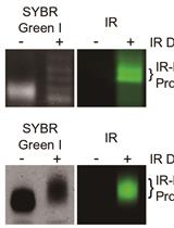
Northern Blot with IR Fluorescent Probes: Strategies for Probe Preparation

利用多种标记方法进行IR标记探针的Northern杂交

Christopher Fields Christopher Fields
Peike  Sheng Peike Sheng
BM Bret Miller
TW Tianqi Wei
MX Mingyi Xie
10516 Views
Apr 20, 2019
Northern blot is a molecular biology technique that can detect, quantify, and determine the molecular weight of RNA. Recently, we published a protocol utilizing near-infrared (IR) fluorescent probes in Northern blot (irNorthern). Our method is as sensitive as other non-radioactive methods but is more straightforward and versatile. Additionally, we found that IR-labeled probes can be used to multiplex or detect different species of RNA at the same time. Here we describe three methods for generating an IR-labeled probe as well as how to perform irNorthern blot. In conclusion, our irNorthern protocol offers a convenient method for RNA detection.

神经科学

急性海马切片中小胶质细胞运动长时间全视野荧光成像及分析

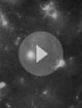
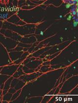
Time-lapse Whole-field Fluorescence Imaging of Microglia Processes Motility in Acute Mouse Hippocampal Slices and Analysis

急性海马切片中小胶质细胞运动长时间全视野荧光成像及分析

Bernadette   Basilico Bernadette Basilico
Barbara  Cortese Barbara Cortese
Patrizia  Ratano Patrizia Ratano
Silvia  Di Angelantonio Silvia Di Angelantonio
Davide   Ragozzino Davide Ragozzino
5860 Views
Apr 20, 2019
Microglia are the resident immune cells of the central nervous system (CNS). In the last year, the improvements in the transgenic mouse technologies and imaging techniques have shed light on microglia functions under physiological conditions. Microglia continuously scan the brain parenchyma with their highly motile processes, maintaining tissue homeostasis and participating in neuronal circuits refinement. Here, we describe a protocol that enables us to perform time-lapse imaging of microglial cells in acute hippocampal slices, making image acquisition possible on an electrophysiology rig equipped with a standard imaging system. Using this ex vivo approach, we investigated microglial processes scanning abilities under physiological condition in hippocampus.
大鼠物体位置再认记忆

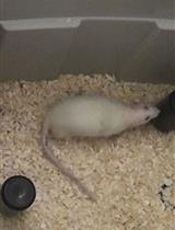
Displaced Object Recognition Memory in Rats

大鼠物体位置再认记忆

AM Ali N. Mclagan
Jena B. Hales Jena B. Hales
5551 Views
Apr 20, 2019
The Displaced Object Recognition (DOR) task, sometimes called the Novel Object Location task, assesses spatial recognition memory without navigational demands, explicit instruction, or the need for multiple days of training. This memory task has two phases. First, the subject is familiarized to an open arena with two objects and is allowed to explore the objects. Following a delay period, the subject returns to the arena, but one of the previous objects has been moved to a new location. Greater exploration of the displaced object is used as the index of memory for the previous object location. An advantage of the DOR task is that subjects can be tested without explicit training, since this task exploits the natural tendency to be more interested in something novel. The spontaneous aspect of this task allows for the testing of animals as well as human populations that are unable to follow verbal instructions, such as babies. Therefore, this powerful test of recognition memory can be administered similarly for many species, including rats and humans, allowing for better translatability.
嗅上皮外植体的制备及培养以用于通过生物膜力学探针测定单个轴突的机械张力

Preparation and Manipulation of Olfactory Epithelium Explant Cultures for Measurement of the Mechanical Tension of Individual Axons Using the Biomembrane Force Probe

嗅上皮外植体的制备及培养以用于通过生物膜力学探针测定单个轴突的机械张力

CF Coralie Fouquet
AT Alain Trembleau
4860 Views
Apr 20, 2019
In this paper, we describe a protocol allowing measurement of the mechanical tension of individual axons grown ex vivo from neural tissue explants. This protocol was developed with primary cultures of olfactory epithelium explants from embryonic (E13.5) mice. It includes a detailed description of explant dissection and culture, as well as the main steps of the procedure for axon tension measurement using the previously established Biomembrane Force Probe.

植物科学

根相互作用系统的示踪实验和质量控制

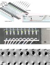
Tracking Root Interactions System (TRIS) Experiment and Quality Control

根相互作用系统的示踪实验和质量控制

HM Hassan Massalha
EK Elisa Korenblum
OS Orr H. Shapiro
AA Asaph Aharoni
6361 Views
Apr 20, 2019
Soil organisms are diverse taxonomically and functionally. This ecosystem experiences highly complex networks of interactions, but may also present functionally independent entities. Plant roots, a metabolically active hotspot in the soil, take an essential part in shaping the rhizosphere. Tracking the dynamics of root-microbe interactions at high spatial resolution is currently limited due to methodological intricacy. In this study, we developed a novel microfluidics-based device enabling direct imaging of root-bacteria interactions in real time.