往期刊物2019

卷册: 9, 期号: 7

left

Dec

20

Dec

5

Nov

20

Nov

5

Oct

20

Oct

5

Sep

20

Sep

5

Aug

20

Aug

5

Jul

20

Jul

5

Jun

20

Jun

5

May

20

May

5

Apr

20

Apr

5

Mar

20

Mar

5

Feb

20

Feb

5

Jan

20

Jan

5

right

微生物学

利用接合生殖将产色细菌荧光蛋白标签导入变形杆菌

Delivering "Chromatic Bacteria" Fluorescent Protein Tags to Proteobacteria Using Conjugation

利用接合生殖将产色细菌荧光蛋白标签导入变形杆菌

Rudolf O Schlechter Rudolf O Schlechter
MR Mitja NP Remus-Emsermann
7464 Views
Apr 5, 2019
Recently, we published a large and versatile set of plasmids, the chromatic bacteria toolbox, to deliver eight different fluorescent protein genes and four combinations of antibiotic resistance genes to Gram-negative bacteria. Fluorescent tags are important tools for single-cell microbiology, synthetic community studies, biofilm, and host-microbe interaction studies. Using conjugation helper strain E. coli S17-1 as a donor, we show how plasmid conjugation can be used to deliver broad host range plasmids, Tn5 transposons delivery plasmids, and Tn7 transposon delivery plasmids into species belonging to the Proteobacteria. To that end, donor and recipient bacteria are grown under standard growth conditions before they are mixed and incubated under non-selective conditions. Then, transconjugants or exconjugant recipients are selected on selective media. Mutant colonies are screened using a combination of tools to ensure that the desired plasmids or transposons are present and that the colonies are not containing any surviving donors. Through conjugation, a wide range of Gram-negative bacteria can be modified without prior, often time-consuming, establishment of competent cell and electroporation procedures that need to be adjusted for every individual strain. The here presented protocol is not exclusive for the delivery of Chromatic bacteria plasmids and transposons, but can also be used to deliver other mobilizable plasmids to bacterial recipients.
qPCR定量分析感染细胞培养模型中乙型肝炎病毒共价闭合环状DNA

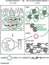
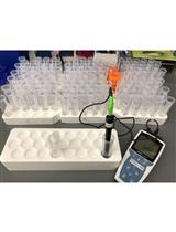
Quantification of Hepatitis B Virus Covalently Closed Circular DNA in Infected Cell Culture Models by Quantitative PCR

qPCR定量分析感染细胞培养模型中乙型肝炎病毒共价闭合环状DNA

Bingqian Qu Bingqian Qu
SU Stephan Urban
7065 Views
Apr 5, 2019
Persistence of the human hepatitis B virus (HBV) requires the maintenance of covalently closed circular (ccc)DNA, the episomal genome reservoir in nuclei of infected hepatocytes. cccDNA elimination is a major aim in future curative therapies currently under development. In cell culture based in vitro studies, both hybridization- and amplification-based assays are currently used for cccDNA quantification. Southern blot, the current gold standard, is time-consuming and not practical for a large number of samples. PCR-based methods show limited specificity when excessive HBV replicative intermediates are present. We have recently developed a real-time quantitative PCR protocol, in which total cellular DNA plus all forms of viral DNA are extracted by silica column. Subsequent incubation with T5 exonuclease efficiently removes cellular DNA and all non-cccDNA forms of viral DNA while cccDNA remains intact and can reliably be quantified by PCR. This method has been used for measuring kinetics of cccDNA accumulation in several in vitro infection models and the effect of antivirals on cccDNA. It allowed detection of cccDNA in non-human cells (primary macaque and swine hepatocytes, etc.) reconstituted with the HBV receptor, human sodium taurocholate cotransporting polypeptide (NTCP). Here we present a detailed protocol of this method, including a work flowchart, schematic diagram and illustrations on how to calculate “cccDNA copies per (infected) cell”.
在单细胞红藻 Cyanidioschyzon merolae中利用URA5.3选择标记对染色体基因进行多重修饰

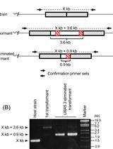
Multiple Modification of Chromosomal Loci Using URA5.3 Selection Marker in the Unicellular Red Alga Cyanidioschyzon merolae

在单细胞红藻 Cyanidioschyzon merolae中利用URA5.3选择标记对染色体基因进行多重修饰

TT Tokiaki Takemura
SI Sousuke Imamura
Yuki Kobayashi Yuki Kobayashi
Kan Tanaka Kan Tanaka
5183 Views
Apr 5, 2019
The unicellular red alga Cyanidioschyzon merolae has been used as a eukaryotic photosynthetic model for various basic and applied studies. Although the nuclear genome of C. merolae can be modified by homologous recombination with exogenously introduced DNA, it has been difficult to modify multiple chromosome loci within the same strain because of the limited number of available positive selection markers. Recently, we reported a modified URA5.3 gene cassette (URA5.3T), which can be used repeatedly for nuclear genome transformation using the pMKT plasmid vectors for epitope tagging (3x FLAG- or 3x Myc-) of nuclear-encoded proteins. In addition, these plasmid vectors can also be used to knock out multiple genes one by one. This report describes the construction of DNA fragments for transformation and the detailed transformation procedure.

分子生物学

利用差异化亲和固定鉴定核糖核酸酶敏感的LINE-1核糖核蛋白间的相互作用

Identification of RNase-sensitive LINE-1 Ribonucleoprotein Interactions by Differential Affinity Immobilization

利用差异化亲和固定鉴定核糖核酸酶敏感的LINE-1核糖核蛋白间的相互作用

HJ Hua Jiang
MT Martin S. Taylor
KM Kelly R. Molloy
IA Ilya Altukhov
John LaCava John LaCava
6760 Views
Apr 5, 2019
Long Interspersed Nuclear Element-1 (LINE-1, L1) constitutes a family of autonomous, self-replicating genetic elements known as retrotransposons. Although most are inactive, copious L1 sequences populate the human genome. L1s proliferate in a ‘copy-and-paste’ fashion through an RNA intermediate; a full-length L1 transcript is ~6,000 nucleotides long and functions as a bicistronic mRNA that encodes and assembles in cis with two main polypeptides, ORF1p and ORF2p, forming a ribonucleoprotein (RNP); L1 RNPs also interact with a wide range of host factors in positive and negative regulatory capacities. The following protocol describes an approach to affinity enrich ectopically expressed L1 RNPs and, using RNases, release the fraction of protein that depends upon the presence of intact RNA for retention in the immobilized macromolecules.

神经科学

大鼠完成连续阴性对照实验

Consummatory Successive Negative Contrast in Rats

大鼠完成连续阴性对照实验

AJ Ana María Jiménez-García
LR Leandro Ruiz-Leyva
AV Ana Vázquez-Ágredos
CT Carmen Torres
MP Mauricio R. Papini
CC Cruz Miguel Cendán
IM Ignacio Morón
4294 Views
Apr 5, 2019
Using animal models in addiction and pain research is pivotal to unravel new pathways and mechanisms for the treatment of these disorders. Reward devaluation through a consummatory successive negative contrast (cSNC) task has shown the ability to reduce physical pain sensitivity (hypoalgesia) and increase oral ethanol consumption in rats. The procedure is based on exposing the experimental animals to a 32% sucrose solution during several sessions (preshift sessions) followed by a devaluation to 4% sucrose during the next few sessions (postshift sessions). The cSNC effect can be monitored by comparing the experimental group to an unshifted control that had access to 4% sucrose throughout the entire experiment (preshift and postshift sessions). The cSNC phenomenon is defined by lower consumption of sucrose in the downshifted group than in the unshifted group during postshfit sessions.

植物科学

拟南芥叶片扁平细胞中胞间连丝渗透性定量测定实验

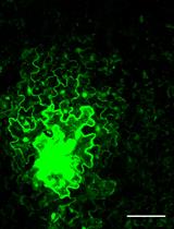
Quantitative Plasmodesmata Permeability Assay for Pavement Cells of Arabidopsis Leaves

拟南芥叶片扁平细胞中胞间连丝渗透性定量测定实验

MD Min Diao
QW Qiannan Wang
Shanjin Huang Shanjin Huang
6610 Views
Apr 5, 2019
Plasmodesmata (PD) are intercellular channels between walled plant cells that enable the transportation of materials between adjacent cells, which are important for plant growth and development. The permeability of PD must be tightly regulated. Assays to determine the permeability of PD are crucial for related studies on the regulation of PD development and permeability. Here we describe an assay for the determination of PD permeability via the observation and quantification of GFP diffusion and cell-to-cell transport of CMV MP-GFP in Arabidopsis leaves.
利用电子显微镜免疫金标记法进行植物细胞壁表位的可视化观察

Visualization of Plant Cell Wall Epitopes Using Immunogold Labeling for Electron Microscopy

利用电子显微镜免疫金标记法进行植物细胞壁表位的可视化观察

MM Mateusz Majda
6251 Views
Apr 5, 2019
Plant cell walls consist of different polysaccharides and structural proteins, which form a rigid layer located outside of the plasma membrane. The wall is also a very dynamic cell composite, which is characterized by complex polysaccharide interactions and various modifications during cell development. The visualization of cell wall components in situ is very challenging due to the small size of cell wall composites (nanometer scale), large diversity of the wall polysaccharides and their complex interactions. This protocol describes immunogold labeling of different cell wall epitopes for high-resolution transmission electron microscopy (TEM). It provides a detailed procedure for collection and preparation of plant material, ultra-thin sectioning, specimen labeling and contrasting. An immunolabeling procedure workflow was optimized to obtain high efficiency of carbohydrates labeling for high-resolution TEM. This method was applied to study plant cell wall characteristics in various plant tissues but could also be applied for other cell components in plant and animal tissues.
水稻茎秆酶的提取与纯化

Extraction and Purification of Laccases from Rice Stems

水稻茎秆酶的提取与纯化

CS Chenna Swetha
PS P. V. Shivaprasad
5721 Views
Apr 5, 2019
Laccases are found in cell walls of plants in very low amounts. This protocol provides an efficient method to purify laccases from rice stems. The method involves three steps: 1) Isolation of total protein from rice stems using buffers with high salt concentration to extract protein from cell walls; 2) Purification of laccases using concanavalin-A beads; and, 3) In-gel staining of laccases with 4-hydroxyindole. Concanavalin-A specifically binds to internal or non-reducing terminal α-D-mannosyl and α-D-glucosyl groups found in glycoproteins and glycolipids. Laccases being glycoproteins binds to concanavalin-A during purification process and eluted with mannose.
离子渗漏实验定量分析湿度对超敏反应的影响

Quantification of the Humidity Effect on HR by Ion Leakage Assay

离子渗漏实验定量分析湿度对超敏反应的影响

MM Musoki Mwimba
XD Xinnian Dong
5345 Views
Apr 5, 2019
We describe a protocol to measure the contribution of humidity on cell death during the effector-triggered immunity (ETI), the plant immune response triggered by the recognition of pathogen effectors by plant resistance genes. This protocol quantifies tissue cell death by measuring ion leakage due to loss of membrane integrity during the hypersensitive response (HR), the ETI-associated cell death. The method is simple and short enough to handle many biological replicates, which improves the power of test of statistical significance. The protocol is easily applicable to other environmental cues, such as light and temperature, or treatment with chemicals.