

往期刊物2018
卷册: 8, 期号: 23

生物化学
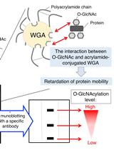
Wheat Germ Agglutinin (WGA)-SDS-PAGE: A Novel Method for the Detection of O-GlcNAc-modified Proteins by Lectin Affinity Gel Electrophoresis
小麦种子凝集素(WGA)-SDS-PAGE:一种利用凝集素亲和凝胶电泳对O-乙酰氨基葡萄糖修饰蛋白进行检测的新方法
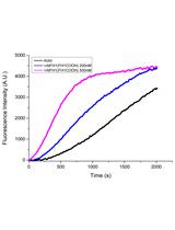
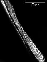
Purification of Globular Actin from Rabbit Muscle and Pyrene Fluorescent Assays to Investigate Actin Dynamics in vitro
从兔肌肉组织中纯化球状肌动蛋白并利用芘荧光分析实验体外检测肌动蛋白的动态变化
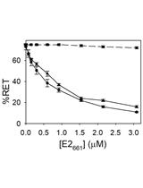
In vitro Membrane Interaction and Liposome Fusion Assays Using Recombinant Hepatitis C Virus Envelope Protein E2
利用重组丙型肝炎病毒包膜蛋白E2体外检测膜互作和脂质体融合
癌症生物学
Cluster Analysis of Endogenous HER2 and HER3 Receptors in SKBR3 Cells
在SKBR3细胞中内源性HER2和HER3受体的聚类分析

细胞生物学
Blinded Visual Scoring of Images Using the Freely-available Software Blinder
使用免费软件Blinder对图像进行盲视觉评分
发育生物学
Notochord Injury Assays that Stimulate Transcriptional Responses in Zebrafish Larvae
诱导幼年期斑马鱼转录响应的脊索损伤实验

免疫学
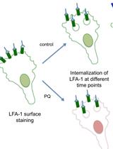
Flow Cytometry Assay for Recycling of LFA-1 in T-lymphocytes
流式细胞术检测T淋巴细胞中LFA-1的再循环
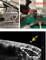
Ultrasound Guided Intra-thymic Injection to Track Recent Thymic Emigrants and Investigate T Cell Development
超声引导胸腺内注射并追踪新近胸腺迁出细胞和T细胞发育观测
Integrin Dependent RhoB Activation Assay Using Leukocytes
白细胞内整合素依赖的RhoB激活实验
微生物学
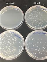
Quantitative Transformation Efficiency Assay for Bacillus subtilis
枯草芽孢杆菌转化效率的定量测定
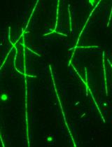
An in vitro Microscopy-based Assay for Microtubule-binding and Microtubule-crosslinking by Budding Yeast Microtubule-associated Protein
利用芽殖酵母微管相关蛋白研究微管结合和微管交联的体外显微镜检测法

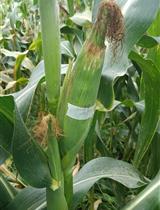
Field Inoculation and Classification of Maize Ear Rot Caused by Fusarium verticillioides
田间接种和轮枝镰孢菌引起的玉米穗粒腐病的分类


植物科学
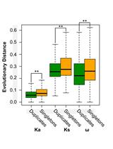
Genome-wide Estimation of Evolutionary Distance and Phylogenetic Analysis of Homologous Genes
进化距离的全基因组评估和同源基因的系统进化分析

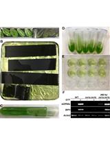
Using Arabidopsis Mesophyll Protoplasts to Study Unfolded Protein Response Signaling
利用拟南芥叶肉细胞原生质体研究未折叠蛋白响应信号
