往期刊物2018

卷册: 8, 期号: 22

left

Dec

20

Dec

5

Nov

20

Nov

5

Oct

20

Oct

5

Sep

20

Sep

5

Aug

20

Aug

5

Jul

20

Jul

5

Jun

20

Jun

5

May

20

May

5

Apr

20

Apr

5

Mar

20

Mar

5

Feb

20

Feb

5

Jan

20

Jan

5

right

生物化学

低丰度泛素化形式蛋白的分离和可视化

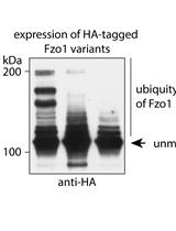
Separation and Visualization of Low Abundant Ubiquitylated Forms

低丰度泛素化形式蛋白的分离和可视化

RS Ramona Schuster
TS Tânia Simões
Fd Fabian den Brave
ME Mafalda Escobar-Henriques
4898 Views
Nov 20, 2018
In this protocol we describe the separation and visualization of ubiquitylated forms of the yeast mitofusin Fzo1 by Western blot. To this aim, we express HA-tagged Fzo1 in Saccharomyces cerevisiae, break the cells to extract a membrane-enriched fraction, solubilize the membranes using detergent and then specifically immunoprecipitate the tagged protein using anti-HA affinity beads. Subsequently, we separate the higher molecular weight (ubiquitylated) forms of Fzo1 via SDS-PAGE. Finally, immunoblotting and immunodecoration are used to detect the protein and its ubiquitylated forms using an HA-specific antibody. By using this protocol, it is possible to separate and visualize higher molecular weight forms of low abundant proteins such as Fzo1 and detect sharp and distinct bands above the unmodified protein by Western blot.

癌症生物学

利用3D细胞培养鉴定耐药相关基因的一种功能性稳健表型筛选

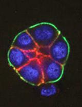
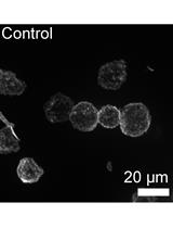
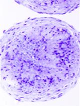
A Functionally Robust Phenotypic Screen that Identifies Drug Resistance-associated Genes Using 3D Cell Culture

利用3D细胞培养鉴定耐药相关基因的一种功能性稳健表型筛选

Sun-Young Lee Sun-Young Lee
Mina J. Bissell Mina J. Bissell
6227 Views
Nov 20, 2018
Drug resistance is a major obstacle in cancer treatment: A case in point is the failure of cancer patients to respond to tyrosine kinase inhibitors (TKI) of EGFR, a receptor that is highly expressed in many cancers. Identification of the targets and delineation of mechanisms of drug resistance remain major challenges. Traditional pharmacological assays of drug resistance measure the response of tumor cells using cell proliferation or cell death as readouts. These assays performed using traditional plastic tissue culture plates (2D) do not translate to in vivo realities. Here, we describe a genetic screen based on phenotypic changes that can be completed over a period of 1-1½ months using functional endpoints in physiologically relevant 3D culture models. This phenotype-based assay could lead to the discovery of previously unknown therapeutic targets and could explain the source of the resistance and relapse. As a proof of principle, we performed our 3D culture assay with a small cDNA library in that yielded five unknown intermediates in EGFR and PI3K signaling pathways. Here, we describe the screening method and the characterization of one of the five molecules, but this approach could be easily expanded for a high-throughput screening to identify or evaluate many more unknown intermediates in oncogenic signaling pathways.

细胞生物学

体内gp100特异的细胞毒CD8+ T 细胞杀伤实验

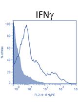
In-vivo gp100-specific Cytotoxic CD8+ T Cell Killing Assay

体内gp100特异的细胞毒CD8+ T 细胞杀伤实验

Mahdia Benkhoucha Mahdia Benkhoucha
NM Nicolas Molnarfi
EB Elodie Belnoue
MD Madiha Derouazi
PL Patrice H Lalive
8623 Views
Nov 20, 2018
Cytotoxic CD8+ T lymphocytes (CTLs) represent a crucial component of the adaptive immune system and play a prominent role in the anti-tumor immune responses of both mice and humans. Cytotoxic CD8+ T cells are responsible for the lysis of cells expressing peptides associated with MHC class I molecules and derived from infection with a pathogen or from mutated antigens. In order to quantify in vivo this antigen-specific CD8+ T cell killing activity, we use the in vivo killing assay (IVKA). Here, we describe the protocol for the lysis of cells expressing a CD8+ T cell melanoma epitope of the hgp10025-33 protein (KVPRNQDWL). C57BL/6 recipient mice, receive first target cells, prepared from naive congenic (CD45.1) C57BL/6 spleen cells pulsed with the hgp10025-33 peptide and labeled with CFSE and of non-pulsed control cells labeled with Brilliant violet. One day later, the spleen cells of recipient mice are isolated and analyzed by FACS to measure the amount of CFSE cells and Brillant Violet (BV) cells. The percentage of lysis is calculated by the difference between CFSE versus BV.Measuring the ability of antigen-specific CD8+ T cells to lyse their antigen in vivo is very important to evaluate the adaptive cytotoxic response induced against a pathogen or a tumor antigen.
利用流式细胞术相对定量分析悬浮细胞中的聚合肌动蛋白

Relative Quantitation of Polymerized Actin in Suspension Cells by Flow Cytometry

利用流式细胞术相对定量分析悬浮细胞中的聚合肌动蛋白

MK Mallory R. Kakley
KV Katrina B. Velle
LF Lillian K. Fritz-Laylin
5674 Views
Nov 20, 2018
The amount of polymerized actin within a cell can vary widely due to natural processes and/or experimentally induced perturbations. We routinely use this protocol to measure relative polymerized actin content between cell populations by staining cells in suspension with fluorescent phalloidin, then measuring total cell fluorescence using flow cytometry. Although developed for human cells, we have easily adapted this method for use with diverse eukaryotic cell types.

发育生物学

利用APEX标记的条件性稳定纳米体电子显微检测哺乳动物细胞中的蛋白和蛋白复合物

Electron Microscopic Detection of Proteins and Protein Complexes in Mammalian Cells Using APEX-tagged, Conditionally Stable Nanobodies

利用APEX标记的条件性稳定纳米体电子显微检测哺乳动物细胞中的蛋白和蛋白复合物

TH Thomas E. Hall
JR James Rae
NA Nicholas Ariotti
RP Robert G. Parton
5498 Views
Nov 20, 2018
The recent development of 3D electron microscopic techniques for cells and tissues has necessitated the development of new methods for the detection of proteins and protein-complexes in situ. The development of new genetic tags, such as the ascorbate peroxidase, APEX, for electron microscopic detection of tagged proteins has expanded the available toolbox and ushered in a new era in biological electron microscopy. Here, we describe methods for combining conditionally-stable nanobodies to fluorescent protein tags with APEX-based detection. These methods are compatible with detection of low levels of expression of fluorescently-tagged proteins and with detection of protein complexes using split GFP-based complementation methods. We describe a simple protocol for applying these methods to the electron microscopic detection of proteins and protein complexes in cultured cells.
斑马鱼左右组织者中单细胞的嵌合标记和3D形态分析

Mosaic Labeling and 3-Dimensional Morphological Analysis of Single Cells in the Zebrafish Left-right Organizer

斑马鱼左右组织者中单细胞的嵌合标记和3D形态分析

AD Agnik Dasgupta
AJ Andrew E. Jacob
JA Jeffrey D. Amack
5445 Views
Nov 20, 2018
A transient epithelial structure called the left-right organizer (LRO) establishes left-right asymmetry in vertebrate embryos. Developmental defects that alter LRO formation result in left-right patterning errors that often lead to congenital heart malformations. However, little is known about mechanisms that regulate individual cell behaviors during LRO formation. To address this, we developed a Cre-loxP based method to mosaically label precursor cells, called dorsal forerunner cells, that give rise to the zebrafish LRO known as Kupffer’s vesicle. This methodology allows lineage tracing, 3-dimensional (3D) reconstruction and morphometric analysis of single LRO cells in living embryos. The ability to visualize and quantify individual LRO cell dynamics provides an opportunity to advance our understanding of LRO development, and in a broader sense, investigate the interplay between intrinsic biochemical mechanisms and extrinsic mechanical forces that drive morphogenesis of epithelial tissues.

环境生物学

海洋脊椎动物消化道,反流物和粪便中塑料微粒的提取:一个适用于所有经验水平研究者的实验方法

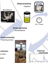
Microplastic Extraction from Marine Vertebrate Digestive Tracts, Regurgitates and Scats: A Protocol for Researchers from All Experience Levels

海洋脊椎动物消化道,反流物和粪便中塑料微粒的提取:一个适用于所有经验水平研究者的实验方法

AL Amy L. Lusher
GH Gema Hernandez-Milian
10134 Views
Nov 20, 2018
It is essential to provide a protocol for the separation and identification of microplastics in marine vertebrates (mammals, birds, turtles and fish) that is easy to follow and adaptable depending on research infrastructure. Digesting organic material is an effective way to analyze samples for microplastics. Presented here is an optimized protocol which uses potassium hydroxide (KOH) for processing samples of digestive tracts, scats and regurgitates. KOH is a cheap, effective and simple alkaline digestant that allows extraction of plastics from the sample matrix. Samples are first digested, then filtered before visual and chemical analysis of remaining particle. This allows size, shape, color and polymer of each particle to be ascertained. This protocol has been harmonized with other protocols for the collection of different samples (e.g., diet, parasites, other pathologies). The implementation of this protocol at different levels of economic and/or laboratory resources make information on microplastic incidence available to the entire research community.

微生物学

利用气相色谱法定量分析小鼠血清、结肠管腔内容物和粪便中短链脂肪酸

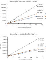
Use of Gas Chromatography to Quantify Short Chain Fatty Acids in the Serum, Colonic Luminal Content and Feces of Mice

利用气相色谱法定量分析小鼠血清、结肠管腔内容物和粪便中短链脂肪酸

WR Willian Rodrigues Ribeiro
MV Marco Aurélio Ramirez Vinolo
Leandro Augusto Calixto Leandro Augusto Calixto
Caroline Marcantonio Ferreira Caroline Marcantonio Ferreira
13505 Views
Nov 20, 2018
Short-Chain Fatty Acids (SCFAs) are a product of the fermentation of resistant starches and dietary fibers by the gut microbiota. The most important SCFA are acetate (C2), propionate (C3) and butyrate (C4). These metabolites are formed and absorbed in the colon and then transported through the hepatic vein to the liver. SCFAs are more concentrated in the intestinal lumen than in the serum. Butyrate is largely consumed in the gut epithelium, propionate in the liver and acetate in the periphery. SCFAs act on many cells including components of the immune system and epithelial cells by two main mechanisms: activation of G-protein coupled receptors (GPCRs) and inhibition of histone deacetylase. Considering the association between changes in SCFA concentrations and the development of diseases, methods to quantify these acids in different biological samples are important. In this study, we describe a protocol using gas chromatography to quantify SCFAs in the serum, feces and colonic luminal content. Separation of compounds was performed using a DB-23 column (60 m x 0.25 mm internal diameter [i.d.]) coated with a 0.15 µm thick layer of 80.2% 1-methylnaphatalene. This method has a good linear range (15-10,000 µg/ml). The precision (relative standard deviation [RSD]) is less than 15.0% and the accuracy (error relative [ER]) is within ± 15.0%. The extraction efficiency was higher than 97.0%. Therefore, this is cost effective and reproducible method for SCFA measurement in feces and serum.

神经科学

利用流式细胞技术进行原代小鼠脑内皮细胞的基于胶原酶的单细胞分离

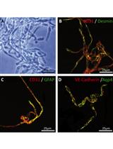
Collagenase-based Single Cell Isolation of Primary Murine Brain Endothelial Cells Using Flow Cytometry

利用流式细胞技术进行原代小鼠脑内皮细胞的基于胶原酶的单细胞分离

CC Cathrin J. Czupalla
Hanadie Yousef Hanadie Yousef
TW Tony Wyss-Coray
EB Eugene C. Butcher
11414 Views
Nov 20, 2018
The brain endothelium is a highly specialized vascular structure that maintains the activity and integrity of the central nervous system (CNS). Previous studies have reported that the integrity of the brain endothelium is compromised in a plethora of neuropathologies. Therefore, it is of particular interest to establish a method that enables researchers to investigate and understand the molecular changes in CNS endothelial cells and underlying mechanisms in conjunction with murine models of disease. In the past, approaches to isolate endothelial cells have either involved the use of transgenic reporter mice or suffered from insufficiently pure cell populations and poor yield. This protocol here is based on well-established protocols that were modified and combined to allow single cell isolation of highly pure brain endothelial cell populations using fluorescence activated cell sorting (FACS). Briefly, after careful removal of the meninges and dissection of the cortex/hippocampus, the brain tissue is mechanically homogenized and enzymatically digested in two steps resulting in a single cell suspension. Cells are stained with a cocktail of fluorochrome-conjugated antibodies identifying not only brain endothelial cells, but also potentially contaminating cell types such as pericytes, astrocytes, and lineage cells. Using flow cytometry, cell populations are separated and sorted directly into either RNA lysis buffer for bulk RNA analyses (e.g., RNA microarray and RNA-Seq) or in pure fetal bovine serum to preserve viability for other downstream applications such as single cell RNA-Seq and Assay for Transposase-Accessible Chromatin using sequencing (ATAC-Seq). The protocol does not require the expression of a transgene to label brain endothelial cells and thus, may be applied to any mouse model. In our hands, the protocol has been highly reproducible with an average yield of 3 x 10⁵ cells from a pool of four adult mice.
利用流式细胞技术进行原代小鼠脑内皮细胞的基于木瓜蛋白酶的单细胞分离

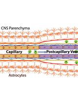
Papain-based Single Cell Isolation of Primary Murine Brain Endothelial Cells Using Flow Cytometry

利用流式细胞技术进行原代小鼠脑内皮细胞的基于木瓜蛋白酶的单细胞分离

Hanadie Yousef Hanadie Yousef
CC Cathrin J Czupalla
DL Davis Lee
EB Eugene C Butcher
TW Tony Wyss-Coray
10542 Views
Nov 20, 2018
Brain endothelial cells (BECs) form the integral component of the blood-brain barrier (BBB) which separates the systemic milieu from the brain parenchyma and protects the brain from pathogens and circulating factors. In order to study BEC biology, it was of particular interest to establish a method that enables researchers to investigate and understand the underlying molecular mechanisms regulating their function during homeostasis, aging and disease. Furthermore, due to the heterogeneity of the cerebrovasculature and different vessel types that comprise the BBB, it is of particular interest to isolate primary BECs for single cell analysis from various subregions of the brain, such as the neurogenic and highly vascularized hippocampus and to enrich for specific vessel types. In the past, approaches to isolate endothelial cells were dependent on transgenic mice and often resulted in insufficiently pure cell populations and poor yield. This protocol describes a technique that allows single-cell isolation of highly pure brain endothelial cell populations using fluorescence activated cell sorting (FACS). Briefly, after perfusion and careful removal of the meninges, and dissection of the cortex/hippocampus, the brain tissue is mechanically homogenized and enzymatically digested resulting in a single cell suspension. Cells are stained with fluorochrome-conjugated antibodies identifying CD31+ brain endothelial cells, as well as CD45+CD11b+ myeloid cells for exclusion. Using flow cytometry, cell populations are separated and CD31+BECs are sorted in bulk into RNA later or as single cells directly into either RNA lysis buffer for single or bulk RNA-Seq analyses. The protocol does not require the expression of a transgene to label brain endothelial cells and thus, may be applied to any mouse model. In our hands, the protocol has been highly reproducible with an average yield of 1 x 10⁵ cells isolated from an adult mouse cortex/hippocampus.
双标签亲和纯化法从细菌中纯化人可溶重组Tau蛋白

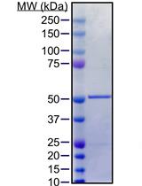
Purification of Soluble Recombinant Human Tau Protein from Bacteria Using Double-tag Affinity Purification

双标签亲和纯化法从细菌中纯化人可溶重组Tau蛋白

Joseph McInnes Joseph McInnes
Lujia Zhou Lujia Zhou
Patrik Verstreken Patrik Verstreken
8249 Views
Nov 20, 2018
Dysfunction of the microtubule-associated protein Tau (encoded by the MAPT gene) has been implicated in more than twenty neurodegenerative diseases, including Alzheimer’s. As such, the physiological and disease-relevant functions of Tau have garnered great interest in the research community. One barrier hampering investigations into the functions of Tau and the generation of pharmacological agents targeting Tau has been the difficulty of obtaining soluble Tau protein in purified form. Here, we describe a protocol that uses dual affinity tag purification to selectively purify soluble recombinant Tau protein from bacteria that is functionally active for downstream applications including immunization, microtubule binding assays, and protein-protein interaction studies.
一种改进的用于幼年大鼠的巴恩斯迷宫

A Modified Barnes Maze for Juvenile Rats

一种改进的用于幼年大鼠的巴恩斯迷宫

NV Nazanin Valibeigi
DM Daniel G. McHail
Carmen L. Kimball Carmen L. Kimball
TD Theodore C. Dumas
5829 Views
Nov 20, 2018
To better understand neural codes for spatial navigation and to address cognitive development questions, the neurobiology of postnatal hippocampal development is receiving increased attention. While the Morris Water Maze is the gold standard for assessing hippocampal integrity, potential confounds can be difficult to control for in juvenile rodents (around three weeks of age, when spatial navigation ability first emerges) and this wet maze is not amenable to electrophysiological recording. A dry maze alternative with minimal training, like the Barnes Maze adapted for juvenile rats, can help overcome these obstacles. This paper describes in detail the experimental procedure and data analyses for the adapted Barnes maze for juvenile animals.

干细胞

人多能干细胞到功能性间充质干细胞的单步派生

One-step Derivation of Functional Mesenchymal Stem Cells from Human Pluripotent Stem Cells

人多能干细胞到功能性间充质干细胞的单步派生

Yan Zhou Yan Zhou
JinQi Liao JinQi Liao
ChongZhou Fang ChongZhou Fang
CM CuiPing Mo
GZ GuangQian Zhou
Yonglun Luo Yonglun Luo
8522 Views
Nov 20, 2018
Mesenchymal stem cells (MSCs) are invaluable cell sources for understanding stem cell biology and potential application in tissue engineering and regenerative medicine. The current issues of MSCs that demand to be further addressed are limited donors, tissue sources and limited capacity of ex vivo expansion. Here, we describe a simple and easy protocol for generating functional mesenchymal stem cells from human pluripotent stem cells (hPSCs) via one-step low glucose medium switch strategy in feeder-free culture system. In this protocol, human induced pluripotent stem cells (hiPSCs) and H9 human embryonic stem cells (hESCs) were successfully differentiated into MSCs, named hiPSC-MSCs and hESC-MSCs, respectively. The derived hiPSC-MSCs and hESC-MSCs exhibited common MSC characteristics as MSCs derived from human bone marrow (hBM-MSCs), including expressing MSC surface markers and possessing capability of tri-lineage differentiation in vitro (adipogenesis, osteogenesis and chondrogenesis). As compared with other available protocols, our protocol can be applied to generate a large number of MSCs from hPSCs with high efficiency, low-cost manner, moreover, not involving embryoid body, mouse feeder-cell, flow sorting, and pathway inhibitors (such as SB203580 and SB431542). We believe that this protocol could provide a robust platform to reach the future demand for producing the industrial scale of MSC from hPSCs for autologous cell-based therapy.