往期刊物2015

卷册: 5, 期号: 8

left

Dec

20

Dec

5

Nov

20

Nov

5

Oct

20

Oct

5

Sep

20

Sep

5

Aug

20

Aug

5

Jul

20

Jul

5

Jun

20

Jun

5

May

20

May

5

Apr

20

Apr

5

Mar

20

Mar

5

Feb

20

Feb

5

Jan

20

Jan

5

right

生物化学

13C 动力标记和提取附着哺乳动物细胞的代谢产物

13C Kinetic Labeling and Extraction of Metabolites from Adherent Mammalian Cells

13C 动力标记和提取附着哺乳动物细胞的代谢产物

PM Parul Mehrotra
NS Najmuddin Saquib
KR Kanury V. S. Rao
10331 Views
Apr 20, 2015
Fluctuations in metabolite levels in mammalian cells are the most direct form of readout of the cellular metabolic state. The current protocol describes a method for pulse labeling and subsequent isolation of metabolites from adherent mammalian cells. The isolated metabolites can be identified and quantified by mass-spectrometry, allowing for estimation of the rates of synthesis and removal of metabolites from the system being analyzed.
通过焦磷酸盐的产生测量核苷三磷酸糖转移酶的活性

Measurement of Nucleotide Triphosphate Sugar Transferase Activity via Generation of Pyrophosphate

通过焦磷酸盐的产生测量核苷三磷酸糖转移酶的活性

Kaisei Maeda Kaisei Maeda
RN Rei Narikawa
MI Masahiko Ikeuchi
9761 Views
Apr 20, 2015
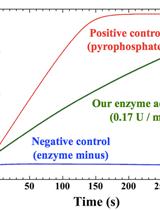
Nucleotide triphosphate (NTP) transferases (EC. 2. 7. 7. X) transfer a nucleoside monophosphate moiety from NTP to another substrate. NTP sugar transferases form a large member of the NTP transferase. There are many variations for the substrate combination of the NTP sugar transferases. It is important to measure the precise enzymatic activity of such NTP sugar transferases by a simple and efficient method. In our method, we measure pyrophosphate as a byproduct of nucleotide diphosphate (NDP)-sugar generation using the pyrophosphate assay kit. The kit reagents include two enzymes that convert pyrophosphate to phosphate, and then phosphorolyze chromogenic substrate to allow color development at 360 nm (see details below). Thus, the NDP-sugar formation can be simply traced as production of pyrophosphate, which is monitored by absorbance at 360 nm. This method is reliable and versatile for measurements of various pyrophosphate-producing enzymes that include NTP sugar transferases.[Principle and overview] NTP transferases catalyze the reversible reaction as follows: NTP + sugar-1P NDP-sugar + PPi The enzyme reaction can be monitored as generation of inorganic pyrophosphate (PPi). The EnzChek Pyrophosphate Assay kit (Molecular Probes, Life Technologies, Carlsbad, CA) includes two enzymes and sufficient materials for color development to quantitate pyrophosphate. The inorganic pyrophosphatase (component E in the kit) degrades pyrophosphate into phosphate. Purine nucleoside phosphorylase (PNP, component B) utilizes phosphate to cleave the colorgenic substrate 2-amino-6-mercapto-7-methylpurine ribonucleoside (MESG, component A) into ribose-1-phosphate and 2-amino-6-mercapto-7-methylpurine. The product 2-amino-6-mercapto-7-methylpurine has the absorption maximum at 360 nm. The component C is the dilution solution that includes minimal MgCl2 sufficient for the inorganic pyrophosphatase. Thus, NTP transferase activity can be monitored at 360 nm as generation of a byproduct pyrophosphate. Typically, the nucleotidyl sugar transferase reactions have been measured by High Performance Liquid Chromatography (HPLC) (Kawano et al., 2014). There are several advantages and disadvantages in HPLC method and our enzymatic method (O: advantage, X: disadvantage).{HPLC method}O) Can measure the reaction in both directions (NDP-sugar formation and degradation)O) Can measure with a small amount of the sample protein X) Can not follow the real-time reactionX) The peaks of substrates and products must be separated in the chromatogram{Pyrophosphate assay method}O) Can observe the reaction in real-timeO) Easy to use many kinds of substrates because of the simple detection at 360 nmO) Can be used in other pyrophosphate or phosphate generating reactions such as adenylate cyclase, diguanylate cyclase ( Enomoto et al., 2014), and DNA polymeraseX) Can not measure the pyrophosphate-consuming direction of the reversible reaction of NDP-sugar pyrophosphorylase. Km and kcat for NDP-sugar and pyrophosphate are not obtained by the pyrophosphate assay method but by HPLC method.X) Need certain amount of the experimental protein. The maximum activity that the kit reagents allow corresponds theoretically to the rate of color development for the positive control is 2 x 10⁻² U in our case. The instruction states the minimum detection of 5 x 10⁻⁵ U. Of course, the minimum activity we can measure may depend on the background activity due to contaminants in the substrates or in the sample preparation.

微生物学

从鱼腥藻属 PCC 7120分离异型胞

Isolation of Heterocysts from Anabaena sp. PCC 7120

从鱼腥藻属 PCC 7120分离异型胞

ME Maria Ermakova
YA Yagut Allahverdiyeva
14966 Views
Apr 20, 2015
During combined nitrogen step-down, filaments of cyanobacterium Anabaena sp. PCC 7120 differentiate about 5-10% of vegetative photosynthetic cells into heterocysts, the specialized cells for N2 fixation (Walk, 1996). Heterocysts have a thick cell wall reducing permeation of O2 and consist of two additional layers composed of glycolipids and polysaccharides. The difference in structure and composition of the cell wall between heterocysts and vegetative cells allows separation and isolation of heterocyst. Heterocysts isolated by this protocol can be subjected to protein analysis and activity measurements, which do not require strict anaerobic conditions.
采用Southern蛋白印迹法进行HBV体外药敏性试验

In vitro Drug Susceptibility Assay for HBV Using Southern Blotting

采用Southern蛋白印迹法进行HBV体外药敏性试验

SA Sung Hyun Ahn
YP Yong Kwang Park
Kyun-Hwan Kim Kyun-Hwan Kim
10718 Views
Apr 20, 2015
Antiviral agents for the suppression of hepatitis B virus (HBV) have been used for treating chronic hepatitis B. However, the emergence of drug-resistant HBV is still a major problem for antiviral treatment. To identify and characterize the drug-resistant HBV, the construction of HBV replicon and in vitro drug susceptibility assay are essential. Here we describe the experimental methods to study drug-resistant HBV.
把亲体分离的HBV RT序列引入HBV 1.2-mer复制子中并测序

Introduction and Sequencing of Patient-isolated HBV RT Sequences into the HBV 1.2-mer Replicon

把亲体分离的HBV RT序列引入HBV 1.2-mer复制子中并测序

SA Sung Hyun Ahn
YP Yong Kwang Park
Kyun-Hwan Kim Kyun-Hwan Kim
9049 Views
Apr 20, 2015
Antiviral agents for the suppression of hepatitis B virus (HBV) have been used for treating chronic hepatitis B. However, the emergence of drug-resistant HBV is still a major problem for antiviral treatment. To identify and characterize the drug-resistant HBV, the construction of HBV replicon and in vitro drug susceptibility assay are essential. Here we describe the experimental methods to study drug-resistant HBV.
表面暴露幽门螺杆菌蛋白的生物素化和纯化

Biotinylation and Purification of Surface-exposed Helicobacter pylori Proteins

表面暴露幽门螺杆菌蛋白的生物素化和纯化

BV Bradley J. Voss
TC Timothy L. Cover
7992 Views
Apr 20, 2015
Interactions between pathogenic bacteria and host cells are often mediated by proteins found on the surfaces of the bacteria. The Gram-negative bacterium Helicobacter pylori is predicted to produce at least 50 surface-exposed outer membrane proteins, but there has been relatively little progress in experimentally analyzing the cell-surface proteome of this organism. Herein, we describe in detail a protocol that allows biotinylation and purification of surface-exposed H. pylori proteins. A comparative analysis of surface-exposed proteins identified by this biotinylation-based approach and by several other independent methods is described in a recent publication (Voss et al., 2014).

分子生物学

DNA斑点杂交修复试验

DNA Slot Blot Repair Assay

DNA斑点杂交修复试验

Jeong-Min Park Jeong-Min Park
TK Tae-Hong Kang
21083 Views
Apr 20, 2015
Ultraviolet (UV) irradiation induces helix distorting photolesions such as cyclobutane pyrimidine dimers (CPD) and pyrimidine-pyrimidone (6-4) photoproducts (6-4PP) which threaten genomic integrity if unrepaired. In mammals, nucleotide excision repair (NER) is the only pathway that removes UV-induced DNA damages. Here we describe DNA slot blot repair assay for quantitative detection of NER activity using DNA damage specific antibodies such as anti-CPD and anti-6-4PP. Briefly, genomic DNA irradiated with UV was isolated from cells, and the genomic DNA was vacuum-transferred to a nitrocellulose membrane using a Bio-Dot SF microfiltration apparatus (Bio-Rad). A monoclonal antibody that recognizes CPD or 6-4PP was applied to detect the remaining amount of photolesions in the genomic DNA. For loading control of even loading, DNA onto the membrane can be further analyzed by SYBR-gold staining.
随机DNA结合选择分析(RDSA)

Random DNA Binding Selection Assay (RDSA)

随机DNA结合选择分析(RDSA)

YW Yafei Wang
ML Meizhong Luo
9532 Views
Apr 20, 2015
Protein-DNA interaction is a very important cellular process, by which regulation of DNA biological function, usually gene expression, is exerted. The method of random DNA binding selection assay (RDSA) can be used to identify DNA elements bound by proteins with DNA-binding activities. This method is based on the enrichment of the target DNA element by the immobilized recombinant protein on special beads and repeated PCR amplification.

植物科学

拟南芥、番茄和大麦叶片中相对含水量(RWC)的测定

Measuring Arabidopsis, Tomato and Barley Leaf Relative Water Content (RWC)

拟南芥、番茄和大麦叶片中相对含水量(RWC)的测定

NS Nir Sade
EG Eyal Galkin
MM Menachem Moshelion
14552 Views
Apr 20, 2015
Measuring leaf relative water content (RWC) is a reliable and simple way to assess the water status of a leaf without any need for special equipment. Similar to leaf water potential, leaf RWC gives a strong indication of the plant’s response to different environmental conditions; yet RWC has been shown to be a more stable parameter than leaf water potential (Sade et al., 2009; Sade et al., 2012). Although measuring RWC is destructive to the leaf, with proper planning, it need not affect the plant’s behavior. This note will focus on three different model plants which are representative of plants with various leaf shapes (e.g., Arabidopsis, tomato and barley). The technique for measuring RWC is the same for all three of these species (as well as for plants with many other types of leaves).
拟南芥DCL1催化miRNA合成的体外实验

In vitro Reconstitution Assay of miRNA Biogenesis by Arabidopsis DCL1

拟南芥DCL1催化miRNA合成的体外实验

Tian Wang Tian Wang
Claudia Castillo-González Claudia Castillo-González
LY Lan You
RL Rui Li
LW Liwei Wen
Hongliang Zhu Hongliang Zhu
XZ Xiuren Zhang
10046 Views
Apr 20, 2015
microRNAs (miRNAs) are small non-coding RNAs, regulating most if not all, biological processes in eukaryotic organisms. miRNAs are initially processed from primary transcripts (pri-miRNAs) to produce miRNA precursors (pre-miRNAs), that are further processed into miRNA and its complementary strands (miRNA/*). In Arabidopsis, and possibly other plants, the processing from pri-miRNAs to pre-miRNAs and from pre-miRNAs to miRNA/* are both implemented through Dicer-like 1 (DCL1) complexes. Recently, we demonstrated isolation of DCL1 complexes of unprecedented quality from in planta. We further successfully reconstituted DCL1 cleavage assays in vitro that were able to fully recapitulate in vivo miRNA biogenesis. Here we provide a detailed protocol of DCL1 reconstitution assays. The protocol comprises three major parts (Figure 1): 1) Preparation of pri- and pre-miRNA transcripts (Procedures A-C); 2) Purification of the recombinant Arabidopsis DCL1 machinery from Nicotiana benthamiana (N. benthamiana) through immunoprecipitation (IP) (Procedures D and E); and 3) in vitro processing of radioisotope-labeled pri- or pre-miRNAs using the isolated DCL1 complexes (Procedure F). It is our desire that the protocol be a powerful tool for the RNAi community to study mechanistic issues or to develop RNA silencing technologies.
水稻不同组织中砷含量的测定和形态分析

Measuring the Arsenic Content and Speciation in Different Rice Tissues

水稻不同组织中砷含量的测定和形态分析

E. Marie Muehe E. Marie Muehe
AK Andreas Kappler
CC Christina Chaban
BD Birgit Daus
9814 Views
Apr 20, 2015
Arsenic (As) plays an important role in rice production as vast soils used for rice cultivation contain As. To understand how rice plants deal with inorganic As (III) and As (V) and organic As in their tissue, it is important to obtain specific information on how much and what species of As are present in which tissue of the rice plant. The protocol presented here allows to analyse the As contents and As speciation in roots, shoots, and husks of rice plants, and thus permits direct comparison of the As contents of these rice plant tissues.
拟南芥侧根器官形成的实时分析

Real-time Analysis of Lateral Root Organogenesis in Arabidopsis

拟南芥侧根器官形成的实时分析

Peter Marhavý Peter Marhavý
Eva Benková Eva Benková
9688 Views
Apr 20, 2015
Plants maintain capacity to form new organs such as leaves, flowers, lateral shoots and roots throughout their postembryonic lifetime. Lateral roots (LRs) originate from a few pericycle cells that acquire attributes of founder cells (FCs), undergo series of anticlinal divisions, and give rise to a few short initial cells. After initiation, coordinated cell division and differentiation occur, giving rise to lateral root primordia (LRP). Primordia continue to grow, emerge through the cortex and epidermal layers of the primary root, and finally a new apical meristem is established taking over the responsibility for growth of mature lateral roots [for detailed description of the individual stages of lateral root organogenesis see Malamy and Benfey (1997)]. To examine this highly dynamic developmental process and to investigate a role of various hormonal, genetic and environmental factors in the regulation of lateral root organogenesis, the real time imaging based analyses represent extremely powerful tools (Laskowski et al., 2008; De Smet et al., 2012; Marhavý et al., 2013; Marhavý et al., 2014). Herein, we describe a protocol for real time lateral root primordia (LRP) analysis, which enables the monitoring of an onset of the specific gene expression and subcellular protein localization during primordia organogenesis, as well as the evaluation of the impact of genetic and environmental perturbations on LRP organogenesis.
植物粗提物(油橄榄)中脂肪酶活性的凝胶检测

In Gel Detection of Lipase Activity in Crude Plant Extracts (Olea europaea)

植物粗提物(油橄榄)中脂肪酶活性的凝胶检测

Agnieszka Zienkiewicz Agnieszka Zienkiewicz
Juan David Rejón Juan David Rejón
Krzysztof Zienkiewicz Krzysztof Zienkiewicz
Antonio Jesus Castro Antonio Jesus Castro
Maria Isabel Rodríguez-García Maria Isabel Rodríguez-García
8017 Views
Apr 20, 2015
Here, we provide a detailed protocol describing a SDS-PAGE based procedure to assay in gel neutral lipase activity. Total protein extracts are separated by SDS-PAGE and gels are treated with lipase substrate-α-naphthyl palmitate. This long-chain fatty acid ester is hydrolysed by lipases present in the gel. The product resulting from this reaction can be then visualized in the gel as yellow-brownish activity bands. This relatively simple and effective method of lipase assay detection can be used for crude protein extracts from different plant tissues.