往期刊物2018

卷册: 8, 期号: 5

left

Dec

20

Dec

5

Nov

20

Nov

5

Oct

20

Oct

5

Sep

20

Sep

5

Aug

20

Aug

5

Jul

20

Jul

5

Jun

20

Jun

5

May

20

May

5

Apr

20

Apr

5

Mar

20

Mar

5

Feb

20

Feb

5

Jan

20

Jan

5

right

发育生物学

末端脱氧核苷酸转移酶介导的用于单分子FISH或RNA捕获的标记探针的生成

Terminal Deoxynucleotidyl Transferase Mediated Production of Labeled Probes for Single-molecule FISH or RNA Capture

末端脱氧核苷酸转移酶介导的用于单分子FISH或RNA捕获的标记探针的生成

Imre   Gaspar Imre Gaspar
Frank   Wippich Frank Wippich
Anne  Ephrussi Anne Ephrussi
12474 Views
Mar 5, 2018
Arrays of short, singly-labeled ssDNA oligonucleotides enable in situ hybridization with single molecule sensitivity and efficient transcript specific RNA capture. Here, we describe a simple, enzymatic protocol that can be carried out using basic laboratory equipment to convert arrays of PCR oligos into smFISH and RAP probesets in a quantitative, cost-efficient and flexible way.

免疫学

肾被膜下小鼠胰岛移植

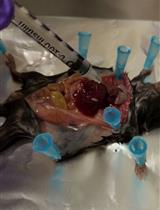
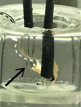
Murine Pancreatic Islets Transplantation under the Kidney Capsule

肾被膜下小鼠胰岛移植

TJ Tatiana Jofra
GG Giuseppe Galvani
FG Fousteri Georgia
GS Gregori Silvia
NG Nicola Gagliani
Manuela  Battaglia Manuela Battaglia
10819 Views
Mar 5, 2018
Type 1 diabetes (T1D) is an autoimmune disease caused by the lack of insulin-producing pancreatic beta cells leading to systemic hyperglycemia. Pancreatic islet transplantation is a valid therapeutic approach to restore insulin loss and to promote adequate glycemic control. Pancreatic islet transplantation in mice is an optimal preclinical model to identify new therapeutic strategies aiming at preventing rejection and optimizing post-transplant immuno-suppressive/-tolerogenic therapies. Islet transplantation in preclinical animal models can be performed in different sites such the kidney capsule, spleen, bone marrow and pancreas. This protocol describes murine islet transplantation under the kidney capsule. This is a widely accepted procedure for research purposes. Stress caused in the animals is minimal and it leads to reliable and reproducible results.
单钠尿酸盐晶体诱导的腹膜炎用于体内评估炎症体激活

Mono Sodium Urate Crystal-induced Peritonitis for in vivo Assessment of Inflammasome Activation

单钠尿酸盐晶体诱导的腹膜炎用于体内评估炎症体激活

Marianne R. Spalinger Marianne R. Spalinger
Michael   Scharl Michael Scharl
9168 Views
Mar 5, 2018
Due to its particulate material, mono-sodium urate (MSU) crystals are potent activators of the NOD-like receptor NLRP3. Upon activation, NLRP3 induces the formation of inflammasome complexes, which lead to the production and release of mature IL-1β. Bioactive IL-1β is a potent activator of innate immune responses and promotes recruitment of inflammatory cells, including neutrophils from the blood into damaged/inflamed tissues. This protocol describes a method to study in vivo inflammasome activation via intraperitoneal injection of MSU crystals. MSU-injection results in a drastic increase of intraperitoneal IL-1β levels, promoting neutrophil infiltration. Early-stage neutrophil numbers correlate with the amount of released IL-1β and can be used as a read-out for the extent of in vivo inflammasome activation. In addition, this protocol might also be used as a sterile peritonitis model, to investigate mechanisms of neutrophil recruitment to the peritoneum, or as a means to obtain large numbers of in vivo activated neutrophils.
组织切片细菌附着定量

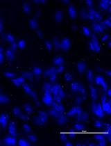
Quantification of Bacterial Attachment to Tissue Sections

组织切片细菌附着定量

BI Batya Isaacson
TH Tehila Hadad
GB Gilad Bachrach
OM Ofer Mandelboim
8069 Views
Mar 5, 2018
Here we describe a method to test bacterial adhesion to paraffin embedded tissue sections. This method allows examining binding of different bacterial strains, transfected with a fluorescent protein reporter plasmid to various tissues, to better understand different mechanisms such as colonization. This assay provides a more physiological context to bacterial binding, than would have been achieved using adhesion assays to cell lines. The sections can be imaged using fluorescent microscopy and adhesion of various bacterial strains can be quantified and tested, simultaneously.
静脉标记和胸腺血管周围间隙含量分析

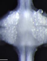
Intravenous Labeling and Analysis of the Content of Thymic Perivascular Spaces

静脉标记和胸腺血管周围间隙含量分析

RR Roland Ruscher
KH Kristin A. Hogquist
7006 Views
Mar 5, 2018
Following development in the thymus, T cells are thought to exit into the periphery predominantly through perivascular spaces (PVS). This exit route is used by conventional T cells, and likely also applies to unconventional T cell subsets, such as precursors of CD8αα and TCRγδ intraepithelial lymphocytes, regulatory T cells and natural killer T cells. Additional cell types might also be found in the PVS and initiate interactions with exiting T cells. The exact content of the PVS, and the processes within, are not well studied. To distinguish vascular from resident cells within various tissues by flow cytometry, intravenous (i.v.) labeling is becoming a commonly employed method. We recently used anti-CD45.2 antibodies and magnetic enrichment to further evaluate this technique, and compared labeled and unlabeled cells in the thymus and blood. This assay can be used to specifically investigate hematopoietic cell subsets within the PVS of the thymus.

微生物学

采用气相色谱法和尼罗红染色法测定富养罗尔斯通菌中聚羟基丁酸酯(PHB)的含量

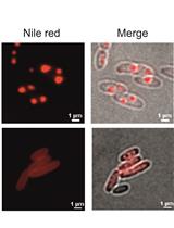
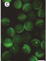
Determination of Polyhydroxybutyrate (PHB) Content in Ralstonia eutropha Using Gas Chromatography and Nile Red Staining

采用气相色谱法和尼罗红染色法测定富养罗尔斯通菌中聚羟基丁酸酯(PHB)的含量

JJ Janina R. Juengert
SB Stephanie Bresan
DJ Dieter Jendrossek
17136 Views
Mar 5, 2018
Ralstonia eutropha H16 produces and mobilizes (re-utilizes) intracellular polyhydroxybutyrate (PHB) granules during growth. This protocol describes the visualization of intracellular Nile red stained PHB granules and the quantification of PHB by gas chromatography. Our first method describes how to analyze PHB granules by fluorescence microscopy qualitatively. Our second approach enables the conversion of PHB to volatile hydroxycarboxylic acid methyl esters by acidic methanolysis and their quantification by gas chromatography. Through this method, it is possible to obtain an absolute quantification of PHB, e.g., per cell dry weight.
3'RACE联合引物延伸法检测大肠埃希氏菌RNA 3'末端

Visualization of RNA 3’ ends in Escherichia coli Using 3’ RACE Combined with Primer Extension

3'RACE联合引物延伸法检测大肠埃希氏菌RNA 3'末端

XW Xun Wang
HJ Heung Jin Jeon
MA Monford Paul Abishek N
JH Jin He
HL Heon M. Lim
8477 Views
Mar 5, 2018
In this assay, 3’ RACE (Rapid Amplification of cDNA 3’ Ends) followed by PE (primer extension), abbreviated as 3’ RACE-PE is used to identify the mRNA 3’ ends. The following protocol describes the amplification of the mRNA 3’ ends at the galactose operon in E. coli and the corresponding visualization of the PCR products through PE. In PE, the definite primer is 5’ end-labeled using [γ-(32) P] ATP and T4 polynucleotide kinase, which anneals to the specific DNA molecules within the PCR product of the 3’ RACE. The conventional PE can only be used to locate the 5’ end of an mRNA transcript since reverse transcriptase (RTase) polymerizes only in the 5’ → 3’ direction. Thus, Taq polymerase is used instead of RTase, PCR is performed. Therefore, we are able to locate the 3’ end of the mRNA using this assay. The relative amount of the 3’ end can be directly visualized and quantified by way of separating DNA products in a denaturing 8% urea-PAGE (Polyacrylamide Gel Electrophoresis) gel. The exact position of the 3’ ends can be sequenced by comparison of these final DNA products with the corresponding DNA sequencing ladder.
使用基于Vc2 Riboswitch的测定法体内分析大肠杆菌中环状di-GMP环化酶和磷酸二酯酶活性

In vivo Analysis of Cyclic di-GMP Cyclase and Phosphodiesterase Activity in Escherichia coli Using a Vc2 Riboswitch-based Assay

使用基于Vc2 Riboswitch的测定法体内分析大肠杆菌中环状di-GMP环化酶和磷酸二酯酶活性

YL Ying Liu
Hyunhee  Kim Hyunhee Kim
Ute   Römling Ute Römling
7314 Views
Mar 5, 2018
Cyclic di-guanosine monophosphate (c-di-GMP) is a ubiquitous second messenger that regulates distinct aspects of bacterial physiology. It is synthesized by diguanylate cyclases (DGCs) and hydrolyzed by phosphodiesterases (PDEs). To date, the activities of DGC and PDE are commonly assessed by phenotypic assays, mass spectrometry analysis of intracellular c-di-GMP concentration, or riboswitch-based fluorescent biosensors. However, some of these methods require cutting-edge equipment, which might not be available in every laboratory. Here, we report a new simple, convenient and cost-effective system to assess the function of DGCs and PDEs in E. coli. This system utilizes the high specificity of a riboswitch to c-di-GMP and its ability to regulate the expression of a downstream β-galactosidase reporter gene in response to c-di-GMP concentrations. In this protocol, we delineate the construction of this system and its use to assess the activity of DGC and PDE enzymes.

神经科学

用于小鼠空间学习和记忆的Barnes迷宫程序

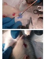
Barnes Maze Procedure for Spatial Learning and Memory in Mice

用于小鼠空间学习和记忆的Barnes迷宫程序

MP Matthew W. Pitts
17839 Views
Mar 5, 2018
The Barnes maze is a dry-land based rodent behavioral paradigm for assessing spatial learning and memory that was originally developed by its namesake, Carol Barnes. It represents a well-established alternative to the more popular Morris Water maze and offers the advantage of being free from the potentially confounding influence of swimming behavior. Herein, the Barnes maze experimental setup and corresponding procedures for testing and analysis in mice are described in detail.
评估果蝇运动功能的飞行和攀爬分析

Flight and Climbing Assay for Assessing Motor Functions in Drosophila

评估果蝇运动功能的飞行和攀爬分析

SM Steffy B Manjila
GH Gaiti Hasan
12148 Views
Mar 5, 2018
Motor control requires the central nervous system to integrate different sensory inputs and convey this information to the relevant central pattern generator for execution of motor function through motor neurons and muscles. Proper motor control is essential for any mobile organism to survive and interact with the external environment. For flying insects, motor control is required for flying, walking, feeding and mating apart from other more advanced behaviours such as grooming and aggression. Any perturbation to the sensory input or malfunctioning of neural connections to the motor output can result in motor defects. Here, we describe simple protocols for assessing flight and climbing ability of fruit flies, which can be used as two general tests to assess their motor function.
用于体内潜在的剪接突变分析的微小基因的构建及克隆

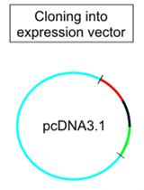
Construction and Cloning of Minigenes for in vivo Analysis of Potential Splice Mutations

用于体内潜在的剪接突变分析的微小基因的构建及克隆

LR Lisa Maria Riedmayr
SB Sybille Böhm
SM Stylianos Michalakis
EB Elvir Becirovic
11464 Views
Mar 5, 2018
Disease-associated mutations influencing mRNA splicing are referred to as splice mutations. The majority of splice mutations are found on exon-intron boundaries defining canonical donor and acceptor splice sites. However, mutations in the coding region (exonic mutations) can also affect mRNA splicing. Exact knowledge of the disease mechanism of splice mutations is essential for developing optimal treatment strategies. Given the large number of disease-associated mutations thus far identified, there is an unmet need for methods to systematically analyze the effects of pathogenic mutations on mRNA splicing. As splicing can vary between cell types, splice mutations need to be tested under native conditions if possible. A commonly used tool for the analysis of mRNA splicing is the construction of minigenes carrying exonic and intronic sequences. Here, we describe a protocol for the design and cloning of minigenes into recombinant adeno-associated virus (rAAV) vectors for gene delivery and investigation of mRNA splicing in a native context. This protocol was developed for minigene-based analysis of mRNA splicing in retinal cells, however, in principle it is applicable to any cell type, which can be transduced with rAAV vectors.
小鼠中膈神经偏侧膈分析(MPN)

Mouse Phrenic Nerve Hemidiaphragm Assay (MPN)

小鼠中膈神经偏侧膈分析(MPN)

Giulia  Zanetti Giulia Zanetti
SN Samuele Negro
Marco  Pirazzini Marco Pirazzini
Paola  Caccin Paola Caccin
9683 Views
Mar 5, 2018
The neuromuscular junction (NMJ) is the specialized synapse by which peripheral motor neurons innervate muscle fibers and control skeletal muscle contraction. The NMJ is the target of several xenobiotics, including chemicals, plant, animal and bacterial toxins, as well as of autoantibodies raised against NMJ antigens. Depending on their biochemical nature, the site they target (either the nerve or the muscle) and their mechanism of action, substances affecting NMJ produce very specific alterations of neuromuscular functionality.Here we provide a detailed protocol to isolate the diaphragmatic muscle from mice and to set up two autonomously innervated hemidiaphragms. This preparation can be used to study bioactive substances like toxins, venoms and neuroactive molecules of various origin, or to measure the force of skeletal muscle contraction.The ‘mouse phrenic nerve hemidiaphragm assay’ (MPN) is an established model of ex vivo NMJ and recapitulates the complexity of neuromuscular transmission in a system easy to control and to manipulate, thus representing a valuable tool to study both NMJ physiology and the mechanism of action of toxins and other molecules acting at this synapse.
水蛭神经节的双面电压敏感染料成像

Dual-sided Voltage-sensitive Dye Imaging of Leech Ganglia

水蛭神经节的双面电压敏感染料成像

YT Yusuke Tomina
Daniel A. Wagenaar Daniel A. Wagenaar
7553 Views
Mar 5, 2018
In this protocol, we introduce an effective method for voltage-sensitive dye (VSD) loading and imaging of leech ganglia as used in Tomina and Wagenaar (2017). Dissection and dye loading procedures are the most critical steps toward successful whole-ganglion VSD imaging. The former entails the removal of the sheath that covers neurons in the segmental ganglion of the leech, which is required for successful dye loading. The latter entails gently flowing a new generation VSD, VF2.1(OMe).H, onto both sides of the ganglion simultaneously using a pair of peristaltic pumps. We expect the described techniques to translate broadly to wide-field VSD imaging in other thin and relatively transparent nervous systems.
适合观察一根或多根大鼠触须的荧光染料方法

A Fluorescent Dye Method Suitable for Visualization of One or More Rat Whiskers

适合观察一根或多根大鼠触须的荧光染料方法

JR Jacopo Rigosa
AL Alessandro Lucantonio
GN Giovanni Noselli
AF Arash Fassihi
EZ Erik Zorzin
FM Fabrizio Manzino
FP Francesca Pulecchi
Mathew E Diamond Mathew E Diamond
6439 Views
Mar 5, 2018
Visualization and tracking of the facial whiskers is critical to many studies of rodent behavior. High-speed videography is the most robust methodology for characterizing whisker kinematics, but whisker visualization is challenging due to the low contrast of the whisker against its background. Recently, we showed that fluorescent dye(s) can be applied to enhance visualization and tracking of whisker(s) (Rigosa et al., 2017), and this protocol provides additional details on the technique.

植物科学

通过电解质渗漏测定法定量植物细胞死亡

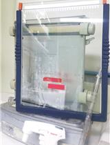
Quantification of Plant Cell Death by Electrolyte Leakage Assay

通过电解质渗漏测定法定量植物细胞死亡

NH Noriyuki Hatsugai
FK Fumiaki Katagiri
23373 Views
Mar 5, 2018
We describe a protocol to measure the electrolyte leakage from plant tissues, resulting from loss of cell membrane integrity, which is a common definition of cell death. This simple protocol is designed to measure the electrolyte leakage from a tissue sample over a time course, so that the extent of cell death in the tissue can be monitored dynamically. In addition, it is easy to handle many tissue samples in parallel, which allows a high level of biological replication. Although the protocol is exemplified by cell death in Arabidopsis in response to pathogen challenge, it is easily applicable to other types of plant cell death.
玉米南方叶斑病及其他叶片真菌病的大面积野外接种与评分

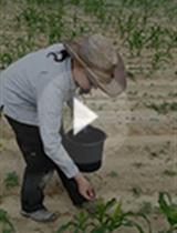
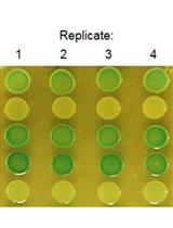
Large Scale Field Inoculation and Scoring of Maize Southern Leaf Blight and Other Maize Foliar Fungal Diseases

玉米南方叶斑病及其他叶片真菌病的大面积野外接种与评分

Shannon M. Sermons Shannon M. Sermons
PB Peter J. Balint-Kurti
8622 Views
Mar 5, 2018
Field-grown maize is inoculated with Cochliobolus heterostrophus, causal agent of southern leaf blight disease, by dropping sorghum grains infested with the fungus into the whorl of each maize plant at an early stage of growth. The initial lesions produce secondary inoculum that is dispersed by wind and rain, causing multiple cycles of infection that assures a high uniform disease pressure over the entire field by the time of disease scoring, which occurs after anthesis. This method, with slight modifications, can also be used to study the maize fungal diseases northern leaf blight (caused by Exserohilum turcicum) and gray leaf spot (Cercospora zeae-maydis).
在非洲爪蟾卵母细胞系统中进行硼的吸收分析实验

Boron Uptake Assay in Xenopus laevis Oocytes

在非洲爪蟾卵母细胞系统中进行硼的吸收分析实验

SW Sheliang Wang
NM Namiki Mitani-Ueno
Junpei  Takano Junpei Takano
7898 Views
Mar 5, 2018
Boron (B) is essential for plant growth and taken up by plant roots as boric acid. Under B limitation, B uptake and translocation in plants are dependent on the boric acid channels located in the plasma membrane. Xenopus leavis oocyte is a reliable heterologous expression system to characterize transport activities of boric acid channels and related major intrinsic proteins (aquaporins). Here, we outline the protocols for expression of boric acid channels and boric acid uptake assay in Xenopus leavis oocytes.
本氏烟草中的组蛋白去泛素化分析

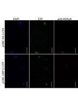
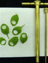
Histone Deubiquitination Assay in Nicotiana benthamiana

本氏烟草中的组蛋白去泛素化分析

Shujing  Liu Shujing Liu
Lars  Hennig Lars Hennig
6049 Views
Mar 5, 2018
Histone modifications are a group of post-translational modifications on histones which can alter chromatin structure and affect gene expression. Histone ubiquitination is a histone modification found in particular on histone H2A and H2B. Histone ubiquitination can be reversed by ubiquitin-specific proteases (UBP). Here, we describe an in vivo assay for histone deubiquitination activity. After infiltrating UBP12 into Nicotiana benthamiana leaves, H2Aub was visualized by immunocytochemistry. Nicotiana benthamiana leaves, which show high agro infiltration efficiency, were used for transient UBP12 expression for a labor- and time-saving protocol. Reduced H2Aub levels indicated histone deubiquitination activity of UBP12. The clear visualization of nuclei of N. benthamiana leaves makes this method able to easily measure the level of histone modification in vivo by using specific antibodies, providing robust clues of protein function. Thus, this protocol is a powerful complementation to in vitro assays of histone deubiquitination activity.

系统生物学

秀丽隐杆线虫中合成遗传相互作用(CRISPR-SGI)分析

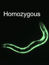
Synthetic Genetic Interaction (CRISPR-SGI) Profiling in Caenorhabditis elegans

秀丽隐杆线虫中合成遗传相互作用(CRISPR-SGI)分析

JC John A. Calarco
AN Adam D. Norris
8230 Views
Mar 5, 2018
Genetic interaction screens are a powerful methodology to establish novel roles for genes and elucidate functional connections between genes. Such studies have been performed to great effect in single-cell organisms such as yeast and E. coli (Schuldiner et al., 2005; Butland et al., 2008; Costanzo et al., 2010), but similar large-scale interaction studies using targeted reverse-genetic deletions in multi-cellular organisms have not been feasible. We developed a CRISPR/Cas9-based method for deleting genes in C. elegans and replacing them with a heterologous fluorescent reporter (Norris et al., 2015). Recently we took advantage of that system to perform a large-scale, reverse genetic screen using null alleles in animals for the first time, focusing on RNA binding protein genes (Norris et al., 2017). This type of approach should be similarly applicable to many other gene classes in C. elegans. Here we detail the protocols involved in generating a library of double mutants and performing medium-throughput competitive fitness assays to test for genetic interactions resulting in fitness changes.
核酸外切酶消化联合选择性化学标记碱基分辨率定位基因组中DNA中5-羟甲基胞嘧啶

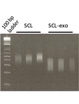
Coupling Exonuclease Digestion with Selective Chemical Labeling for Base-resolution Mapping of 5-Hydroxymethylcytosine in Genomic DNA

核酸外切酶消化联合选择性化学标记碱基分辨率定位基因组中DNA中5-羟甲基胞嘧啶

Aurélien  A. Sérandour Aurélien A. Sérandour
SA Stéphane Avner
GS Gilles Salbert
7123 Views
Mar 5, 2018
This protocol is designed to obtain base-resolution information on the level of 5-hydroxymethylcytosine (5hmC) in CpGs without the need for bisulfite modification. It relies on (i) the capture of hydroxymethylated sequences by a procedure known as ‘selective chemical labeling’ (see Szulwach et al., 2012) and (ii) the digestion of the captured DNA by exonucleases. After Illumina sequencing of the digested DNA fragments, an ad hoc bioinformatic pipeline extracts the information for further downstream analysis.