

往期刊物2015
卷册: 5, 期号: 7
生物化学
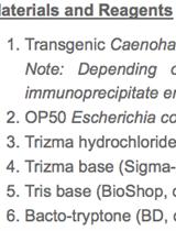
Immunoprecipitation of Proteins in Caenorhabditis elegans
免疫沉淀法检测秀丽隐杆线虫蛋白质
免疫学
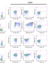
Intracellular Cytokine Staining (ICS) on Human Lymphocytes or Peripheral Blood Mononuclear Cells (PBMCs)
人淋巴细胞或外周血单核细胞(PBMC)的细胞因子胞内染色(ICS)

微生物学
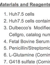
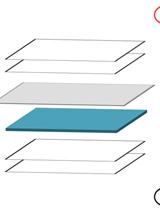
Membrane Flotation Assay
膜漂浮分析
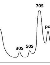
Purification of 70S Ribosomes from Bacillus subtilis
从枯草杆菌中纯化70S核糖体
Cyst Detection in Toxoplasma gondii Infected Mice and Rats Brain
刚地弓形虫感染小鼠和大鼠脑部囊肿检测


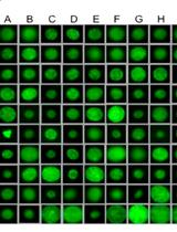
Lectin Binding Analysis of Streptococcus mutans Glycoproteins
变异链球菌糖蛋白的凝集素结合分析


神经科学

Nictation Assays for Caenorhabditis and Other Nematodes
杆状线虫和其它线虫的Nictation行为分析

植物科学
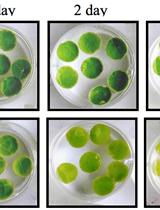
Leaf Disc Stress Tolerance Assay for Tobacco
烟草叶盘抗逆性分析


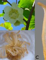
TUNEL Assay in Kiwifruit Stigmatic Arms
奇异果柱头臂的TUNEL检测


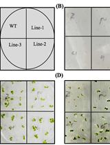
Stress Tolerance Assay at the Seed Germination Stage for Tobacco
烟草种子萌芽阶段的抗逆性分析
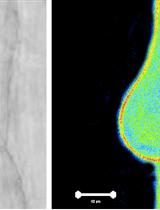
Arabidopsis thaliana Root Hair Cell Cytoplasmic pH (pHc) Imaging
拟南芥根毛细胞质pH(pHc)的成像
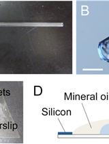
Microscopic Observation, Three-dimensional Reconstruction, and Volume Measurements of Sperm Nuclei
精子细胞核的显微观察、三维重建和体积测量

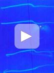
Gravitropic Analysis of Tomato Seedlings using Time Lapse Video Imaging
延时视频成像法监测番茄幼苗的向地性


