

往期刊物2017
卷册: 7, 期号: 20
细胞生物学
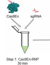
Cytosolic and Nuclear Delivery of CRISPR/Cas9-ribonucleoprotein for Gene Editing Using Arginine Functionalized Gold Nanoparticles
使用精氨酸功能化金纳米粒子进行CRISPR/Cas9-核糖核蛋白的细胞溶质和细胞核递送以用于基因编辑

发育生物学

Protocol for Notch-ligand Binding Assays Using Dynabeads
使用免疫磁珠进行Notch配体结合分析的实验方案
微生物学
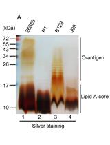
Crude Preparation of Lipopolysaccharide from Helicobacter pylori for Silver Staining and Western Blot
粗制备幽门螺杆菌脂多糖用于银染和蛋白质印迹

A Method for Radioactive Labelling of Hebeloma cylindrosporum to Study Plant-fungus Interactions
一种用于研究植物-真菌相互作用的圆形孢子粘花菇放射性标记方法


分子生物学
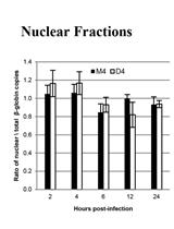
NP-40 Fractionation and Nucleic Acid Extraction in Mammalian Cells
NP-40 提取分选哺乳动物细胞核酸
Uptake Assays in Xenopus laevis Oocytes Using Liquid Chromatography-mass Spectrometry to Detect Transport Activity
非洲爪蟾卵母细胞联合液相色谱-质谱法的吸收实验检测转运活性
神经科学
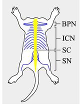
Isolation and Purification of Schwann Cells from Spinal Nerves of Neonatal Rat
从新生大鼠脊髓神经中分离和纯化施万细胞
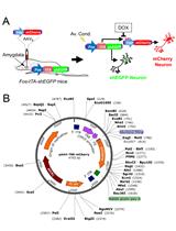
Labeling Aversive Memory Trace in Mouse Using a Doxycycline-inducible Expression System
使用多西环素诱导表达系统标记小鼠厌恶记忆痕迹


Touchscreen-based Visual Discrimination and Reversal Tasks for Mice to Test Cognitive Flexibility
采用基于触摸屏的小鼠视觉辨别和颠倒任务测试认知灵活性
植物科学
Trimolecular Fluorescence Complementation (TriFC) Assay for Direct Visualization of RNA-Protein Interaction in planta
三分子荧光互补(TriFC)实验直接观察植物中RNA-蛋白质的相互作用

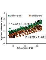
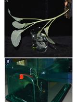
Monitoring Xylem Hydraulic Pressure in Woody Plants
木本植物中木质部液压的监测

Capturing Z-stacked Confocal Images of Living Bacteria Entering Hydathode Pores of Cauliflower
采集活细菌进入花椰菜泌水孔的Z-栈共聚焦图像

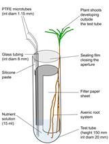
Establishing a Symbiotic Interface between Cultured Ectomycorrhizal Fungi and Plants to Follow Fungal Phosphate Metabolism
在培养的外生菌根真菌和植物之间建立共生界面以示踪真菌磷酸盐代谢
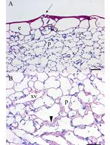
Histochemical Preparations to Depict the Structure of Cauliflower Leaf Hydathodes
用于研究花椰菜叶泌水孔结构的组织化学制备
干细胞

In vitro Co-culture of Mesenchymal Stem Cells and Endothelial Colony Forming Cells
间充质干细胞和内皮集落形成细胞的体外共培养
