往期刊物2014

卷册: 4, 期号: 21

left

Dec

20

Dec

5

Nov

20

Nov

5

Oct

20

Oct

5

Sep

20

Sep

5

Aug

20

Aug

5

Jul

20

Jul

5

Jun

20

Jun

5

May

20

May

5

Apr

20

Apr

5

Mar

20

Mar

5

Feb

20

Feb

5

Jan

20

Jan

5

right

免疫学

小鼠小肠派尔结的淋巴结细胞分离和体外激活

Isolation and in vitro Activation of Mouse Peyer’s Patch Cells from Small Intestine Tissue

小鼠小肠派尔结的淋巴结细胞分离和体外激活

CP Claudia Pastori
LL Lucia Lopalco
20487 Views
Nov 5, 2014
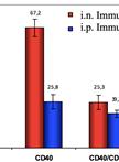
The lumen of gastrointestinal tract is exposed to several potentially pathogenic microorganisms, thus it is extremely relevant to understand how immunosurveilance can be established. Peyer’s Patches (PPs) are oval or round lymphoid nodules that protrude from the outer wall of the ileum portion of small intestine. PPs contain a high percentage of B and T lymphocytes, macrophages and dendritic cells. Here we summarize a protocol for isolation and culture of mouse PP cells, which can be used to get a better insight into immunopathologies of microbes and to evaluate immune responses elicited by mucosal vaccines.
表面等离子共振法测定原初杂交瘤样本中抗体与抗原的结合亲和力

Binding Affinity Measurement of Antibodies from Crude Hybridoma Samples by SPR

表面等离子共振法测定原初杂交瘤样本中抗体与抗原的结合亲和力

DN Dorin Mlaki Ndao
DH David T. Hickman
ML María Pilar López-Deber
AD Aurélien Davranche
AP Andrea Pfeifer
AM Andreas Muhs
16705 Views
Nov 5, 2014
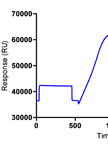
Surface Plasmon Resonance (SPR) is widely used to generate kinetic and affinity information on specific interactions between biomolecules. This technique is label-free and monitors the binding event in real-time. It is generally used for characterization of monoclonal antibody - antigen interactions. This protocol describes specifically the use of SPR with a Biacore T100 instrument to measure the affinity of crude hybridoma samples to a protein. For that purpose an anti-IgG antibody was firstly covalently immobilized onto a CM5 chip by amide coupling (Canziani et al., 2004; Schraml and Biehl, 2012). Then the antibodies from hybridoma supernatants were captured non-covalently onto the surface via their Fc region providing an optimal analyte-binding orientation. Finally, the resulting complex was stabilized by crosslinking with EDC/NHS to avoid baseline drift during measurement and regeneration (Pope et al., 2009). Then the interaction with the protein was monitored at several concentrations and its affinity towards the immobilized antibodies was determined with the corresponding KD obtained from classical kinetics analysis. This set-up avoids the avidity effects of the bivalent antibodies, allows the use of non-purified analytes with unknown concentrations and the specific capture of the antibodies in a similar stable covalent-orientated manner.

微生物学

超速离心法分级分离革兰氏阴性菌细胞质和细胞膜蛋白

Fractionation by Ultracentrifugation of Gram Negative Cytoplasmic and Membrane Proteins

超速离心法分级分离革兰氏阴性菌细胞质和细胞膜蛋白

SS Sara M Sandrini
RH Richard Haigh
PF Primrose P. E. Freestone
20087 Views
Nov 5, 2014
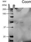
Protein fractionation is a useful separation process which divides membrane proteins (including those located in the outer and inner membrane) and cytoplasmic proteins into discrete fractions. Fractionation of proteins can simplify analysis of the numbers of proteins present, and therefore make easier to characterize any environmentally or mutation induced changes in expression profiles, or changes in protein strucutre resulting from post-translational modification. This protocol is derived from Haigh et al. (2013) and it is specific to Gram negative bacteria.
集胞藻细胞的稳定转化

Stable Transformation of Cyanobacterium Synechocystis sp.

集胞藻细胞的稳定转化

Reinhard K Proels Reinhard K Proels
13752 Views
Nov 5, 2014
Cyanobacteria are prokaryotes, which perform oxygenic photosynthesis. Among them, the unicellular cyanobacterium Synechocystis sp. PCC 6803 (hereafter Synechocystis) is a well characterized model system for studies on oxygenic photosynthesis, light signal transduction etc. Moreover, Synechocystis is applied in biotechnological applications (Desai and Atsumi, 2013). Stable transformation of Synechocytis is achieved via the uptake of DNA and incorporation into the host genome by homologous double recombination. This allows for the generation of gene knock-outs (KO) by replacing the coding sequence of the gene of interest by a KO-cassette (comprising of a selection marker flaked by sequences of the gene of interest) or stable overexpression of certain genes of interest after insertion of a corresponding overexpression cassette at a neutral insertion site on the host genome. Stable transformation of Synechocystis was reported by Grigorieva and Shestakov (1982). Since then, variants of the initial protocol have been applied successfully to transform Synechocystis sp. Here we describe a lab-protocol that was applied successfully for stable transformation of Synechocystis (Schwarzkopf et al., 2014).
大肠杆菌质子逆向运输蛋白活性的测定

Measurement of Proton-driven Antiport in Escherichia coli

大肠杆菌质子逆向运输蛋白活性的测定

SH Scarlett R. Holdsworth
Christopher J. Law Christopher J. Law
9256 Views
Nov 5, 2014
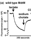
Secondary active transport of substrates across the inner membrane is vital to the bacterial cell. Of the secondary active transporter families, the ubiquitous major facilitator superfamily (MFS) is the largest and most functionally diverse (Reddy et al., 2012). Recently, it was reported that the MFS multidrug efflux protein MdtM from Escherichia coli (E. coli) functions physiologically in protection of bacterial cells against bile salts (Paul et al., 2014). The MdtM transporter imparts bile salt resistance to the bacterial cell by coupling the exchange of external protons (H+) to the efflux of bile salts from the cell interior via an antiport reaction. This protocol describes, using fluorometry, how to detect the bile salt/H+ antiport activity of MdtM in inverted membrane vesicles of an antiporter-deficient strain of E. coli TO114 cells by measuring transmembrane ∆pH. This method exploits the changes that occur in the intensity of the fluorescence signal (quenching and dequenching) of the pH-sensitive dye acridine orange in response to changes in [H+] in the vesicular lumen. Due to low levels of endogenous transporter expression that would normally make the contribution of individual transporters such as MdtM to proton-driven antiport difficult to detect, the method typically necessitates that the transporter of interest be overexpressed from a multicopy plasmid. Although the first section of the protocol described here is very specific to the overexpression of MdtM from the pBAD/Myc-His A expression vector, the protocol describing the subsequent measurement of bile salt efflux by MdtM can be readily adapted for measurement of antiport of other substrates by any other antiporter that exchanges protons for countersubstrate.
纯化和检测诺尔曼氏极小单孢菌中的PDGA解聚酶

Purification and Detection of a PDGA Depolymerase from Pusillimonas noertemannii

纯化和检测诺尔曼氏极小单孢菌中的PDGA解聚酶

DN David Negus
PT Peter W. Taylor
9014 Views
Nov 5, 2014
The purification of a target protein from a complex mixture of proteins is a challenging undertaking. If the target protein has been previously characterised, then information such as subcellular location, function, molecular weight and pI can be used for the design of a purification strategy. However, if the target protein is uncharacterised or little information regarding its characteristics is available, a generic purification protocol can be employed that is optimised as additional characteristics of the target protein are determined during subsequent purification steps. Herein, we describe the protocol for the purification and detection of a poly-γ-D-glutamic acid (PDGA) depolymerase from a consortium culture of two Gram-negative bacteria using a combination of chromatography, 2D-electrophoresis and zymography.
具有抗炭疽杆菌潜力的EnvD储藏稳定性提高的检测

Accelerated Storage Stability Testing of a Potential Anti-Anthrax Therapeutic, EnvD

具有抗炭疽杆菌潜力的EnvD储藏稳定性提高的检测

DN David Negus
PT Peter W. Taylor
8369 Views
Nov 5, 2014
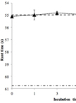
The purpose of stability testing is to determine how the properties of a particular therapeutic vary with time under the influence of specific environmental factors. Information regarding the long-term stability of therapeutics can be extrapolated by performing an accelerated storage stability study. Here, we describe an accelerated storage stability study for the potential anti-anthrax therapeutic, EnvD, a poly-γ-D-glutamic acid (PDGA) depolymerase. Storage conditions were based on those recommended by the International Conference on Harmonisation of Technical Requirements for Registration of Pharmaceuticals for Human Use (ICH). Owing to the high-molecular-weight and associated viscosity of PDGA in solution, loss of enzyme activity on storage can be determined as a reduction in the capacity of the depolymerase to reduce the viscosity of the polymer. This work supported by a Medical Research Council Capacity Building Studentship award.
大肠杆菌中胆汁盐/H+反向协同运输蛋白的电性活力测定

Measurement of the Electrogenicity of Bile Salt/H+ Antiport in Escherichia coli

大肠杆菌中胆汁盐/H+反向协同运输蛋白的电性活力测定

SH Scarlett R. Holdsworth
Christopher J. Law Christopher J. Law
7623 Views
Nov 5, 2014
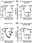
The transmembrane proton gradient (ΔpH) is the primary source of energy exploited by secondary active substrate/H+ antiporters to drive the electroneutral transport of substrates across the Escherichia coli (E. coli) inner membrane. Such electroneutral transport results in no net movement of charges across the membrane. The charge on the transported substrate and the stoichiometry of the exchange reaction, however, can result in an electrogenic reaction which is driven by both the ΔpH and the electrical (∆Ψ) components of the proton electrochemical gradient, resulting in a net movement of electrical charges across the membrane. We have shown that the major facilitator superfamily transporter MdtM - a multidrug efflux protein from E. coli that functions physiologically in protection of bacterial cells against bile salts - imparts bile salt resistance to the bacterial cell by coupling the exchange of external protons (H+) to the efflux of bile salts from the cell interior via an electrogenic antiport reaction (Paul et al., 2014). This protocol describes, using fluorometry, how to detect electrogenic antiport activity of MdtM in inverted membrane vesicles of an antiporter-deficient strain of E. coli TO114 cells by measuring transmembrane ∆Ψ. The method exploits changes that occur in the intensity of the fluorescence signal (quenching and dequenching) of the probe Oxonol V in response to changes in membrane potential due to the MdtM-catalysed sodium cholate/H+ exchange reaction. The protocol can be adapted to detect activity of any secondary active antiporter that couples the electrogenic translocation of H+ across a biological membrane to that of its counter-substrate, and may be used to unmask otherwise camouflaged transport activities and physiological roles.

神经科学

采用大鼠和小鼠福尔马林试验进行疼痛评估

Pain Assessment Using the Rat and Mouse Formalin Tests

采用大鼠和小鼠福尔马林试验进行疼痛评估

Nian Gong Nian Gong
Qian Huang Qian Huang
Yuan Chen Yuan Chen
Meng Xu Meng Xu
Shuai Ma Shuai Ma
Yong-Xiang Wang Yong-Xiang Wang
27013 Views
Nov 5, 2014
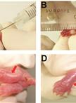
The formalin test was originally developed by Dubuisson and Dennis (1977), and has since been extensively used to assess pain-related responses. Rats and mice are the most frequently used animal models, though other species including cats, rabbits, guinea pigs, Octodon degus, domestic fowls, crocodiles, tortoises, toads and primates have also been employed. The injection of formalin into the skin of rodent hindpaws to cause spontaneous pain-related flinch behaviors is the most commonly used procedure. The resulting nociceptive response can be divided into two phases differing in timing, duration and underlying mechanisms, and is responsive to many classes of analgesic drugs (Coderre et al., 1990; Hunskaar and Hole, 1987; Rosland et al., 1990; Taylor et al., 1995). The behavioral and electrophysiological responses to formalin consist of an acute phase (Phase I) of a short-lasting response, which is believed to reflect the activity of C-fiber afferent nociceptors. After a short quiescent period, the acute phase is followed by a continuous prolonged response (Phase II), which is believed to be due to central sensitization of the spinal dorsal horn neurons as a result of the initial barrage of input from C-fiber nociceptive afferents during the early phase (Coderre et al., 1993; Dickenson and Sullivan, 1987; Raboisson et al., 1995; Shibata et al., 1989). In this respect, the formalin test has been regarded as a more satisfactory model of pain than tests producing phasic pain like the hot-plate and tail-flick tests (Abbott et al., 1981). Here, we describe the procedures for generating an efficient and reproducible formalin test in rats and mice.
新生大鼠伤害性刺激的实验模型

An Experimental Model of Neonatal Nociceptive Stimulation in Rats

新生大鼠伤害性刺激的实验模型

JM Jackeline Moraes Malheiros
CA Cristiane Amaral
AL Ana Teresa S Leslie
RG Ruth Guinsburg
Luciene Covolan Luciene Covolan
10265 Views
Nov 5, 2014
In order to survive, preterm and/or sick neonates need diagnostic and therapeutic measures that may cause discomfort, stress and pain during a critical period of intense growing and modeling of the central nervous system (Anand et al., 2013). Scientific interest in the long lasting effects of the Neonatal Intensive Care (NIC) experience, which provides a sensory experience completely different from the natural uterine environment, is growing (Jobe, 2014). The follow-up of critically ill newborn infants until adulthood indicated an association of early noxious stimuli with long lasting alterations in somatosensory and cognitive processing (Doesburg, 2013; Vinall et al., 2014; Vinall et al., 2013). However, one major limitation of the clinical studies is the difficulty to distinguish between long-term effects of pain suffered during neonatal intensive care and other confounding factors such as the presence of non-painful stress during hospital stay, the occurrence of acute and chronic morbidities, the post-natal environmental influences and family care. In this context, the understanding of the roles played by each factor and the interplay between these diverse variables require the use of animal models. The protocol described here is used to model the noxious stimulation in which premature newborns are subjected during treatment in the NIC. The current protocol models inflammatory nociceptive stimulation in neonatal rats, as previously demonstrated (Leslie et al., 2011; Lima et al., 2014; Malheiros et al., 2014). Complete Freund's adjuvant (CFA) is a solution of antigen emulsified in mineral oil and used as an immunopotentiator, causing a painful reaction that lasts 7-8 days after subcutaneous injection. It is effective in stimulating cell-mediated immunity. The rodent model of neonatal inflammatory stimulation with CFA is advantageous because at birth the formation of the central nervous system is incomplete in rat pups and corresponds to that of 24 week intra-uterine human preterm neonates (Anand et al., 1999), following similar patterns in the development of the pain system (Fitzgerald and Anand,1993). The first postnatal week in newborn rat pups corresponds to human premature infants from 24-36 weeks of gestation (Kim et al., 1996; Wilson, 1995), offering a suitable condition to model and compare preterm (rat pups on P1) to full term (rat pups on P8) infants subjected to noxious stimulation. In this paper, we present our methods to induce nociceptive inflammatory stimulation in neonatal rat pups as an attractive approach to study short- or long-term effects and the mechanisms underlying the behavioral repertoire of ex-premature infants or adolescents.

植物科学

从水稻植株中提取总蛋白

Extraction of Total Proteins from Rice Plant

从水稻植株中提取总蛋白

DL Da-Gin Lin
CW Chang-Sheng Wang
44074 Views
Nov 5, 2014
This protocol provides an efficient method for preparation of high-quality proteins from rice leaves and grains. The method involves phenol extraction to separate proteins from the non-protein components such as polysaccharides, lipids and phenolic compounds that are commonly enriched in plant tissues. Following isolation, proteins are precipitated with ammonium acetate/methanol and then solubilized for proteomic analysis. As the protocol is simple, universal, and most importantly compatible with silver staining, it has been applied to our routine protein extraction from rice and many other plant tissues and it even works fine in animal tissues for the requirement of electrophoretic separation.
制备植物复用小型RNA库

Preparation of Multiplexed Small RNA Libraries from Plants

制备植物复用小型RNA库

KG Kerrigan B. Gilbert
NF Noah Fahlgren
KK Kristin D. Kasschau
EC Elisabeth J. Chapman
JC James C. Carrington
Alberto Carbonell Alberto Carbonell
13756 Views
Nov 5, 2014
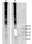
High-throughput sequencing is a powerful tool for exploring small RNA populations in plants. The ever-increasing output from an Illumina Sequencing System allows for multiplexing multiple samples while still obtaining sufficient data for small RNA discovery and characterization. Here we describe a protocol for generating multiplexed small RNA libraries for sequencing up to 12 samples in one lane of an Illumina HiSeq System single-end, 50 base pair run. RNA ligases are used to add the 3’ and 5’ adaptors to purified small RNAs; ligation products that lack a small RNA molecule (adaptor-adaptor products) are intentionally depleted. After cDNA synthesis, a linear PCR step amplifies the DNA fragments. The 3’ PCR primers used here include unique 6-nucleotide sequences to allow for multiplexing up to 12 samples.
异源表达植物蛋白的糖基转移酶活性的体外检测

In vitro Assay of the Glycosyltransferase Activity of a Heterologously Expressed Plant Protein

异源表达植物蛋白的糖基转移酶活性的体外检测

CA Carsten P Ade
FB Felix Bemm
JD James M J Dickson
CW Christian Walter
PH Philip J Harris
11844 Views
Nov 5, 2014
Glycosyltransferases are carbohydrate active enzymes containing catalytic modules involved in catalysing the biosynthesis of glycosidic bonds in oligo- and polysaccharides and glycoconjugates. One of the most comprehensive collections of Carbohydrate Active enZYmes is the CAZy database (http://www.cazy.org) comprising 120,000 glycosyltransferases allocated to 96 families based mainly on sequence homologies of their conserved and catalytically active domains (Cantarel et al., 2009). Interestingly, the glycosyltransferase activities of only about 1.6% of these proteins have been experimentally characterized (Lombard et al., 2014). In recent years, membrane-bound glycosyltransferases of a number of families have been shown to play a key role in the biosynthesis of plant cell-wall polysaccharides (Doblin et al., 2010; Scheller and Ulvskov, 2010; Driouich et al., 2012). They catalyze the transfer of glycosyl residues from donor nucleotide sugars to acceptors, forming the glycosidic bonds between adjacent glycosyl residues. Family 34 contains glycosyltransferases that have been shown to be involved in the biosynthesis of xyloglucans and transfer xylosyl residues to (1→4)-β-glucan chains (Keegstra and Cavalier, 2011). Our previous work suggests that Pinus radiata protein PrGT34B is a xyloglucan (1→6)-α-xylosyltransferase (Ade et al., 2014). Here, we describe a procedure for determining the xylosyltransferase activity of PrGT34B in vitro. We measured the transfer of xylose from the donor substrate UDP-xylose to different cello-oligosaccharide acceptor substrates under controlled reaction conditions. The assays include quantification of radioactively labeled reaction products and their identification by mass spectrometry. We also describe the purification, identification and quantification of the heterologously expressed recombinant protein PrGT34B in preparation for its use in the assays. This procedure may be applied to a wide range of glycosyltransferases in many different plant species.
过氧化物酶体数量的测量

Measurement of the Number of Peroxisomes

过氧化物酶体数量的测量

MS Michitaro Shibata
Kazusato Oikawa Kazusato Oikawa
SM Shoji Mano
Mikio Nishimura Mikio Nishimura
10122 Views
Nov 5, 2014
This is the detailed protocol for the measurement of the number of peroxisomes described by Shibata et al. (2013). It is difficult to count the number of organelles in a cell because of the thickness of plant leaves. To overcome this challenge, protoplasts were isolated from leaves, and the number of peroxisomes per protoplast was counted. This method can be applied to other organelles such as mitochondria that are labeled with GFP or its derivatives.