- Protocols

- Articles and Issues
- For Authors
- About
- Become a Reviewer


A Versatile In Vitro Quantitative Assay for Macrophage Efferocytosis in Diverse Research Applications
Macrophage efferocytosis is a previously unrecognized key pathogenic event, engulfing apoptotic targets, preventing inflammation and necrosis, and maintaining immune homeostasis. The phagocytic function can be disrupted by harmful factors and toxic substances. This protocol describes a versatile visualized in vitro method that can be used for the detection of general efferocytosis. This method is applicable to a wide range of research scenarios. As a representative application, it can be used to evaluate macrophage efferocytosis dysfunction in diseases linked to harmful exposures, including atherosclerosis, chronic inflammation, and malignant tumors. Among them, the detection of the effects of oxidized low-density lipoprotein (ox-LDL) and arsenite on macrophage efferocytosis capacity is an exemplary application of this protocol. Primary macrophages collected from mice were labeled with a cell-tracking dye and exposed to ox-LDL or arsenite, then co-cultured with apoptotic thymocytes or hepatocytes (labeled with another cell-tracking dye) for 2 h at a ratio of 5:1. Macrophage efferocytosis was visualized using a laser confocal microscope. The results indicate that arsenite impaired macrophage efferocytosis, leading to insufficient clearance of apoptotic thymocytes or hepatocytes. This method can be extended to subsequent studies, including those involving different types of phagocytes, apoptotic cell models, and research related to exposure to various factors.
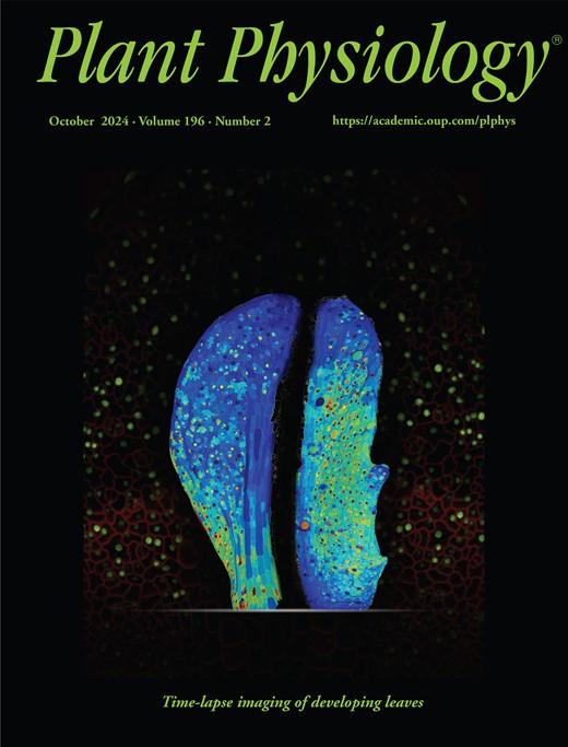
Analysis of Cauline Leaf Development in Arabidopsis thaliana Using Time-Lapse Confocal Microscopy
Understanding cellular growth dynamics in plants requires precise, long-term imaging of developing tissues. Cauline leaves are produced during the transition from vegetative to reproductive development and provide a useful system for studying how laminar organs diversify in form and function. While other laminar organs, such as rosette leaves and sepals, have been extensively studied, early cauline leaf development remains technically challenging to capture due to their concealed position, curved morphology, and the presence of dense trichomes. Here, we provide a complete pipeline for the dissection, confocal imaging, 2.5D segmentation, and image analysis of initiating cauline leaves in Arabidopsis thaliana. This method enables reproducible, high-resolution imaging of cauline leaves, supporting robust quantitative analysis of growth across developmental stages at cellular scale resolution.

An In Vitro A-431 Epithelial Cell Infection Model for Studying Fungal Pathogenicity and Immune Responses Associated With Vulvovaginal Candidiasis
Vulvovaginal candidiasis (VVC), also known as vaginal thrush, is an infection of the vulvovaginal mucosa caused by fungi of the Candida genus. Particularly for patients suffering from recurrent infection, the disease has a significant impact on their quality of life. The still unknown aspects of disease pathogenesis, as well as factors driving the development of infections and recurrence, represent a challenge for both clinical practitioners and patients. Mouse models and patient studies have suggested important roles of the microbiome, deployment of fungal pathogenicity mechanisms in the vagina, and dysregulated immune responses for VVC pathology. Dissecting their individual contributions can reveal specific processes associated with infection and may inspire novel therapeutic strategies. Epithelial in vitro infection models have been playing a key role in dissecting a crucial interaction during VVC, the invasion and infection of the vaginal mucosa. They have been instrumental in characterizing candidalysin as a fungal toxin that damages epithelial cells and elicits initial inflammatory responses to catalyze downstream inflammation. Moreover, they have also revealed potential protective immune pathways. Such a standardized epithelial cell infection model offers high versatility and compatibility with different downstream assays to link epithelial responses with other processes during VVC. This protocol describes a general A-431 vulvovaginal epithelial cell–Candida infection model in detail and provides several adaptations, such as live-cell imaging and mRNA silencing, as well as possible follow-up readouts, like the quantification of cytokine release, cytotoxicity, and neutrophil recruitment to study diverse processes relevant to VVC research.
3D Reconstruction of Mature Arabidopsis Ovules Using FIB-SEM to Study Filiform Apparatus Morphology
Volume electron microscopy based on serial sectioning allows for three-dimensional (3D) visualization and analysis of the internal structures of tissues, cells, and organelles. One such technique, focused ion beam (FIB) scanning electron microscopy (SEM), has the advantages of nanoscale sectioning and high z-resolution, but the disadvantage of limited volume processing. Because of this limitation, targeting localized objects by FIB-SEM is difficult. Here, we developed a FIB-SEM observation workflow that enables the analysis of the filiform apparatus of synergid cells enclosed in the Arabidopsis ovule. In this protocol, plant samples are stained, embedded, trimmed, and carbon-coated while maintaining their orientation within the tissue. Then, sequential observations are performed using Cut & See function of FIB-SEM, followed by image processing for 3D reconstruction. Utilization of multi-scanning and image cropping from high-resolution data helps to identify localized targets within plant tissue. The filiform apparatus, which is an invaginated cell wall structure of the synergid cells, shows distinct contrast in each image, allowing for segmentation using brightness-based binarization. Such segmentation avoids the need to manually trace complex structures and facilitates 3D reconstruction by volume electron microscopy.
Quantification of Spatial Patterns of Microtubule Transport by Kinesin-1 Head and Tail
The conventional kinesin-1 is a plus-end-directed microtubule-dependent motor protein with distinct motor head, stalk, and tail domains. Along with the motor head, which binds and walks along microtubules in an adenosine 5’-triphosphate (ATP) dependent manner, kinesin also contains a C-terminal microtubule binding tail. Motor-driven collective motility is well characterized using in vitro gliding assays, which show uninterrupted, smooth trajectories of transport. However, gliding assays driven by the full-length Drosophila kinesin-1 with both head and tail resulted in the emergence of spontaneous spatial microtubule patterns and stop-and-go motion. This was reproduced by an equimolar ratio of the active head and passive tail. Here, we describe the detailed protocol to reconstitute these microtubule gliding assays using multiple motor types: the full-length kinesin-1, the motor head or tail, mixtures of both head and tail, and a rigor mutant of the kinesin. We provide details of the approach taken to acquire the image time-series, to then quantify the spatial patterns that result from these motor combinations. Our approach provides a framework to systematically characterize the spatiotemporal effects of molecular motor-driven collective microtubule transport.

Isolation and Biophysical Characterization of Extracellular Vesicles Released by Myocytes
Extracellular vesicles (EVs) are lipid bilayer–enclosed vesicles released by diverse cell types and found in various body fluids. Because their composition and cargo dynamically respond to physiological and environmental cues, EVs hold promise both as biomarkers and as carriers for therapeutic delivery. Skeletal muscle functions as an endocrine organ, secreting myokines and EVs that modulate a wide range of cellular processes. The murine C2C12 cell line is a widely used in vitro model for investigating muscle biology. Here, we describe a protocol for isolating EVs from differentiated C2C12 myocytes. The isolated EVs are characterized and validated using western blotting, transmission electron microscopy (TEM), and dynamic light scattering (DLS) analysis. This workflow provides a robust platform for studying the molecular composition and functional roles of muscle-derived EVs.
Extraction and Isolation of Extracellular Vesicles From Piper betle Leaves Using the Apoplastic Fluid Washing and Size Exclusion Chromatography Method

Plant-derived extracellular vesicles (PDEVs) have emerged as important mediators of intercellular communication and hold growing potential in therapeutic applications. However, standardized methods for their isolation, particularly from Piper betle leaves (PBL), remain unexplored. Existing apoplastic fluid washing (AFW) extraction techniques typically rely on manual syringe infiltration, which often leads to inconsistent pressure control, variable yields, and increased risk of tissue damage. This protocol describes a vacuum-assisted AFW extraction method optimized for the recovery of intact extracellular vesicles (EVs) from PBL. The workflow features controlled negative pressure using a vacuum pump and chamber to achieve more efficient leaf infiltration compared to infiltration using the syringe method and reproducible apoplastic fluid (AF) collection with subsequent low-speed centrifugation steps, to ensure minimal contamination and preservation of vesicle integrity. Piper betle–derived extracellular vesicle (PBdEV) isolation and purification steps are performed using size exclusion chromatography (SEC). The size and concentration of PBdEVs were confirmed using nanoparticle tracking analysis (NTA), whereas the cup-shaped and lipid bilayer morphology of the EVs were confirmed using transmission electron microscopy (TEM). The method is scalable and adaptable to various leaf morphologies and physiological states, making it suitable for both exploratory and high-throughput studies. Overall, this protocol provides a more consistent, efficient, and tissue-preserving alternative to traditional syringe-based AF extraction methods, offering higher-quality EV preparations for plant EV research.
Detecting Touch-Induced Calcium Dynamics With Live-Cell Imaging in Torenia Stigma
Calcium ions serve as a universal secondary messenger, integrating diverse external signals, such as light, herbivory, and mechanical stimuli, within plant cells. However, the visualization and mechanistic dissection of calcium signaling specifically in response to mechanical stimulation remain technically challenging and underexplored in most plants. Previous studies have been largely confined to a few model systems, including Arabidopsis; here, we introduce a live-cell imaging approach using the stigmas of Torenia fournieri. This in vitro system enables multiscale observation of calcium signal patterns following controlled mechanical stimulation. This versatile platform not only simplifies the design of calcium imaging assays but also provides a tractable system for functionally validating other key molecular components in this signaling pathway.
A Cell-Based Protocol to Assess Manganese Content and Relative Transport Activity of Manganese Transporters
Manganese (Mn) is an essential trace element whose intracellular homeostasis is tightly controlled by specialized membrane transporters. Dysregulation of Mn transport leads to pathological Mn accumulation and severe human disease; however, efficient and quantitative cell-based methods for assessing Mn2+ transporter activity remain limited. Here, we present an optimized cellular Fura-2 manganese extraction assay (CFMEA) that enables robust quantification of cellular Mn content and provides a normalized framework for assessing relative Mn2+ transport activity in a high-throughput format. This protocol integrates Fura-2-based fluorescence detection of Mn2+ at the Ca2+ isosbestic excitation wavelength with dsDNA quantification to normalize dsDNA levels in cell extracts and immunoblotting to account for transporter protein expression levels. Cells expressing Mn2+ transporters are exposed to MnCl2 in 96-well plates, washed to remove extracellular Mn2+, and lysed in a Fura-2-containing extraction buffer. Fluorescence quenched by Mn2+ is quantified and converted to cellular Mn content using a cell-free Mn-Fura-2 standard curve and then normalized to dsDNA content and protein abundance to determine relative transporter activity. This workflow provides a relatively sensitive, reproducible, and low-cost approach for comparative analysis of Mn2+ transporters and their variants across multiple cell types. The protocol is demonstrated using the Mn2+ efflux transporter SLC30A10 in HEK293T cells and is readily adaptable for studying other Mn2+ transport pathways.
An Advanced Single-Cell RNA Sequencing (scRNA-seq) Protocol Utilizing Custom-Designed Multiplexing
While cell hashing enhances single-cell RNA sequencing (scRNA-seq) efficiency and minimizes batch effects, commercial mouse hashtags often fail in FVB/N and several other strains due to antibody-epitope incompatibility. We describe a robust alternative utilizing biotinylated antibody cocktails and streptavidin-conjugated oligos to enable reliable sample multiplexing. This approach was validated in FVB/N lung tissues, yielding high-quality single-cell libraries. Our protocol offers a practical solution for researchers requiring strain-specific or custom-designed multiplexing strategies for single-cell transcriptomics.
Reconstitution of Active Plant H+-ATPase AHA2 in Giant Unilamellar Vesicles
Membrane transporters mediate the selective movement of ions and molecules across biological membranes and are essential for cellular homeostasis. However, their functional characterization in living cells is often complicated by the complexity of the native membrane environment. Reconstitution into model membrane systems provides a powerful alternative by enabling precise control over lipid composition and experimental conditions. Giant unilamellar vesicles (GUVs) are particularly well suited for transporter studies, as their cell-sized dimensions allow direct microscopic observation and fluorescence-based measurements of protein activity. Here, we describe a two-step reconstitution protocol in which transport proteins are first incorporated into large unilamellar vesicles and then used to generate protein-containing giant unilamellar vesicles (proteo-GUVs) via the poly(vinyl alcohol) swelling method. This two-step approach enhances protein incorporation efficiency and preserves transporter functionality. The method is exemplified using the P3-type ATPase Arabidopsis thaliana plasma membrane H+-ATPase isoform 2 (AHA2). We further describe a fluorescence-based assay to assess proton transport activity in proteo-GUVs. Our approach provides a versatile and controlled platform for biochemical, biophysical, and single-molecule analysis of membrane transporters.
TALENs and Related Technologies for Editing Nuclear and Organellar Genomes in a Model Plant, Arabidopsis thaliana
Plant genome editing is a powerful approach for modifying plant DNA to investigate gene function and to engineer desirable traits. Several genome-editing technologies have been developed, among which CRISPR/Cas systems and transcription activator-like effector nucleases (TALENs) are widely used to introduce targeted double-stranded DNA breaks. While CRISPR/Cas systems are highly efficient for nuclear genome editing, their application to plant organellar genomes remains limited, largely due to difficulties in guide RNA delivery into mitochondria and chloroplasts. Here, we present a detailed and reproducible protocol for constructing TALEN-based binary vectors for targeted genome editing in Arabidopsis thaliana. This protocol describes the assembly of TALE repeat arrays, the generation of nuclear-, mitochondrial-, and plastid-targeted TALEN expression vectors using MultiSite Gateway cloning, and subsequent Agrobacterium-mediated plant transformation and genotyping. The workflow enables the production of nTALENs, mitoTALENs, and ptpTALENs using a unified vector design strategy. In addition, the protocol briefly outlines the construction principles of TALE-based cytidine deaminases (TALECDs) for targeted C-to-T base editing in plant organellar genomes. The protocol provides a flexible and robust framework for plant nuclear and organellar genome editing and can be readily adapted to different target genes and experimental purposes. Its modular design and compatibility with standard molecular cloning techniques make it accessible to laboratories aiming to perform precise genome manipulation in plants.
Assessing Mitochondrial Respiratory Complex-Associated Function From Previously Frozen Mouse Placental Tissue
The placenta is a metabolically active organ whose mitochondrial activity is tightly linked to fetal growth, oxygenation, and nutrient transport, mediating fetal susceptibility to environmental exposures. Accordingly, aberrant mitochondrial function has been implicated in the progression of placental dysfunction. However, existing respirometry platforms require primarily fresh or cryopreserved placental tissue and offer limited throughput, rendering these platforms impractical in the context of large-scale placental dissections. Here, we describe and validate a Seahorse XF approach for measuring mitochondrial respiration in previously frozen placentae, enabling the functional interrogation of placental mitochondria in prenatal studies. Our protocol fundamentally relies on the restoration of matrix substrates that are depleted due to increased mitochondrial membrane permeability following freeze-thaw cycles. We provide a strategy to assess complex I and II-associated respiration adapted for the Seahorse XFe24 Analyzer and further demonstrate comparable oxygen consumption readouts between fresh and frozen placentae. We further demonstrate distinct differences in the magnitude of oxygen consumption between fresh and frozen placentae in the absence of exogenous NADH. Taken together, we present a simplified and convenient protocol for the assessment of respiratory enzyme complex-associated respiration from archived placental tissue.
Quantitative Assessment of Heat Shock-Induced Ferroptosis-Like Cell Death via Electrolyte Leakage in Arabidopsis thaliana Seedlings
We present a protocol to allow continuous assessment of cell death in Arabidopsis thaliana (L.) seedlings by measuring the release of electrolytes from dying cells upon heat shock. The electrolyte leakage assay is a well-established method to quantify the extent of cell death of plant tissues exposed to pathogen infection, since the activation of the immune response leads to compromised membrane integrity and to the release of ions from the dying cell. This prolonged release of electrolytes is considered a hallmark of regulated cell death in plants. Heat shock in plants induces ferroptosis-like cell death, which can be suppressed either pharmacologically, using inhibitors such as ferrostatin, or genetically through knockout of ferroptosis-related genes. Here, we have adapted the electrolyte leakage assay to quantify cell death in young Arabidopsis seedlings exposed to a heat shock previously shown to induce ferroptosis-like cell death. We also illustrate how this method can be used to assess activation of ferroptosis-like cell death in whole Arabidopsis seedlings using ferrostatin or knockout mutants of potential gene candidates involved in ferroptosis-like cell death.
Assessment of Epithelial Barrier Integrity by TEER and FITC-Dextran Permeability Assays
The integrity of epithelial barriers is essential for maintaining tissue homeostasis, particularly in the intestinal tract, where it separates the host from the complex luminal environment. Two complementary, standard methods for assessing this barrier are transepithelial electrical resistance (TEER), which provides a rapid, non-destructive measure of ionic conductance across tight junctions, and the fluorescein isothiocyanate (FITC)-dextran assay, which directly quantifies paracellular macromolecule flux. This protocol details a robust and reproducible method for performing both assays using intestinal epithelial cell monolayers (e.g., Caco-2, T84) cultured on permeable Transwell supports. We outline the procedure from cell culture and monolayer differentiation to TEER measurement with an Epithelial Volt/Ohm Meter 3 (EVOM3) and the subsequent FITC-dextran permeability assay. By combining these techniques, this protocol provides a comprehensive assessment of barrier function, making it ideal for studying tight junction dynamics and regulation under various experimental conditions, such as cytokine stimulation, drug screening, or microbial challenges.
Accessible STORM Imaging: An Optimized Workflow for Conventional Widefield Epifluorescence/TIRF Setups
Stochastic optical reconstruction microscopy (STORM) is a single-molecule localization microscopy technique that enables visualization of cellular structures beyond the diffraction limit. This approach has revealed previously inaccessible ultrastructural details in a wide range of cellular components, including the actin cytoskeleton, clathrin-coated pits, mitochondria, and bacterial nucleoid-associated proteins. STORM relies on the sequential emission of single photons from photosensitive fluorophores, which are precisely localized before entering a dark state or undergoing photobleaching. By activating fluorophores individually and fitting their point spread functions (PSFs), the center of mass can be calculated with a localization precision of up to ~20 nm. The parallel detection of thousands of single-molecule events, each assigned to distinct spatial coordinates, enables the reconstruction of a high-resolution image. Here, we describe a simple and efficient STORM workflow—including sample preparation, image acquisition, and quality control measurements—that we used to visualize various subcellular structures, such as mitochondria, microtubules, and lysosomes labeled with the commonly employed cyanine dye Alexa Fluor 647, as well as the actin cytoskeleton stained with Alexa Fluor 488–conjugated phalloidin. Image acquisition was performed using a conventional epifluorescence/total internal reflection (TIRF) microscope adapted for STORM imaging. Key adaptations included the use of a 160×/1.43 NA oil-immersion objective and a high-power mode, which concentrates the laser beam onto a small region of the sample, ensuring sufficient light intensity to drive fluorophores into the dark state. In addition, implementing a 1.6× magnification lens and a 4×4 binning camera mode allowed us to achieve a 100-nm pixel size optimal for reliable molecule detection. We believe that this protocol will be highly valuable to the microscopy community, as it lowers technical barriers to performing STORM on widely available microscopy platforms, thereby facilitating broader implementation of this powerful super-resolution technique.
- 1
- 2
- 3
- 4
- 5
- 6
- 200