Immunology


Categories

Protocols in Current Issue
Protocols in Past Issues
0 Q&A 555 Views Jul 20, 2023

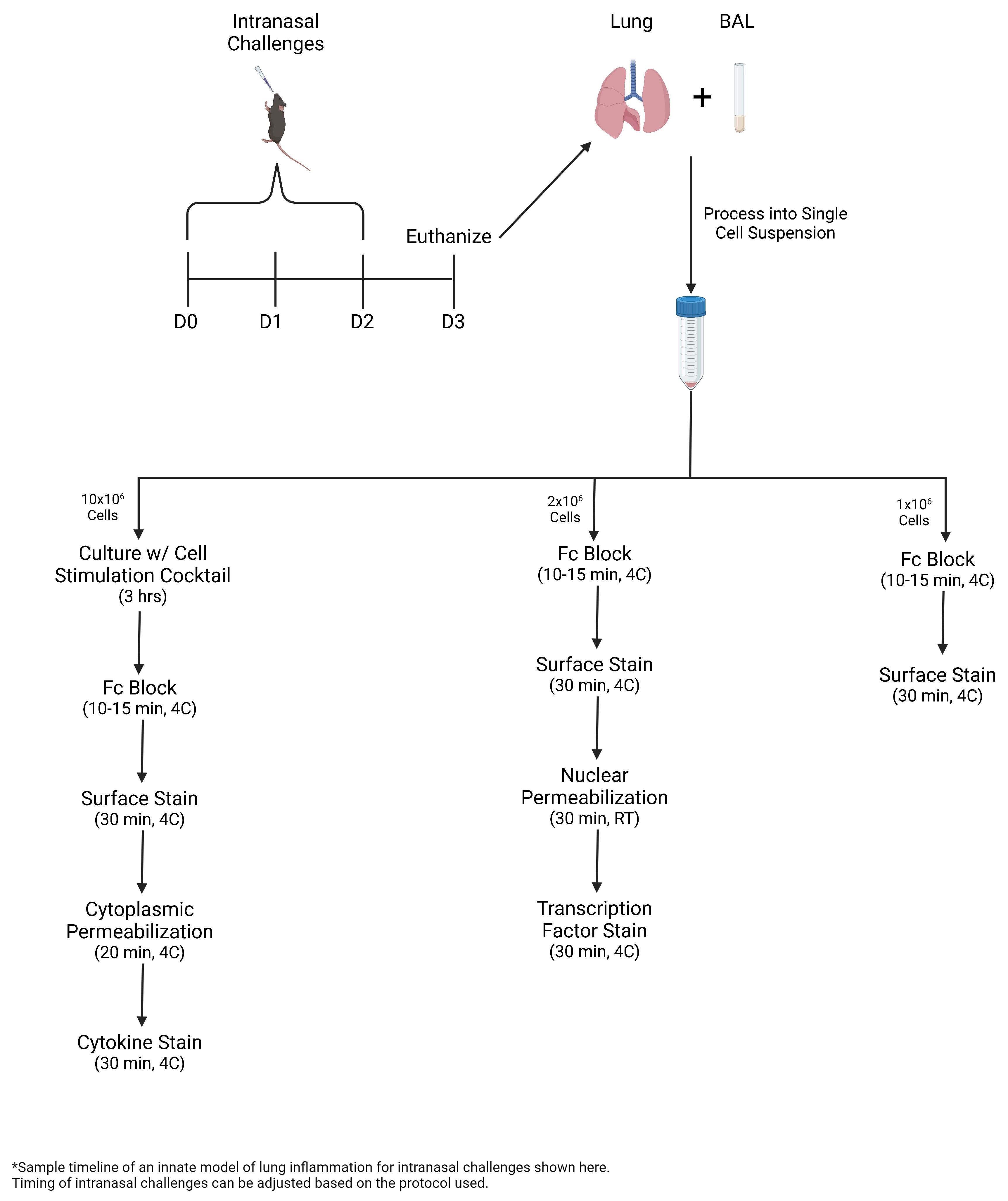
Innate lymphoid cells (ILCs) are a rare cell population subdivided into ILC1s, ILC2s, and ILC3s, based on transcription factor expression and cytokine production. In models of lung inflammation, the release of alarmins from the epithelium activates ILC2s and promotes the production of Th2-cytokines and the proliferation and migration of ILC2s within the lung. ILC2s are the innate counterpart to CD4+ Th2s and, as such, express Gata-3 and produce IL-4, IL-5, and IL-13. Due to the low number of ILCs and the lack of specific surface markers, flow cytometry is the most reliable technique for the identification and characterization of ILCs. In this protocol, multicolor flow cytometry is utilized to identify LineageThy1.2+ ILCs. Intracellular cytokine staining further identifies ILC2s within the lung. This protocol presents a reliable method for promoting ILC2-mediated lung inflammation and for monitoring ILC2 biology.


Key features

• In this protocol, ILC2s are expanded via intranasal challenges with Alternaria alternata, a fungal allergen, or recombinant IL-33.

• Bronchoalveolar lavage (BAL) and lung are collected and processed into single-cell suspension for multicolor flow cytometric analysis, including intracellular staining of transcription factors and cytokines.

• During lung inflammation, the percentage of ILC2s and eosinophils increases. ILC2s express greater levels of Gata-3 and Ki-67 and produce greater amounts of IL-5 and IL-13.


Graphical overview

0 Q&A 1961 Views Jul 20, 2022

Employing a novel mouse model of immune related adverse events (irAEs) induced by combination of anti-PD1 and anti-CTLA-4 antibodies, we visualized immune infiltration into the liver, lung, pancreas, and colon. Here, we describe the avidin-biotin conjugate (ABC) method used to stain T cells (CD4 and CD8), B cells (CD19), macrophages (F4/80), and cells bound by the in vivo administered rat anti-mouse antibodies for chromogenic immunohistochemistry (IHC). Using a biotinylated goat anti-rat antibody, we detected the localization of cells bound to the in vivo antibodies for PD-1 and CTLA-4. IHC has advantages over other techniques, namely antibody availability, resistance to photobleaching, and greater sensitivity. Additionally, detection and localization of in vivo antibodies can be used in mice models to infer their therapeutic efficacy, stability, and function.


Graphical abstract:


0 Q&A 1250 Views Jun 5, 2022

During adaptive immune responses, germinal centers (GC) appear as transient microstructures, in which antigen-specific B and T cells interact with each other. Because only the antigen-activated B and T cells, such as Plasmablasts or follicular T helper (Tfh) cells, are present in GC, the in depth-analysis of GC is of great interest. To identify the cells that reside within GC, the majority of studies use the expression of specific surface molecules for analysis by flow cytometry. To do so, the tissue has to be disrupted for the preparation of single-cell suspensions. Thereby, the local information regarding neighborhoods of B cells and T cells and their potential interaction is lost. To study GC in vivo within their original microenvironment, we established a protocol for the isolation of GC by laser microdissection. To enable the identification of GC for subsequent transcriptomic analysis, the degradation of mRNA was diminished by using frozen tissues and by establishing a rapid staining protocol. This procedure enables histological and transcriptomic analysis of individual GC even within one lymphoid organ.

0 Q&A 1601 Views May 20, 2022

Autoreactive T cells in autoantibody-mediated autoimmune diseases can be divided into two major subsets: (i) follicular T helper cells (Tfh) that provide T cell help in germinal centers (GC) and (ii) effector T (Teff) cells that immigrate into peripheral tissue sites such as the skin and mediate local inflammation. To study the sequence of events leading to the loss of tolerance in autoantibody-mediated autoimmune diseases it is required to investigate both T cell subsets simultaneously. This approach is hampered mainly because the appearance of skin inflammation in mouse models is a random process, which makes it difficult to define the location of inflammation at the right time point. To overcome this problem, we developed a scratching technique for ear skins that leads to the establishment of chronic autoimmune wounds in the mouse model for the pemphigoid-like disease epidermolysis bullosa acquisita. By defining the exact place where the skin wounds should form, this protocol enables a detailed analysis of skin-immigrating Teff cells. Of note, this protocol induces GC in draining lymph nodes in parallel so that Tfh cells in GC can be investigated concurrently. This protocol is not restricted to T cells and can be used for any other skin-immigrating inflammatory cells.

0 Q&A 3268 Views Sep 20, 2021

Neutrophils are one of the first innate immune cells recruited to tissues during inflammation. An important function of neutrophils relies on their ability to release extracellular structures, known as Neutrophil Extracellular Traps or NETs, into their environment. Detecting such NETs in humans has often proven challenging for both biological fluids and tissues; however, this can be achieved by quantitating NET components (e.g., DNA or granule/histone proteins) or by directly visualizing them by microscopy, respectively. Direct visualization by confocal microscopy is preferably performed on formalin-fixed paraffin-embedded (FFPE) tissue sections stained with a fluorescent DNA dye and antibodies directed against myeloperoxidase (MPO) and citrullinated histone 3 (Cit-H3), two components of NETs, following paraffin removal, antigen retrieval, and permeabilization. NETs are defined as extracellular structures that stain double-positive for MPO and Cit-H3. Here, we propose a novel software-based objective method for NET volume quantitation in tissue sections based on the measurement of the volume of structures exhibiting co-localization of Cit-H3 and MPO outside the cell. Such a technique not only allows the unambiguous identification of NETs in tissue sections but also their quantitation and relationship with surrounding tissues.


Graphic abstract:


Graphical representation of the methodology used to stain and quantitate NETs in human lung tissue.


0 Q&A 2765 Views Sep 20, 2019
Immune cell infiltration, particularly cytotoxic CD8α lymphocyte infiltration, plays an important role in development of diabetic nephropathy. Although CD8α infiltration can be evaluated by its production of cytokines, its localization in the kidney is of particular importance. The current protocol describes CD8α immunostaining using a Vectastain ABC kit. This protocol works well with most commercially available antibodies, including CD8α antibodies in kidneys of diabetic mice.
0 Q&A 15242 Views Jun 20, 2018
Macrophages have well-characterized roles in skeletal muscle repair and regeneration. Relatively little is known regarding the role of resident macrophages in skeletal muscle homeostasis, extracellular matrix remodeling, growth, metabolism and adaptation to various stimuli including exercise and training. Despite speculation into macrophage contributions during these processes, studies characterizing macrophages in non-injured muscle are limited and methods used to identify macrophages vary. A standardized method for the identification of human resident skeletal muscle macrophages will aide in the characterization of these immune cells and allow for the comparison of results across studies. Here, we present an immunohistochemistry (IHC) protocol, validated by flow cytometry, to distinctly identify resident human skeletal muscle macrophage populations. We show that CD11b and CD206 double IHC effectively identifies macrophages in human skeletal muscle. Furthermore, the majority of macrophages in non-injured human skeletal muscle show a ‘mixed’ M1/M2 phenotype, expressing CD11b, CD14, CD68, CD86 and CD206. A relatively small population of CD11b+/CD206- macrophages are present in resting skeletal muscle. Changes in the relative abundance of this population may reflect important changes in the skeletal muscle environment. CD11b and CD206 IHC in muscle also reveals distinct morphological features of macrophages that may be related to the functional status of these cells.
0 Q&A 8222 Views Jul 5, 2017
T cell receptor (TCR) recognition of foreign peptide fragments, presented by peptide major histocompatibility complex (pMHC), governs T-cell mediated protection against pathogens and cancer. Many factors govern T-cell sensitivity, including the affinity of the TCR-pMHC interaction and the stability of pMHC on the surface of antigen presenting cells. These factors are particularly relevant for the peptide vaccination field, in which more stable pMHC interactions could enable more effective protection against disease. Here, we discuss a method for the determination of pMHC stability that we have used to investigate HIV immune escape, T-cell sensitivity to cancer antigens and mechanisms leading to autoimmunity.
0 Q&A 23764 Views May 20, 2017
Our protocol describes immunofluorescent staining, hematoxylin and eosin staining and Masson’s trichrome staining on lung sections.
0 Q&A 10426 Views Aug 5, 2016
Efficient staining methods to identify Plasmodium-infected red blood cells (iRBCs) are crucial to discriminate precisely the immune cells responsible for their elimination from circulation. Here, we describe the protocol for the purification of iRBCs and their subsequent staining with the vital dyes Cell Tracer Violet (CTV) or CellTracker Red CMTPX (CMTPX), both of which readily diffuse into cells and bind covalently to intracellular amines. The iRBCs stained by using this protocol were used in ex vivo phagocytosis assays, to determine the ability of splenic dendritic cells of phagocytizing these parasites (Borges da Silva et al., 2015).


We use cookies on this site to enhance your user experience. By using our website, you are agreeing to allow the storage of cookies on your computer.