Cell Biology


Categories

Protocols in Current Issue
Protocols in Past Issues
0 Q&A 566 Views Mar 5, 2024

Here, we describe immunofluorescent (IF) staining assay of 3D cell culture colonoids isolated from mice colon as described previously. Primary cultures developed from isolated colonic stem cells are called colonoids. Immunofluorescence can be used to analyze the distribution of proteins, glycans, and small molecules—both biological and non-biological ones. Four-day-old colonoid cell cultures grown on Lab-Tek 8-well plate are fixed by paraformaldehyde. Fixed colonoids are then subjected to antigen retrieval and blocking followed by incubation with primary antibody. A corresponding secondary antibody tagged with desired fluorescence is used to visualize primary antibody–marked protein. Counter staining to stain actin filaments and nucleus to assess cell structure and DNA in nucleus is performed by choosing the other two contrasting fluorescences. IF staining of colonoids can be utilized to visualize molecular markers of cell behavior. This technique can be used for translation research by isolating colonoids from colitis patients’ colons, monitoring the biomarkers, and customizing their treatments.


Key features

• Analysis of molecular markers of cell behavior.

Protocol to visualize proteins in 3D cell culture.

• This protocol requires colonoids isolated from mice colon grown on matrigel support.

• Protocol requires at least eight days to complete.


Graphical overview


0 Q&A 543 Views Oct 5, 2023

B cells play a critical role in host defense, producing antibodies in response to microbial infection. An inability to produce an effective antibody response leaves affected individuals prone to serious infection; therefore, proper B-cell development is essential to human health. B-cell development begins in the bone marrow and progresses through various stages until maturation occurs in the spleen. This process involves several sequential, complex events, starting with pre- and pro-B cells, which rearrange the heavy and light chain genes responsible for producing clonally diverse immunoglobulin (Ig) molecules. These cells then differentiate into immature B cells, followed by mature B cells. The bone marrow is a complex ecological niche of supporting stromal cells, extracellular matrix components, macrophages, and hematopoietic precursor cells influencing B-cell development, maturation, and differentiation. Once fully mature, B cells circulate in peripheral lymphoid organs and can respond to antigenic stimuli. As specific cell surface markers are expressed during each stage of B-cell development, researchers use flow cytometry as a powerful tool to evaluate developmental progression. In this protocol, we provide a step-by-step method for bone marrow isolation, cell staining, and data analysis. This tool will help researchers gain a deeper understanding of the progression of B-cell development and provide a pertinent flow gating strategy.

0 Q&A 555 Views Jul 20, 2023

Innate lymphoid cells (ILCs) are a rare cell population subdivided into ILC1s, ILC2s, and ILC3s, based on transcription factor expression and cytokine production. In models of lung inflammation, the release of alarmins from the epithelium activates ILC2s and promotes the production of Th2-cytokines and the proliferation and migration of ILC2s within the lung. ILC2s are the innate counterpart to CD4+ Th2s and, as such, express Gata-3 and produce IL-4, IL-5, and IL-13. Due to the low number of ILCs and the lack of specific surface markers, flow cytometry is the most reliable technique for the identification and characterization of ILCs. In this protocol, multicolor flow cytometry is utilized to identify LineageThy1.2+ ILCs. Intracellular cytokine staining further identifies ILC2s within the lung. This protocol presents a reliable method for promoting ILC2-mediated lung inflammation and for monitoring ILC2 biology.


Key features

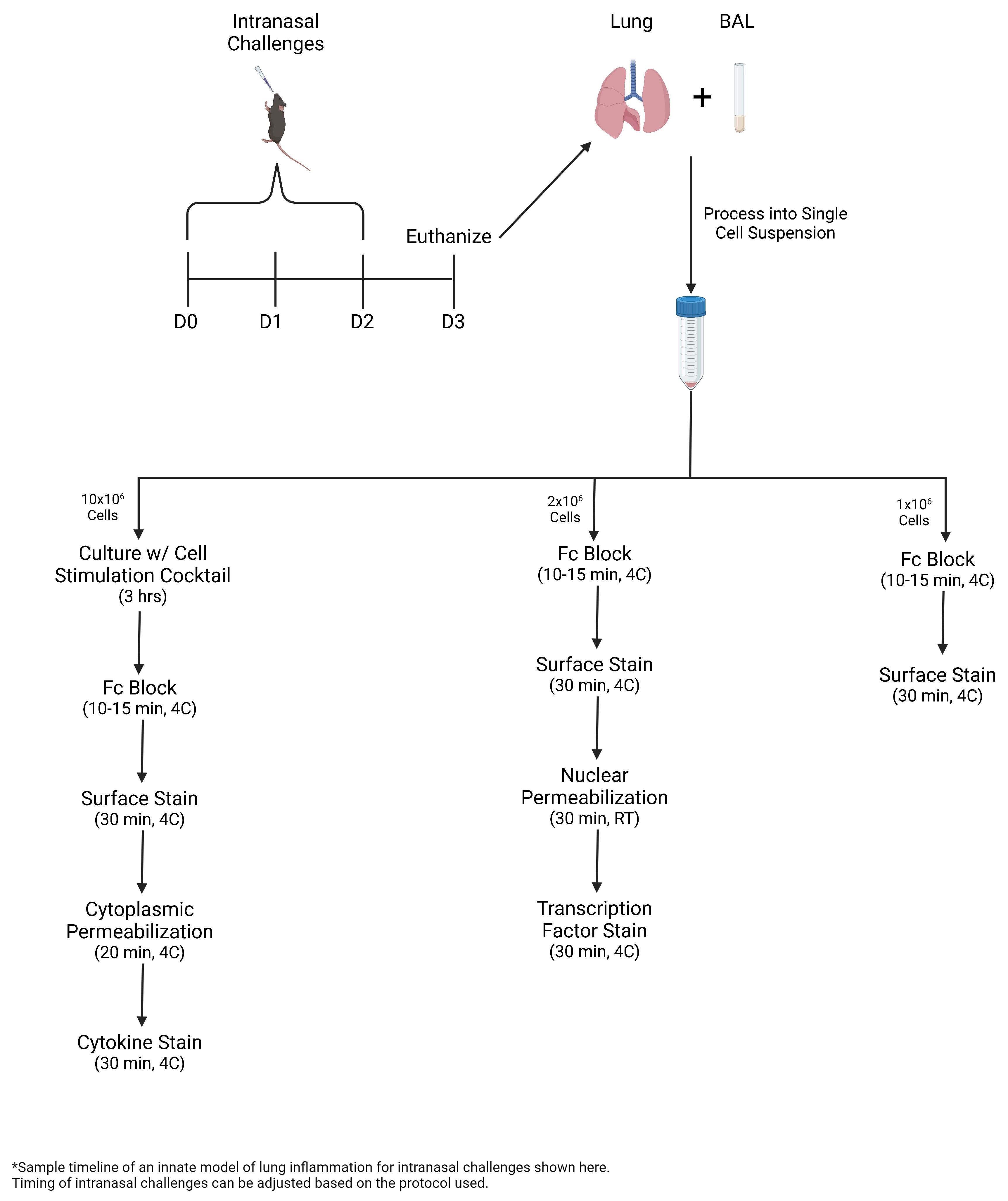
• In this protocol, ILC2s are expanded via intranasal challenges with Alternaria alternata, a fungal allergen, or recombinant IL-33.

• Bronchoalveolar lavage (BAL) and lung are collected and processed into single-cell suspension for multicolor flow cytometric analysis, including intracellular staining of transcription factors and cytokines.

• During lung inflammation, the percentage of ILC2s and eosinophils increases. ILC2s express greater levels of Gata-3 and Ki-67 and produce greater amounts of IL-5 and IL-13.


Graphical overview


0 Q&A 493 Views Feb 5, 2023

In addition to cytosolic protein synthesis, mitochondria also utilize another translation system that is tailored for mRNAs encoded in the mitochondrial genome. The importance of mitochondrial protein synthesis has been exemplified by the diverse diseases associated with in organello translation deficiencies. Various methods have been developed to monitor mitochondrial translation, such as the classic method of labeling newly synthesized proteins with radioisotopes and the more recent ribosome profiling. However, since these methods always assess the average cell population, measuring the mitochondrial translation capacity in individual cells has been challenging. To overcome this issue, we recently developed mito-fluorescent noncanonical amino acid tagging (FUNCAT) fluorescence-activated cell sorting (FACS), which labels nascent peptides generated by mitochondrial ribosomes with a methionine analog, L-homopropargylglycine (HPG), conjugates the peptides with fluorophores by an in situ click reaction, and detects the signal in individual cells by FACS equipment. With this methodology, the hidden heterogeneity of mitochondrial translation in cell populations can be addressed.

0 Q&A 570 Views Jan 20, 2023

Lysosomes play a central role in signaling, nutrient sensing, response to stress, and the degradation and recycling of cellular content. Defects in lysosomal digestive enzymes or structural components can impair lysosomal function with dire consequences to the cell, such as neurodegeneration. A number of methods exist to assess lysosomal stress in the model Drosophila, such as specific driver and reporter strains, transmission electron microscopy, and the investigation of gene expression. These methods, however, can be time consuming and, in some cases, costly. The procedure described here provides a quick, reliable, and low-cost approach to measure lysosomal stress in the Drosophila brain. Using fluorescence confocal microscopy and the LysoTracker staining, this protocol allows for the direct measurement of lysosome size and number. This method can be used to assess lysosomal stress under a number of different genetic and environmental scenarios in the Drosophila brain.

0 Q&A 857 Views Nov 20, 2022

Sphingolipids are important structural components of cellular membranes. They also function as prominent signaling molecules to control a variety of cellular events, such as cell growth, differentiation, and apoptosis. Impaired sphingolipid metabolism, particularly defects in sphingolipid degradation, has been associated with many human diseases. Fluorescence sphingolipid analogs have been widely used as efficient probes to study sphingolipid metabolism and intracellular trafficking in living mammalian cells. Compared with nitrobenzoxadiazole fluorophores (NBD FL), the boron dipyrromethene difluoride fluorophores (BODIPY FL) have much higher absorptivity and fluorescence quantum. These features allow more intensive labeling of cells for fluorescence microscopy imaging and flow cytometry analysis. Here, we describe a protocol employing BODIPY FL-labeled sphingolipid analogs to elucidate sphingolipid internalization, trafficking, and endocytosis in mouse embryonic stem cells.


Graphical abstract:


0 Q&A 1961 Views Jul 20, 2022

Employing a novel mouse model of immune related adverse events (irAEs) induced by combination of anti-PD1 and anti-CTLA-4 antibodies, we visualized immune infiltration into the liver, lung, pancreas, and colon. Here, we describe the avidin-biotin conjugate (ABC) method used to stain T cells (CD4 and CD8), B cells (CD19), macrophages (F4/80), and cells bound by the in vivo administered rat anti-mouse antibodies for chromogenic immunohistochemistry (IHC). Using a biotinylated goat anti-rat antibody, we detected the localization of cells bound to the in vivo antibodies for PD-1 and CTLA-4. IHC has advantages over other techniques, namely antibody availability, resistance to photobleaching, and greater sensitivity. Additionally, detection and localization of in vivo antibodies can be used in mice models to infer their therapeutic efficacy, stability, and function.


Graphical abstract:


0 Q&A 1523 Views Jun 5, 2022

Macrophages are important for host defense against intracellular pathogens like Salmonella and can be differentiated into two major subtypes. M1 macrophages, which are pro-inflammatory and induce antimicrobial immune effector mechanisms, including the expression of inducible nitric oxide synthase (iNOS), and M2 macrophages, which exert anti-inflammatory functions and express arginase 1 (ARG1). Through the process of phagocytosis, macrophages contain, engulf, and eliminate bacteria. Therefore, they are one of the first lines of defense against Salmonella. Infection with Salmonella leads to gastrointestinal disorders and systemic infection, termed typhoid fever. For further characterization of infection pathways, we established an in vitro model where macrophages are infected with the mouse Salmonella typhi correlate Salmonella enterica serovar Typhimurium (S.tm), which additionally expresses red fluorescent protein (RFP). This allows us to clearly characterize macrophages that phagocytosed the bacteria, using multi-color flow cytometry.


In this protocol, we focus on the in vitro characterization of pro- and anti-inflammatory macrophages displaying red fluorescent protein-expressing Salmonella enterica serovar Typhimurium, by multi-color flow cytometry.

0 Q&A 4901 Views Mar 5, 2022

The ability to stain lipid stores in vivo allows for the facile assessment of metabolic status in individuals of a population following genetic and environmental manipulation or pharmacological treatment. In the animal model Caenorhabditis elegans, lipids are stored in and mobilized from intracellular lipid droplets in the intestinal and hypodermal tissues. The abundance, size, and distribution of these lipids can be readily assessed by two staining methods for neutral lipids: Oil Red O (ORO) and Nile Red (NR). ORO and NR can be used to quantitatively measure lipid droplet abundance, while ORO can also define tissue distribution and lipid droplet size. C. elegans are a useful animal model in studying pathways relating to aging, fat storage, and metabolism, as their transparent nature allows for easy microscopic assessment of lipid droplets. This is done by fixation and permeabilization, staining with NR or ORO, image capture on a microscope, and computational identification and quantification of lipid droplets in individuals within a cohort. To ensure reproducibility in lipid measurements, we provide a detailed protocol to measure intracellular lipid dynamics in C. elegans.


Graphic abstract:


Flow chart depicting the preparation of C. elegans for fat staining protocols.


0 Q&A 2463 Views Oct 5, 2021

Although the advent of genetically-encoded fluorescent markers, such as the green fluorescent protein (GFP; Chalfie et al., 1994), has enabled convenient visualization of gene expression in vivo, this method is generally not effective for detecting post-translational modifications because they are not translated from DNA sequences. Genetically-encoded, fluorescently-tagged transgene products can also be misleading for observing expression patterns because transgenes may lack endogenous regulatory DNA elements needed for precise regulation of expression that could result in over or under expression. Fluorescently-tagged proteins created by CRISPR genome editing are less prone to defective expression patterns because the loci retain endogenous DNA elements that regulate their transcription (Nance and Frøkjær-Jensen, 2019). However, even CRISPR alleles encoding heritable fluorescently-tagged protein markers can result in defects in function or localization of the gene product if the fluorescent tag obstructs or otherwise interferes with important protein interaction domains or affects the protein structure.


Indirect immunofluorescence is a method for detecting endogenous gene expression or post-translational modifications without the need for transgenesis or genome editing. Here, we present a reliable protocol in which C. elegans nematodes are fixed, preserved, and permeabilized for staining with a primary antibody to bind proteins or post-translational modifications, which are then labeled with a secondary antibody conjugated to a fluorescent dye. Use of this method may be limited by the availability of (or ability to generate) a primary antibody that binds the epitope of interest in fixed animals. Thousands of animals are simultaneously subjected to a series of chemical treatments and washes in a single centrifuge tube, allowing large numbers of identically-treated stained animals to be examined. We have successfully used this protocol (O’Hagan et al., 2011 and 2017; Power et al., 2020) to preserve and detect post-translational modifications of tubulin in C. elegans ciliated sensory neurons and to detect non-modified endogenous protein (Topalidou and Chalfie, 2011).


We use cookies on this site to enhance your user experience. By using our website, you are agreeing to allow the storage of cookies on your computer.